. 2012;7(4):e34854.
doi: 10.1371/journal.pone.0034854. Epub 2012 Apr 6.

Genomic clustering and homology between HET-S and the NWD2 STAND protein in various fungal genomes

Affiliations

Genomic clustering and homology between HET-S and the NWD2 STAND protein in various fungal genomes

Asen Daskalov et al. PLoS One. 2012.

Abstract

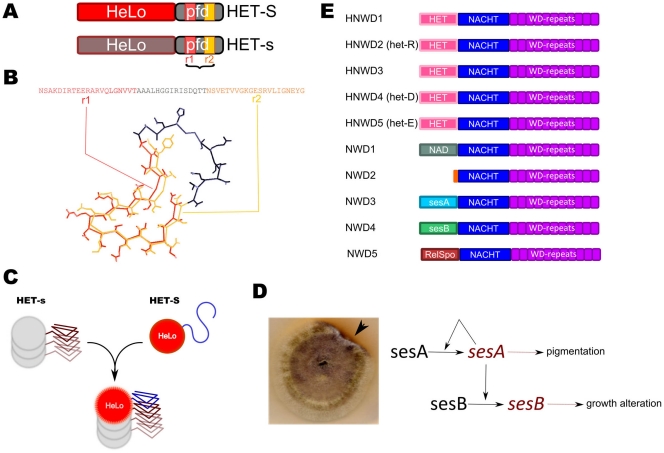
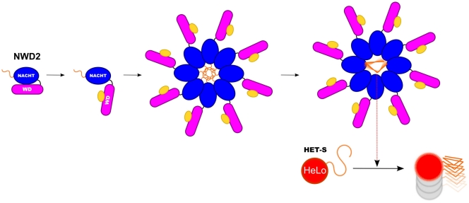
Background: Prions are infectious proteins propagating as self-perpetuating amyloid polymers. The [Het-s] prion of Podospora anserina is involved in a cell death process associated with non-self recognition. The prion forming domain (PFD) of HET-s adopts a β-solenoid amyloid structure characterized by the two fold repetition of an elementary triangular motif. [Het-s] induces cell death when interacting with HET-S, an allelic variant of HET-s. When templated by [Het-s], HET-S undergoes a trans-conformation, relocates to the cell membrane and induces toxicity.

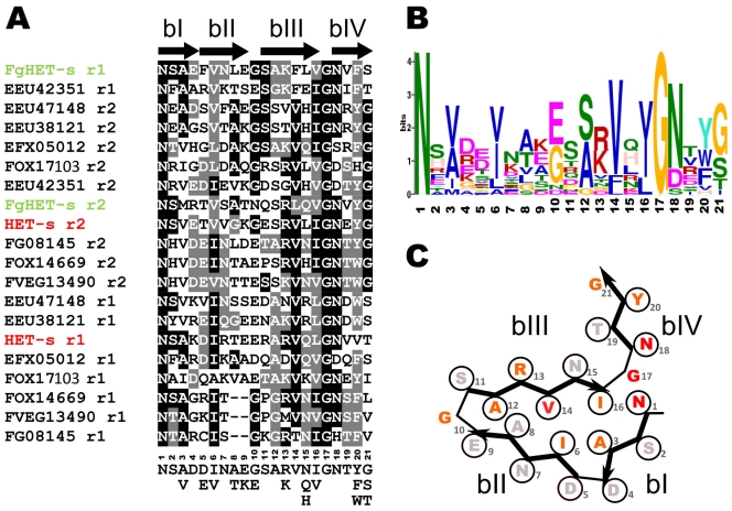
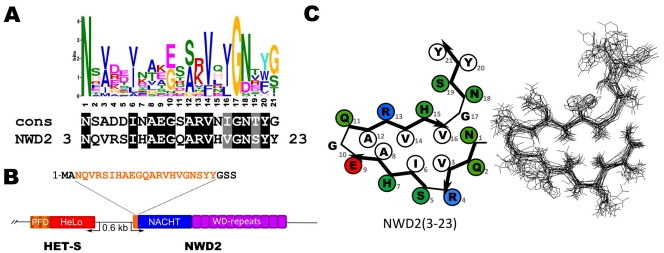
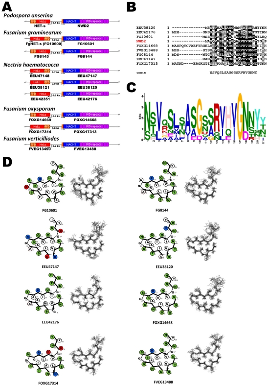
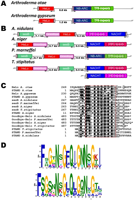
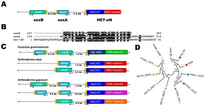
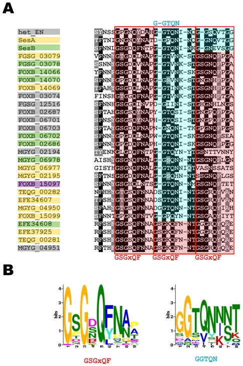
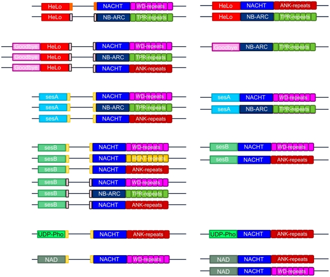
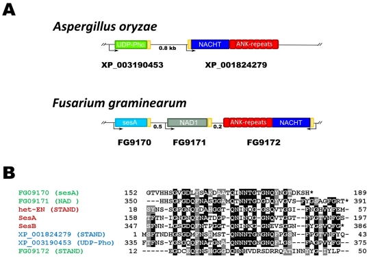
Methodology/principal findings: Here, comparing HET-s homologs from different species, we devise a consensus for the HET-s elementary triangular motif. We use this motif to screen genomic databases and find a match to the N-terminus of NWD2, a STAND protein, encoded by the gene immediately adjacent to het-S. STAND proteins are signal transducing ATPases which undergo ligand-induced oligomerisation. Homology modelling predicts that the NWD2 N-terminal region adopts a HET-s-like fold. We propose that upon NWD2 oligomerisation, these N-terminal extensions adopt the β-solenoid fold and template HET-S to adopt the amyloid fold and trigger toxicity. We extend this model to a putative prion, the σ infectious element in Nectria haematococca, because the s locus controlling propagation of σ also encodes a STAND protein and displays analogous features. Comparative genomic analyses indicate evolutionary conservation of these STAND/prion-like gene pairs, identify a number of novel prion candidates and define, in addition to the HET-s PFD motif, two distinct, novel putative PFD-like motifs.

Conclusions/significance: We suggest the existence, in the fungal kingdom, of a widespread and evolutionarily conserved mode of signal transduction based on the transmission of an amyloid-fold from a NOD-like STAND receptor protein to an effector protein.


Conflict of interest statement

Competing Interests: The authors have declared that no competing interests exist.

Figures

Figure 1
Figure 1. Introductory figure. A.
Diagram of the HET-s and HET-S domain organization with a N-terminal HeLo domain and a C-terminal PFD comprising two pseudo-repeats (r1 and r2). B. Structure of the HET-s PFD (pdb: 2RNM), only the two pseudo-repeats and the connecting loop are shown. C. Mechanism of HET-S/[Het-s] incompatibility, when the HET-S PFD region interacts with the prion form of HET-s, transconformation of the C-terminal region induces refolding and activation of the HeLo toxicity domain. D. The “secteur” phenotype caused by the σ infectious element in Nectria haematococca. The arrow points to a mycelia sector in which the σ infectious element is present and leads to an alteration of the growth margin and to the secretion of a red pigment. The diagram on the right is the working model proposed by Daboussi and co-workers to account for the role of the sesA and sesB proteins. The model proposes that sesA and sesB can exist in a normal and modified form (italicized) and that the modification of sesA is autocatalytic and can also induce modification of sesB. Modified sesA leads to the red pigmentation phenotype, modification of sesB to the growth alteration characteristic of the “secteur” (redrawn after Graziani et al. 2004). D. Domain organization of the members of NWD-gene family of P. anserina. All family members share a central NACHT nucleotide binding oligomerisation domain and a C-terminal WD repeat domain but differ by their N-terminal effector domains. This effector domain is a HET cell death inducing domain (PF06985) in NWD1 to NWD5 (see text for description of the other effector domains). NWD2 lacks a N-terminal effector domain.
Figure 2
Figure 2. Defining a consensus sequence for the elementary unit of HET-s β-solenoid fold.
A. alignment of the 21 amino acid repeat units of HET-s, FgHET-s and 9 other HET-s homologs from various fungal species, r1 and r2 designate the first and second repeat unit respectively. The arrows above the alignment represent the 4 β-strands defining the β-solenoid fold (bI to bIV). Below the alignment the corresponding consensus sequence is given. FOX1713 and FOX14669 are from Fusarium oxysporum, FG08145 is from Fusarium graminearum, FVEG13490 is from Fusarium verticillioides, EEU42351, EEU47148, EEU38121 are from Nectria haematococca, EFX055012 is from Grosmannia clavigera. B. Weighted consensus of the HET-s elementary repeat unit generated using the MEME algorithm. C. Fitting of the consensus sequence for the elementary HET-s repeat into the β-solenoid fold model. The colour coding reflects the weight of the residue in the MEME consensus (red, very strong conservation, informational content >2 bits; orange, strong conservation, >1 bit; grey, weak conservation, <0.5 bit).
Figure 3
Figure 3. The N-terminal end of the NWD2 protein matches the HET-s PFD consensus.
A. Comparison of the NWD2 sequence with the HET-s consensus sequence. B. Diagram showing the genome organization of het-s and nwd2 as two divergently transcribed adjacent genes. C. Fitting of the NWD2 (3-23) sequence into the β-solenoid fold model (left) and homology model of the NWD2(3-23) sequence (right). Polar residues are given in green, hydrophobic residues are given in white and positively and negatively charged residues in blue and red, respectively.
Figure 4
Figure 4. Evolutionary conservation of the nwd2/het-S genomic association and homology. A.
Genomic organization of the het-s homologs found in various filamentous fungi. Note that in all cases the het-S and nwd2 homologs are encoded by adjacent genes but gene orientation differs between species. B. Sequence alignment of the N-terminal end of the NWD2-homologs from various species. C. Weighted consensus based on the alignment in Figure 4B and generated with the MEME algorithm. D. Fitting of the N-terminal sequences of the NWD2-homologs into the β-solenoid fold model (left) and homology model of the same sequences (right), polar residues or given in green, hydrophobic residues are given in white and positively and negatively charged residues in blue and red, respectively.
Figure 5
Figure 5. Proposed model for the NWD2/HET-S interaction.
Based on the APAF-1 paradigm for STAND protein activation mechanism, it is proposed that NWD2 exists in an inactive closed state in the absence of its cognate ligand and undergoes a transition to an open state upon ligand binding to the WD-repeat region, which allows for oligomerization via the NOD domains. This oligomerization step is proposed to bring the N-terminal extensions of NWD2 molecules into close proximity and to allow their cooperative folding into the β-solenoid fold. In this model, this amyloid-like fold then serves as a template to nucleate HET-S transconformation and activation of the HeLo toxicity domain.
Figure 6
Figure 6. Distant HET-S homologs are also associated to genes encoding STAND proteins.
A. Genome organization of two distant HET-S homologs with a HeLo domain from Arthroderma species. The pink box depicts the region of homology between the C-terminal region of the HET-S homolog and the N-terminal region of the STAND protein. B. Genome organization HET-S homologs with a HeLo domain and Goodbye domain in various species. The pink box depicts the region of homology between the C-terminal region of the HET-s homolog and sesB homologs the N-terminal region of the STAND protein. C. Alignment of the HeLo, HeLo/Goodbye, sesB and STAND proteins depicted in A and B. D. Weighted consensus based on the alignment in Figure 6C and generated with the MEME algorithm.
Figure 7
Figure 7. The s locus controlling propagation of the σ infectious element of Nectria haematococca.
A. Genome organization of the sesA and sesB genes controlling formation of the σ element. Note that sesA and sesB are adjacent to the het-eN gene encoding a STAND protein. B. Sequence alignment of the C-terminal regions of sesA and sesB and the N-terminal region of HET-eN. C. Genome organization of the sesA and sesB loci in different fungal species. Note that sesA and/or sesB homologs are found adjacent to a gene encoding a STAND protein in various fungal species. Gene order varies from species to species. D. Phylogenetic tree of the sesA (triangle), sesB (square) and STAND (circle) extension from F. graminearum (blue), F. oxysporum (yellow), N. haematococca (red) and Arthroderma gypsem (green). Note that extension group by gene cluster rather than by orthologous groups. The F. oxysporum STAND homolog was omitted from the analysis because gene annotation is ambiguous. The evolutionary tree was constructed using the Neighbor-Joining method. The optimal tree is shown. The analysis involved 11 amino acid sequences. All positions containing gaps and missing data were eliminated. There were a total of 32 positions in the final dataset. Evolutionary analyses were conducted in MEGA5. Bootstrap values are given for each node.
Figure 8
Figure 8. Alignment of sesA, sesB and het-eN homologs from various species and definition of the consensus σ-motif.
A. PFD-like extension of STAND, sesA and sesB homologs (boxed in grey, yellow and green respectively; the sequence with a purple box is a NAD domain protein) from various species have been aligned using ClustalW. The A (GSGxQF) and B (GGTQN) motifs have been boxed in red and blue respectively. B. Weighted consensus of the A and B motifs generated with the MEME algorithm.
Figure 9
Figure 9. Alternate gene architectures of effector domain/STAND associations.
The figure gives a side-by-side comparison of the alternate gene architecture of fungal STAND prioteins. The genes encoding the effector domain described here can be found adjacent to genes encoding STAND proteins and alternatively associated directly at the N-terminus of the STAND protein. The type of superstructure forming repeats is also variable. The HET-s motif, PP-motif and σ-motif PFD or PFD-like extension are represented as orange, pink and yellow boxes respectively.
Figure 10
Figure 10. Identification of additional prion candidates. A.
In A. oryzae, a gene encoding a protein with a UDP-Phosphorylase domain lies adjacent to a gene encoding a STAND protein and there is a region of homology between the C-terminus of that protein and the N-terminus of the STAND protein. In F. graminearum, a protein with a domain termed NAD lies adjacent to a gene encoding a STAND protein and adjacent to a gene encoding a sesA homolog. There is a region of homology between the C-terminus of that protein and the N-terminus of the STAND protein and the C-terminus of the sesA homolog. B. Alignment of the regions of homology depicted as yellow boxes in A with the sesA and sesB and het-eN proteins from Nectria haematococca.

Similar articles

Cited by

References

    1. Aguzzi A, Baumann F, Bremer J. The prion’s elusive reason for being. Annu Rev Neurosci. 2008;31:439–477. - PubMed
    1. Prusiner SB. Prions. Proc Natl Acad Sci U S A. 1998;95:13363–13383. - PMC - PubMed
    1. Wickner RB. [URE3] as an altered URE2 protein: evidence for a prion analog in Saccharomyces cerevisiae [see comments]. Science. 1994;264:566–569. - PubMed
    1. Tuite MF, Serio TR. The prion hypothesis: from biological anomaly to basic regulatory mechanism. Nat Rev Mol Cell Biol. 2010;11:823–833. - PMC - PubMed
    1. Crow ET, Li L. Newly identified prions in budding yeast, and their possible functions. Semin Cell Dev Biol. 2011;22:452–459. - PMC - PubMed

MeSH terms

LinkOut - more resources