. 2012 May 7;209(5):947-54.
doi: 10.1084/jem.20111760. Epub 2012 Apr 9.

Proinflammatory cytokine signaling required for the generation of natural killer cell memory

Affiliations

Proinflammatory cytokine signaling required for the generation of natural killer cell memory

Joseph C Sun et al. J Exp Med. .

Abstract

Although natural killer (NK) cells are classified as innate immune cells, recent studies demonstrate that NK cells can become long-lived memory cells and contribute to secondary immune responses. The precise signals that promote generation of long-lived memory NK cells are unknown. Using cytokine receptor-deficient mice, we show that interleukin-12 (IL-12) is indispensible for mouse cytomegalovirus (MCMV)-specific NK cell expansion and generation of memory NK cells. In contrast to wild-type NK cells that proliferated robustly and resided in lymphoid and nonlymphoid tissues for months after MCMV infection, IL-12 receptor-deficient NK cells failed to expand and were unable to mediate protection after MCMV challenge. We further demonstrate that a STAT4-dependent IFN-γ-independent mechanism contributes toward the generation of memory NK cells during MCMV infection. Understanding the full contribution of inflammatory cytokine signaling to the NK cell response against viral infection will be of interest for the development of vaccines and therapeutics.


Figures
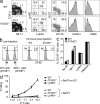
Figure 1.
Figure 1.
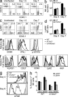
NK cells from IL-12R–deficient mice are phenotypically and functionally similar to NK cells from WT mice. (a) Percentages of TCR-β NK1.1+ NK, TCR-β+ NK1.1+ NKT, and TCR-β+ NK1.1 T cell populations are shown for uninfected WT and Il12rb2−/− mice (left plot). The right plots are gated on TCR-β NK1.1+ cells and analyzed for expression of Ly49D, Ly49H, CD27, CD11b, KLRG1, and CD69. (b) WT and B2m−/− splenocytes were labeled with low or high concentrations of CFSE, respectively, and co-injected into WT or Il12rb2−/− mice. Transferred cells were analyzed in spleen of recipient mice 72 h after injection. (c) NK cells from WT or Il12rb2−/− mice were stimulated with plate-bound antibodies against NK1.1 and Ly49H, or with PMA and ionomycin. Uncoated wells (containing no Abs) served as negative control and background staining. Percentages of TCR-β NK1.1+ cells expressing CD107 (LAMP-1) are shown for each condition. Error bars show SEM (n = 2–3 for each condition). (d) Varying numbers of NK cells from WT, Il12rb2−/−, or Ly49h−/− mice were incubated with Ba/F3 and Ba/F3-m157 target cells labeled with 51Cr. Percentage of killing was calculated based on release of 51Cr into supernatant by lysed target cells. All data are representative of at least three independent experiments.
Figure 2.
Figure 2.
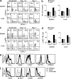
IL-12R–deficient NK cells exhibit defective proliferation during MCMV infection. (a) Mixed bone marrow chimeric mice were infected with MCMV and percentages of splenic WT (CD45.1+) and Il12rb2−/− (CD45.2+) NK cells are shown (gated on CD3 NK1.1+) in uninfected mice and various time points PI. (b) The absolute numbers of splenic WT and Il12rb2−/− NK cells on day 0 and 7 PI are graphed. (c) Percentages of Ly49H+ cells within the WT and Il12rb2−/− NK cell population (TCR-β NK1.1+) are shown for uninfected mice and various time points PI. (d) The absolute numbers of WT and Il12rb2−/− Ly49H+ NK cells on day 0 and 7 PI are graphed. (e) Expression of CD69, production of IFN-γ, and phosphorylation of STAT4 are shown for WT and Il12rb2−/− NK cells (compared with uninfected mice) at day 1.5 PI. (f) Expression of KLRG1, CD27, CD90 (Thy-1), Ly6C, and Ki67 on WT and Il12rb2−/− Ly49H+ NK cells (compared with uninfected mice) at day 7 PI. (g) WT or Il12rb2−/− NK cells (CD45.2+) were labeled with 5 µM CFSE and transferred into Ly49H-deficient hosts (CD45.1+). After MCMV infection, dividing NK cells were analyzed at days 4 and 6 PI (compared with uninfected control mice). (h) Adoptively transferred WT and Il12rb2−/− NK cells were stained for Annexin V at days 0 and 4 PI. Percentages of Ly49H+ and Ly49H NK cells positive for Annexin V are shown. Error bars for all graphs show SEM (n = 3–5 for each time point) and all data are representative of five independent experiments.
Figure 3.
Figure 3.
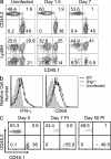
NK cells from Il12rb2−/− mice fail to become long-lived memory cells and mediate protection after MCMV infection. (a and b) A total of 105 WT or Il12rb2−/− Ly49H+ NK cells (both CD45.2+) were transferred into DAP12-deficient mice (CD45.1+) and infected with MCMV. (a) Plots are gated on total NK cells and percentages of adoptively transferred CD45.2+ Ly49H+ NK cells are shown for each time point PI. (b) Absolute numbers of adoptively transferred WT and Il12rb2−/− Ly49H+ NK cells in the spleen of recipient mice are shown. Error bars show SEM (n = 3–5). ND, not detectable (or below the limits of detection). (c) WT (CD45.1+) and Il12rb2−/− (CD45.2+) Ly49H+ NK cells were co-adoptively transferred into Ly49H-deficient mice and infected with MCMV. Plots are gated on transferred NK cells and percentages of WT and Il12rb2−/− NK cells are shown for spleen and liver at days 0, 7, and 50 PI. All data are representative of five experiments with three to five mice per time point. (d) WT (CD45.1+) and Il12rb2−/− (CD45.2+) mice were infected with MCMV and splenic Ly49H+ NK cells on day 7 PI were purified, mixed at a 1:1 ratio, and co-transferred into Ly49H-deficient mice. Plots are gated on transferred NK cells and percentages of WT and Il12rb2−/− NK cells are shown for at various time points PI and post transfer (PT). All data are representative of three experiments with two to four mice per time point. The graph shows the percentage of adoptively transferred WT and Il12rb2−/− Ly49H+ NK cells within the total NK cell population, and error bars show SEM (n = 3–4). (e) DAP12-deficient neonatal mice received 105 WT or Il12rb2−/− NK cells (or PBS as control) followed by MCMV infection. The graph shows the percentage of surviving mice for each group, and data were pooled from three experiments.
Figure 4.
Figure 4.
WT NK cells outcompete Stat4−/− NK cells during MCMV infection. (a) Mixed bone marrow chimeric mice were infected with MCMV and percentages of WT (CD45.1+) and Stat4−/− (CD45.2+) NK cells in spleen and liver are shown for uninfected mice and various time points PI. (b) The graph shows absolute numbers of WT and Stat4−/− NK cells in spleen and liver on day 0 and 7 PI. Error bars show SEM (n = 3–5). (c) Percentages of Ly49H+ cells within the WT and Stat4−/− NK cell population in spleen and liver are shown for uninfected mice and various time points PI. (d) The graph shows absolute numbers of WT and Stat4−/− Ly49H+ NK cells in spleen and liver on day 0 and 7 PI. Error bars show SEM (n = 3–5). (e) Expression of CD69, CD27, CD11b, and CD90, and production of IFN-γ are shown for WT and Stat4−/− NK cells (compared with uninfected mice) at day 1.5 PI. (f) Expression of KLRG1, CD90, Ly49C/I, and CD27 are shown for WT and Stat4−/− Ly49H+ NK cells (compared with uninfected mice) at day 7 PI. All data are representative of three experiments with three to five mice per time point.
Figure 5.
Figure 5.
Defective memory NK cell generation in the absence of STAT4. (a) A total of 105 WT (CD45.1+) and Stat4−/− (CD45.2+) Ly49H+ NK cells were co-transferred into Ly49H-deficient mice (CD45.2+) and infected with MCMV. Plots are gated on total NK cells and percentages of adoptively transferred Ly49H+ NK cells (WT in top left quadrant and Stat4−/− in top right quadrant) are shown for each time point PI. (b) Percentage of adoptively transferred WT and Stat4−/− Ly49H+ NK cells within the total NK cell population are shown. Error bars show SEM (n = 3). All data are representative of three experiments with three to four mice per time point.
Figure 6.
Figure 6.
WT and Ifngr−/− NK cells expand equally and generate memory cells after MCMV infection. (a) Mixed bone marrow chimeric mice consisting of a 1:1 mixture of WT (CD45.1+) and Ifngr−/− (CD45.2+) hematopoietic cells were infected with MCMV. Top row: percentages of total WT and Ifngr−/− NK cells (gated on CD3 NK1.1+) are shown for uninfected mice and at day 1.5 and 7 PI. Bottom row: percentages of Ly49H+ cells within the WT and Ifngr−/− NK cell populations are shown. (b) Production of IFN-γ and expression of CD69 are shown for WT and Ifngr−/− NK cells in chimeric mice (compared with uninfected chimeras) at day 1.5 PI. (c) WT (CD45.1+) and Ifngr−/− (CD45.2+) Ly49H+ NK cells were co-adoptively transferred into Ly49H-deficient mice and infected with MCMV. Plots are gated on transferred NK cells and percentages of WT and Ifngr−/− NK cells are shown for day 0, 7, and 50 PI. All data are representative of three experiments with three to five mice per time point.

Similar articles

Cited by

References

    1. Bakker A.B., Hoek R.M., Cerwenka A., Blom B., Lucian L., McNeil T., Murray R., Phillips L.H., Sedgwick J.D., Lanier L.L. 2000. DAP12-deficient mice fail to develop autoimmunity due to impaired antigen priming. Immunity. 13:345–353 10.1016/S1074-7613(00)00034-0 - DOI - PubMed
    1. Berg R.E., Forman J. 2006. The role of CD8 T cells in innate immunity and in antigen non-specific protection. Curr. Opin. Immunol. 18:338–343 10.1016/j.coi.2006.03.010 - DOI - PubMed
    1. Björkström N.K., Lindgren T., Stoltz M., Fauriat C., Braun M., Evander M., Michaëlsson J., Malmberg K.J., Klingström J., Ahlm C., Ljunggren H.G. 2011. Rapid expansion and long-term persistence of elevated NK cell numbers in humans infected with hantavirus. J. Exp. Med. 208:13–21 10.1084/jem.20100762 - DOI - PMC - PubMed
    1. Cooper M.A., Elliott J.M., Keyel P.A., Yang L., Carrero J.A., Yokoyama W.M. 2009. Cytokine-induced memory-like natural killer cells. Proc. Natl. Acad. Sci. USA. 106:1915–1919 10.1073/pnas.0813192106 - DOI - PMC - PubMed
    1. Cousens L.P., Peterson R., Hsu S., Dorner A., Altman J.D., Ahmed R., Biron C.A. 1999. Two roads diverged: interferon α/β– and interleukin 12–mediated pathways in promoting T cell interferon γ responses during viral infection. J. Exp. Med. 189:1315–1328 10.1084/jem.189.8.1315 - DOI - PMC - PubMed

Publication types