. 2012 May 25;287(22):18130-41.
doi: 10.1074/jbc.M111.311852. Epub 2012 Apr 9.

Tudor staphylococcal nuclease (Tudor-SN) participates in small ribonucleoprotein (snRNP) assembly via interacting with symmetrically dimethylated Sm proteins

Affiliations

Tudor staphylococcal nuclease (Tudor-SN) participates in small ribonucleoprotein (snRNP) assembly via interacting with symmetrically dimethylated Sm proteins

Xingjie Gao et al. J Biol Chem. .

Abstract

Human Tudor staphylococcal nuclease (Tudor-SN) is composed of four tandem repeats of staphylococcal nuclease (SN)-like domains, followed by a tudor and SN-like domain (TSN) consisting of a central tudor flanked by two partial SN-like sequences. The crystal structure of the tudor domain displays a conserved aromatic cage, which is predicted to hook methyl groups. Here, we demonstrated that the TSN domain of Tudor-SN binds to symmetrically dimethylarginine (sDMA)-modified SmB/B' and SmD1/D3 core proteins of the spliceosome. We demonstrated that this interaction ability is reduced by the methyltransferase inhibitor 5-deoxy-5-(methylthio)adenosine. Mutagenesis experiments indicated that the conserved amino acids (Phe-715, Tyr-721, Tyr-738, and Tyr-741) in the methyl-binding cage of the TSN domain are required for Tudor-SN-SmB interaction. Furthermore, depletion of Tudor-SN affects the association of Sm protein with snRNAs and, as a result, inhibits the assembly of uridine-rich small ribonucleoprotein mediated by the Sm core complex in vivo. Our results reveal the molecular basis for the involvement of Tudor-SN in regulating small nuclear ribonucleoprotein biogenesis, which provides novel insight related to the biological activity of Tudor-SN.


Figures

FIGURE 1.
FIGURE 1.
Analysis of Tudor-SN and U snRNA profiles by glycerol gradient sedimentation. A, U snRNP complexes from HeLa cells were analyzed by glycerol gradient sedimentation. The total lysates of HeLa cells were sedimented through 10–30% glycerol gradients. The distribution of Tudor-SN, Prp8, and U1–70 in each fraction was analyzed by SDS-PAGE and immunoblotted with specific antibodies as indicated. Fraction (Fr.) 1 is at the top of the gradient. The extracted snRNAs from fractions were analyzed by Northern blotting with U1, U2, U4, U5, and U6 snRNA probes and visualized by autoradiography. Positions of snRNAs are indicated on the left and right. B, Tudor-SN and SmB associate with U1, U2, U4, U5, and U6 snRNA in vivo. RNA-binding protein immunoprecipitation assay was performed with total cell lysates of HeLa cells with formaldehyde and glycine treatment. The TCLs were incubated with bead-bound anti-Tudor-SN, anti-SmB, anti-TMG-cap, and rabbit IgG antibody as indicated. The co-precipitated snRNAs was extracted and reverse-transcribed to cDNA with random hexamer primers. The U1, U2, U4, U5, and U6 snRNAs were generated by PCR analysis. C, U1, U2, U4, U5, and U6 snRNAs in the TCLs of HeLa cells for RIP assays were extracted and reverse-transcribed to cDNA with random hexamer primers and then were generated by PCR assay.
FIGURE 2.
FIGURE 2.
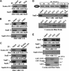
Tudor-SN interacts with SmB/B′ and SmD1/D3 via the tudor domain. A, ectopically expressed Tudor-SN interacts with SmB. COS-7 cells were transfected with the pSG5-Tudor-SN-FLAG plasmid. The total cell lysates of the transfected COS-7 cells were used in immunoprecipitation (IP) with anti-FLAG or anti-His-agarose beads as control. The co-precipitated proteins were subjected to SDS-PAGE and blotted with anti-FLAG (upper panel) or anti-SmB (lower panel) antibody. Approximately 10% of the TCLs were included as input. B and C, endogenous Tudor-SN or SMN interacts with SmB/B′ and SmD1/D3. TCLs of HeLa cells were immunoprecipitated with rabbit anti-Tudor-SN, anti-SMN, or anti-IgG as control. Bound proteins were subjected to SDS-PAGE and blotted with mouse anti-Tudor-SN, anti-SMN, anti-SmB, or Y12 antibody. Approximately 10% of the TCLs were included as input. D, mapping the interaction domain of Tudor-SN with SmB. TCLs of HeLa cells were incubated with equal amounts of GST and GST fusion proteins containing the isolated SN domains or the TSN domain as indicated in the upper panel. Bound proteins were separated by SDS-PAGE and immunoblotted with anti-SmB antibody (middle panel). The expression levels of the GST fusion proteins were visualized by Coomassie Blue staining (lower panel). Approximately 10% of the TCLs were included as input. E, GST pulldown assay was also performed with GST fusion proteins containing TSN or tudor domain alone. Bound proteins were detected with anti-SmB (upper panel) or Y12 antibody (2 middle panels), and the GST fusion proteins were visualized by Coomassie Blue staining (lower panel).
FIGURE 3.
FIGURE 3.
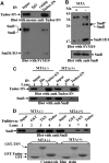
Tudor-SN interacts with SmB/B′ and SmD1/D3 in RNA-free lysates. A, TCLs of HeLa cells were incubated in the presence (+) or absence (−) of RNase mixture (120 μg of RNase A; 300 units of RNase Ti; 50 units of RNase H; 300 units of RNase I) or 1% BSA (Sigma) for 30 min at 37 °C. The RNAs were extracted and separated by EB-agarose gel electrophoresis. The 28 S, 18 S, and 5 S rRNAs are shown. rRNAs (5S and 5.8S), snRNAs (U1, U2, and U5), and GAPDH mRNAs were also detected using normal PCR assay. The primer sequences of rRNAs and GAPDH are shown in the supplemental Table S2. B, Tudor-SN interacts with SmB/B′ and SmD1/D3 in an RNase-resistant manner in vivo. The RNase treated or nontreated TCLs were immunoprecipitated with rabbit anti-Tudor-SN or anti-IgG as control. Bound proteins were subjected to SDS-PAGE and blotted with mouse anti-Tudor-SN (upper panel), anti-SmB (middle panel), or Y12 antibody (2 lower panels). 10% of the TCLs was included as input. C, TSN domain binds endogenous SmB/B′ and SmD1/D3 in an RNase-resistant manner. TCLs of HeLa cells (±RNase) were incubated with equal amounts of GST or GST-TSN fusion protein. Bound proteins were resolved by SDS-PAGE and blotted with anti-SmB (upper panel) or Y12 antibody (2 middle panels). The expression levels of the GST fusion proteins were visualized by Coomassie Blue staining (lower panel). Approximately 10% of the TCLs were also included as a control. IP, immunoprecipitated.
FIGURE 4.
FIGURE 4.
Tudor-SN interacts with sDMA-modified SmB/B′ and SmD1/D3 proteins. A, endogenous Tudor-SN interacts with sDMA-SmB/B′ and SmD1/D3. TCLs of HeLa cells were immunoprecipitated (IP) with rabbit anti-Tudor-SN or anti-IgG as control. Bound proteins were subjected to SDS-PAGE and blotted with mouse anti-Tudor-SN (upper panel) or anti-sDMA antibody (SMY10) (lower panel). Approximately 10% of the TCLs were included as input. B, total cell lysates were prepared from HeLa cells cultured in the presence (+) or absence (−) of the methylation inhibitor MTA (250 μm) for 20 h and then immunoblotted with SMY10 antibody. The filter was stripped and re-blotted with anti-SmB to assess the reduction in methylation level. C, same HeLa TCLs (+/− MTA) were immunoprecipitated with anti-Tudor-SN antibody or anti-His- agarose beads as negative control. The precipitated proteins were separated by SDS-PAGE and blotted with either anti-Tudor-SN (upper panel) or anti-SmB (lower panel) antibody. D, total cell lysates from +/− MTA-treated HeLa cells were incubated with GST, GST-TSN, or GST-Tudor fusion proteins. The amount of bound SmB protein was visualized by immunoblotting with anti-SmB antibody (upper panel). The GST fusion proteins were visualized by Coomassie Blue staining (lower panel).10% of the TCLs was included as input.
FIGURE 5.
FIGURE 5.
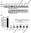
TSN mutants affect the SmB binding. A, mutations of TSN affect the binding ability to SmB. TCLs of HeLa cells were incubated with GST and GST fusion proteins containing wild-type (WT) TSN or different mutants of TSN as indicated. The precipitated proteins were resolved by SDS-PAGE and immunoblotted with anti-SmB antibody (upper panel). The expression levels of GST and different GST fusion proteins were determined by Coomassie Blue staining (lower panel). 10% of the TCLs were included as input. B, band density was digitized with TotalLab software and then analysis of variance was used for statistical analysis. Significant differences were indicated as follows: #, p < 0.001 versus control (n = 3). The level of bound SmB proteins was normalized against the corresponding GST fusion proteins.
FIGURE 6.
FIGURE 6.
TSN proteins with mutations have no ability to promote the kinetics of in vitro spliceosome formation. A, in vitro splicing reactions were performed at different time points with the addition of indicated purified proteins followed by native gel analysis of spliceosome complex formation. The gel was visualized by autoradiography. The bands corresponding to the H, A, B, and C complexes as well as the gel origin are indicated on the right. B, quantitative analysis of spliceosome formation. The vertical axis of coordinate represents the density value of complex A and B. The horizontal axis of coordinate represents the different time points (0, 5, 10, 30, and 60 min) for the spliceosome assembly. The intensities of complex A and B were determined by PhosphorImager and normalized by setting the highest value of complex A to 1.
FIGURE 7.
FIGURE 7.
Knockdown of TSN reduces the recruitment of Sm to the U snRNP in vivo. A, HeLa cells were transfected with the Tudor-SN siRNA or scramble siRNA (control). The total cell lysates of different samples were blotted with anti-Tudor-SN (upper panel), anti-SmB (1st middle panel), Y12 (2nd middle panel), or anti-β-actin antibody (lower panel) to detect the protein level of corresponding proteins. B, band density was digitized with TotalLab software and then Independent-Samples Student's t Test was performed. Significant difference was indicated: #, p < 0.01 versus control (n = 5); *, p > 0.05 (n = 5). The expression level of targeting protein (Tudor-SN, SmB, and SmD1/D3) in HeLa cells was normalized against the β-actin. C, total lysates of different samples were immunoprecipitated (IP) with anti-TMG-cap antibody or anti-IgG coupled with protein G Dynabeads as negative control. The co-precipitated proteins were subjected to SDS-PAGE and blotted with anti-SmB (upper panel) or Y12 antibody (lower panel). D, amount of SmB and SmD1/D3 proteins immunoprecipitated with anti-TMG-cap antibody was normalized against the total input Sm proteins. Band density was measured and then Independent-Samples Student's t Test was used for statistical analysis. Significant difference was indicated: &, p < 0.05 versus control group (n = 3). E, RNAs in the total cell lysates of different samples were isolated and reverse-transcribed to cDNA with random hexamer primers, and we then performed the quantitative real time PCR assay to detect the relative fold changes of U1, U2, U4, U5, and U6 snRNA. The fold changes were analyzed with Independent-Samples Student's t test. *, p > 0.05 (n = 3). F, total lysates of different samples were immunoprecipitated with Y12 Dynabeads or anti-IgG Dynabeads as control. The bound RNAs were extracted and reverse-transcribed to cDNA, and we then performed the quantitative real time PCR assay to detect the relative fold changes of precipitated U1, U2, U4, U5, and U6 snRNA. The RIP fold changes were analyzed with Independent-Samples Student's t test. Significant difference was indicated as follows: #, p < 0.01; &, p < 0.05, versus control (n = 3).
FIGURE 8.
FIGURE 8.
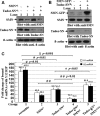
Ectopically expressed SMN restored the reduced U snRNP assembly caused by depletion of endogenous Tudor-SN. A and B, HeLa cells were transfected with the Tudor-SN siRNA and SMN siRNA (A) or mammalian expression plasmids containing full-length SMN tagged with GFP epitope (SMN-GFP) as indicated (B). The total cell lysates of different samples were blotted with anti-SMN (upper panel), anti-Tudor-SN (middle panel), or anti-β-actin antibody (lower panel) to detect the protein level of corresponding proteins. C, total lysates of different samples were immunoprecipitated with Y12 Dynabeads or anti-IgG Dynabeads as control. The bound RNAs were isolated and reverse-transcribed to cDNA with random hexamer primers, and we then performed the quantitative real time PCR assay to detect the relative fold changes of precipitated U1 and U2 snRNA. The RIP fold changes were analyzed with analysis of variance. Significant difference was indicated as follows: &, <0.001; #, p < 0.01 versus nontreatment group (n = 3), *, p < 0.05; # #, p < 0.01 versus Tudor-SNsi or SMNsi group (n = 3).
FIGURE 9.
FIGURE 9.
Model demonstrates two roles of Tudor-SN in regulating pre-mRNA splicing, particularly the formation of spliceosomal complex A and the transition from complex A to B. Tudor-SN preferentially interacts with symmetrically dimethylated SmB/B′, SmD1, and SmD3 proteins, which facilitates the recruitment of Sm proteins to the U snRNAs and the formation of spliceosomal complex A. However, Tudor-SN takes part in U5 snRNP via the association with Prp8 and U5–116, which may facilitate the transition from complex A to complex B.

Similar articles

Cited by

References

    1. Broadhurst M. K., Lee R. S., Hawkins S., Wheeler T. T. (2005) The p100 EBNA-2 co-activator. A highly conserved protein found in a range of exocrine and endocrine cells and tissues in cattle. Biochim. Biophys. Acta 1681, 126–133 - PubMed
    1. Rodríguez L., Ochoa B., Martínez M. J. (2007) NF-Y and Sp1 are involved in transcriptional regulation of rat SND p102 gene. Biochem. Biophys. Res. Commun. 356, 226–232 - PubMed
    1. Zhao C. T., Shi K. H., Su Y., Liang L. Y., Yan Y., Postlethwait J., Meng A.M. (2003) Two variants of zebrafish p100 are expressed during embryogenesis and regulated by Nodal signaling. FEBS Lett. 543, 190–195 - PubMed
    1. Howard-Till R. A., Yao M. C. (2007) Tudor nuclease genes and programmed DNA rearrangements in Tetrahymena thermophila. Eukaryot. Cell 6, 1795–1804 - PMC - PubMed
    1. Sami-Subbu R., Choi S. B., Wu Y., Wang C., Okita T. W. (2001) Identification of a cytoskeleton-associated 120-kDa RNA-binding protein in developing rice seeds. Plant Mol. Biol. 46, 79–88 - PubMed

Publication types

LinkOut - more resources