. 2012;7(4):e34623.
doi: 10.1371/journal.pone.0034623. Epub 2012 Apr 2.

Interaction of chandipura virus N and P proteins: identification of two mutually exclusive domains of N involved in interaction with P

Affiliations

Interaction of chandipura virus N and P proteins: identification of two mutually exclusive domains of N involved in interaction with P

Arindam Mondal et al. PLoS One. 2012.

Abstract

The nucleocapsid protein (N) and the phosphoprotein (P) of nonsegmented negative-strand (NNS) RNA viruses interact with each other to accomplish two crucial events necessary for the viral replication cycle. First, the P protein binds to the aggregation prone nascent N molecules maintaining them in a soluble monomeric (N(0)) form (N(0)-P complex). It is this form that is competent for specific encapsidation of the viral genome. Second, the P protein binds to oligomeric N in the nucleoprotein complex (N-RNA-P complex), and thereby facilitates the recruitment of the viral polymerase (L) onto its template. All previous attempts to study these complexes relied on co-expression of the two proteins in diverse systems. In this study, we have characterised these different modes of N-P interaction in detail and for the first time have been able to reconstitute these complexes individually in vitro in the chandipura virus (CHPV), a human pathogenic NNS RNA virus. Using a battery of truncated mutants of the N protein, we have been able to identify two mutually exclusive domains of N involved in differential interaction with the P protein. An unique N-terminal binding site, comprising of amino acids (aa) 1-180 form the N(0)-P interacting region, whereas, C-terminal residues spanning aa 320-390 is instrumental in N-RNA-P interactions. Significantly, the ex-vivo data also supports these observations. Based on these results, we suggest that the P protein acts as N-specific chaperone and thereby partially masking the N-N self-association region, which leads to the specific recognition of viral genome RNA by N(0).


Conflict of interest statement

Competing Interests: The authors have declared that no competing interests exist.

Figures

Figure 1
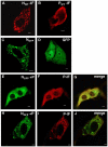
Figure 1. CHPV N and P proteins interact differentially in transfected cells depending on their stoichiometric availabilities.
(A) Vero-76 cells were transfected with 2 µg pCDNA 3.1 (+) N and immunofluorescence performed with N-Ab, 24 hours post transfection. N exhibits a punctate distribution in the cytoplasm. (B) Immunofluorescence of Vero-76 cells transfected with pCDNA 3.1 (+) P, with P-Ab. P exhibits a smooth distribution in the cytoplasm. (C) GFP fluorescence of Vero-76 cells transfected with pEGFP-C1 N. GFP-tagged N maintains its punctuated distribution. (D) GFP fluorescence of Vero-76 cells transfected with pEGFP-C1 vector alone. GFP alone shows characteristic smooth fluorescence throughout the cell. (E to G) Vero-76 cells co-transfected with pEGFP-C1 N and pCDNA 3.1 (+) P in a 1∶1 ratio. P was detected by immunofluorescence (F). Colocalization of GFP-N with P is shown in the merged image (G). Co-expression with P redistributes the otherwise punctuated N into a more homogenous fluorescence. (H to J) Co-transfection in a 1∶0.5 ratio. The lower abundance of P is insufficient to homogenise the punctuated distribution of N (H). Immunofluorescence against P reveals colocalization of P with oligomeric forms of N (I and J). All data were captured on a laser scanning confocal microscope (Carl Zeiss). All Immunofluorescence were performed with anti-rabbit TRITC conjugated secondary antibody. 2 µg of DNA was used for all transfection, except for H, I and J where 1 µg of pCDNA 3.1 (+) P was used. The bar represents 5 µm.
Figure 2
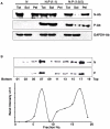
Figure 2. Soluble-insoluble fractionation and sucrose density gradient centrifugation.
Stoichiometry of N and P ratio is important for the N specific chaperone activity of P. (A) Total (Tot), Soluble (Sol) and Insoluble (Pel) fractionation of Vero-76 cells transfected with different ratios of pEGFP-C1 N and pCDNA 3.1 (+) P at 24 hours post-transfection. N and P proteins were detected by immunoblotting with N and P Ab respectively. It is evident that a 1∶0.5 N-P ratio is incapable of solubilising the otherwise insoluble N; however, a 1∶1 ratio can do so. GAPDH was used as a loading control. (B) Oligomerization status of soluble N. Sucrose density gradient centrifugation of the soluble fraction of cells transfected with 1∶1 ratio of GFP-N and P constructs. Fractions were collected from the bottom of the tube, and alternative fractions were immunobloted with N and P Abs. The curve shows the band intensities representing distribution of GFP-N and P against the fraction number. While majority of the soluble fraction of N is found in the monomeric form (fraction 17), a substantial amount is also found in fraction 7, indicating decameric forms. P is found to interact with both the populations of N. However, other stoichiometries of homo-oligomerization cannot be ruled out (fractions 13 through 17).
Figure 3
Figure 3. N forms distinct complexes with P in vitro.
Size exclusion chromatography of bacterially expressed purified N and P proteins, visualised by Coomasie brilliant blue staining. (A) N alone shows higher oligomeric distribution, suggesting decameric species. (B) P alone shows characteristic dimeric forms. (C) N and P incubated together at 4°C for 30 minutes. Interaction between N (oligomer) and P is evident, as they co-elute just after the void volume fraction. (D) N treated with 1% DOC for 30 minutes and subsequently dialysed to remove DOC. Though DOC treatment dissociated the oligomeric N into monomeric forms, removal of DOC by dialysis allows them to re-oligomerize. (E) Similar to D, except for the fact that DOC was removed in the presence of equimolar concentration of P protein. Presence of P protein during DOC removal retains the monomeric population of N, and subsequently, monomer N-P complexes elute at around 12 ml. Elution profiles of gel filtration standard are shown for molecular weight estimation. (F) Plot representing densitometric scans of N bands with respect to elution volume in ml.
Figure 4
Figure 4. Schematic representation of the wild-type N protein and the mutant N proteins used in this study.
Prokaryotic clones were made in pET3a vector for bacterial expression. Eukaryotic clones were made in pEGFP-C1 vector as N terminally GFP fused proteins. Different functionally relevant domains are also shown . The numbers represents amino acid positions.
Figure 5
Figure 5. N protein utilizes two separate domains for interacting with P in its monomeric and oligomeric forms.
N-terminally His-tagged P protein (His-P) was allowed to interact with either wild-type N or different N mutants in 100 mM NaCl TET buffer containing 10 mM Imidazole for 30 minutes at 4°C. Reaction mixtures were applied to Ni-NTA column and elution profile assayed by silver staining. L- loading; F- flow through; W- 10 mM Imidazole wash; E- 250 mM Imidazole elution. (A) In the absence of 1% DOC treatment. Bovine Serum Albumin (BSA) was used as negative control. (B) Wild-type N or N mutants were pre-incubated with 1% DOC for 30 minutes, followed by dialysis in presence of His-P, before applying to Ni-NTA column. Samples were resolved in 12% SDS-PAGE and visualised by Coomasie brilliant blue staining.
Figure 6
Figure 6. Ex vivo expression and immunoprecipitation of different N mutants with P.
(A) Intra-cellular distribution of different mutants of N used in this study. Vero-76 cells were transfected with 2 µg of pEGFP-C1 constructs of each mutant. N-terminal deletants N(48–422) and N(180–422) exhibits smooth distribution. N(1–47) also exhibits smooth distribution, probably because of the large GFP-tag, which interferes with its oligomerization. The bar represents 5 µm. (B) Co-expression of wild-type N and different N mutants with P protein in Vero-76 cells. Co-expression was confirmed by immunobloting with N and P Abs (upper and lower panels, respectively). All of the mutants used for this study expresses satisfactorily, and is of the right relative size. (C) Co-immunoprecipitation of wild-type N and different N mutants with P protein. Vero-76 cells were co-transfected with 2 µg of both plasmids, labelled with L-Methionine-35S 24 hours post-transfection followed by immunoprecipitation with P Ab. Except for N(180–422), all mutants co-immunoprecipitate with P.
Figure 7
Figure 7. Monomers of N(1–320) binds to P, but oligomers do not.
Size exclusion chromatography of bacterially expressed purified N(1–320) and P proteins, visualised by Coomasie brilliant blue staining. (A) N(1–320) alone. (B) N(1–320) treated with 1% DOC (to dissociate the oligomers into monomers) for 30 minutes and subsequently dialysed to remove DOC in the presence of equimolar concentrations of P protein. Co-elution of the two proteins confirms that N(1–320) can bind to P in monomeric state. (C) Plot representing densitometric scans of N(1–320) bands with respect to elution volume in ml. (D) GFP fluorescence of Vero-76 cells co-transfected with 1∶1 ratio of pEGFP-C1 N(1–320) and pCDNA 3.1 (+) P. Smooth distribution of N(1–320) is observed. (E) Immunofluorescence of the cells in D with P Ab. P also shows smooth distribution. (F) Merge of D and E. (G) GFP fluorescence of Vero-76 cells co-transfected with 1∶0.5 ratio of pEGFP-C1 N(1–320) and pCDNA 3.1 (+) P. N(1–320) shows punctuated distribution. (H) Immunofluorescence of the cells in G with P Ab. Distribution of P is smooth. (I) Merge of G and H. All data were captured on a laser scanning confocal microscope (Carl Zeiss). P Immunofluorescence were performed with anti-rabbit TRITC conjugated secondary antibody. 2 µg of DNA was used for all transfection, except for G, H and I where 1 µg of pCDNA 3.1 (+) P was used. The bar represents 5 µm.
Figure 8
Figure 8. Schematic representation of the domains of CHPV N involved in interaction with P and their functional importance.
Binding of P to nascent N masks the N-N self association region of CHPV N (N0-P complex formation) and also blocks non-specific RNA binding (upper panel). N0 is capable of specifically recognizing the viral leader sequence and the C-terminal 102 amino acids are essential for this recognition. Therefore, in the monomeric form, N specifically recognizes the leader RNA, to form the nucleation complex. Subsequently, the process of N-N self-association begins and P is released. Upon oligomerization, a new RNA binding cavity is formed utilizing the N-terminal arm (1–47 aa) and the central region of N (lower panel). Thus, the phase of non-specific encapsidation begins. Once nucleocapsids have formed, P can again interact with N, this time with the C-terminal region of oligomeric N, to usher the viral polymerase (L) onto its template.

Similar articles

Cited by

References

    1. Bhatt PN, Rodrigues FM. Chandipura: a new Arbovirus isolated in India from patients with febrile illness. Indian J Med Res. 1967;55:1295–1305. - PubMed
    1. Chadha MS, Arankalle VA, Jadi RS, Joshi MV, Thakare JP, et al. An outbreak of Chandipura virus encephalitis in the eastern districts of Gujarat state, India. Am J Trop Med Hyg. 2005;73:566–570. - PubMed
    1. Gurav YK, Tandale BV, Jadi RS, Gunjikar RS, Tikute SS, et al. Chandipura virus encephalitis outbreak among children in Nagpur division, Maharashtra, 2007. Indian J Med Res. 2010;132:395–399. - PubMed
    1. Rao BL, Basu A, Wairagkar NS, Gore MM, Arankalle VA, et al. A large outbreak of acute encephalitis with high fatality rate in children in Andhra Pradesh, India, in 2003, associated with Chandipura virus. Lancet. 2004;364:869–874. - PMC - PubMed
    1. Rao NS, Wairagkar NS, Mohan MV, Khetan M, Somavathi S. Brain stem encephalitis associated with Chandipura virus in Andhra Pradesh outbreak. . J Trop Pediatr. 2008;54:25–30. - PMC - PubMed

Publication types

MeSH terms