. 2012 Jul;37(8):1867-78.
doi: 10.1038/npp.2012.34. Epub 2012 Apr 4.

Aquaporin-4 deficiency impairs synaptic plasticity and associative fear memory in the lateral amygdala: involvement of downregulation of glutamate transporter-1 expression

Affiliations

Aquaporin-4 deficiency impairs synaptic plasticity and associative fear memory in the lateral amygdala: involvement of downregulation of glutamate transporter-1 expression

Yan-Kun Li et al. Neuropsychopharmacology. 2012 Jul.

Abstract

Astrocytes are implicated in information processing, signal transmission, and regulation of synaptic plasticity. Aquaporin-4 (AQP4) is the major water channel in adult brain and is primarily expressed in astrocytes. A growing body of evidence indicates that AQP4 is a potential molecular target for the regulation of astrocytic function. However, little is known about the role of AQP4 in synaptic plasticity in the amygdala. Therefore, we evaluated long-term potentiation (LTP) in the lateral amygdala (LA) and associative fear memory of AQP4 knockout (KO) and wild-type mice. We found that AQP4 deficiency impaired LTP in the thalamo-LA pathway and associative fear memory. Furthermore, AQP4 deficiency significantly downregulated glutamate transporter-1 (GLT-1) expression and selectively increased NMDA receptor (NMDAR)-mediated EPSCs in the LA. However, low concentration of NMDAR antagonist reversed the impairment of LTP in KO mice. Upregulating GLT-1 expression by chronic treatment with ceftriaxone also reversed the impairment of LTP and fear memory in KO mice. These findings imply a role for AQP4 in synaptic plasticity and associative fear memory in the amygdala by regulating GLT-1 expression.


Figures
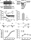
Figure 1
Figure 1
Aquaporin-4 (AQP4) deficiency impairs long-term potentiation (LTP) in the thalamo-LA pathway with no effect on basal synaptic transmission. (a) Expression of AQP4 in the LA from wild-type (WT) (AQP4+/+) and knockout (KO) (AQP4−/−) mice. Reverse transcriptase-polymerase chain reaction (RT-PCR) (upper) and western blot analysis (lower) revealed that the expression of Aqp4 mRNA and AQP4 protein was readily detected in LA of WT mice (n=3) but not KO mice (n=3). Glyceraldehyde 3-phosphate dehydrogenase (GAPDH) and β-actin were used as internal control. (b) Schematic representation of an amygdala slice showed location of recording and stimulation electrodes in the thalamo-LA pathway. LA, lateral amygdala; BLA, basolateral amygdala; CeA, central amygdala; IC, internal capsule; EC, external capsule. (c) Time course of the field excitatory postsynaptic potential (fEPSP) evoked by stimulation of thalamic inputs recorded in amygdala slices from WT (n=11 slices from 6 mice) and KO mice (n=13 slices from 7 mice). (Inset) Schematic representation of fEPSP recorded in individual slices before (1) and 60 min after (2) the LTP-inducing stimulation in either WT (left) or KO (right) mice. (d) The histogram showed the level of LTP 60 min after high-frequency stimulation (HFS) (five trains at 100 Hz for 1 s with 90 s interval between trains) in the thalamo-LA pathway in WT and KO mice. Each point was the normalized mean±SEM of slices. **P<0.01 vs WT. (e) Typically superimposed fEPSP recorded in the thalamo-LA pathway in WT (left) and KO (right) mice with gradually increased stimulation intensity. (f) Typical fEPSP recorded in the thalamo-LA pathway from individual experiment at 50 ms interpulse interval before HFS stimulation. (g) Input–output curves in the thalamo-LA pathway illustrating the relationship between the stimulation intensity and evoked response for fEPSP recorded in brain slices from WT (n=10 slices from 5 mice) and KO mice (n=11 slices from 6 mice). No significant differences were observed between the two genotypes. (h) Paired-pulse facilitation in the thalamo-LA pathway was measured by varying the intervals (25, 50, 75, and 100 ms) between pairs of stimuli before HFS stimulation. No significant differences were observed between WT (n=10 slices from 5 mice) and KO mice (n=11 slices from 6 mice).
Figure 2
Figure 2
Aquaporin-4 (AQP4) deficiency impairs the associative fear memory of mice. (a) Experimental design of fear conditioning tasks. (b) Freezing percentage before conditioned stimulus (pre-CS), during conditioning (training) and 24 h after conditioning (test). Wild-type (WT) and knockout (KO) mice exhibited similar freezing in conditioning chamber before CS and during conditioning training. However, freezing behavior was significantly reduced in KO mice (n=8) 24 h after conditioning training compared with WT mice (n=8). The data are expressed by mean±SEM. **P<0.01 vs WT. (c) Total distance animals moved during 10 min intervals in open field. No difference was observed between KO (n=8) and WT (n=8) mice. (d) Average speed animals moved during 10 min intervals in open field. No difference was observed between KO (n=8) and WT (n=8) mice. (e) The number of animal entries to the open arm as the percentage of the total number of arm entries in the elevated plus maze. No difference was observed between KO (n=8) and WT (n=8) mice. (f) The total number of animal entries to open and closed arms in the elevated plus maze. No difference was observed between KO (n=8) and WT (n=8) mice. (g) Thresholds of shock intensities for vocalizing and jumping are shown. No difference was detected between KO (n=8) and WT (n=7) mice.
Figure 3
Figure 3
Aquaporin-4 (AQP4) deficiency downregulates glutamate transporter-1 (GLT-1) expression without altering GLAST level in the lateral amygdala (LA). (a) Representative images of immunoblots using antibodies against GLAST-1 and β-actin. (b) Representative images of immunoblots using antibodies against GLT-1 and β-actin. (c) Quantitative analyses of GLAST by densitometry are summarized. Data were expressed as a percentage of value of wild-type (WT). No significant differences in GLAST expression were observed between AQP4 knockout (KO) and WT mice. (d) Quantitative analyses of GLT-1 by densitometry are summarized. Data were expressed as percentage of value of WT. The expression of GLT-1 was dramatically reduced in AQP4 KO mice compared with those in WT mice (n=6, **P<0.01 vs WT).
Figure 4
Figure 4
Aquaporin-4 (AQP4) deficiency selectively increases N-methyl-D-aspartate receptor (NMDAR)-mediated currents in the lateral amygdala (LA). (a) Representative traces of AMPA receptor (AMPAR) (lower traces)- and NMDAR (upper traces)-mediated excitatory postsynaptic currents (EPSCs) in AQP4 wild-type (WT) and knockout (KO) mice. Stimulus artifacts were omitted for clarity. (b) Summary histogram for the ratio of NMDAR- to AMPAR-mediated EPSC amplitudes in WT and KO mice. The NMDAR/AMPAR ratio was significantly larger in KO mice (0.95±0.09, n=4 cells from 4 mice) than in WT mice (0.59±0.06, n=4 cells from 4 mice), *P<0.05 vs WT. (c) Summary histograms for AMPAR-mediated EPSCs in WT and KO mice. The mean amplitudes of AMPAR-mediated EPSCs were not significantly different among groups. (d) Summary histograms for NMDAR-mediated EPSCs in WT and KO mice. The mean amplitudes of NMDAR-mediated EPSCs of KO mice (119.1±23.4 pA) were significantly increased compared with those in WT mice (68.5±16.0 pA). *P<0.05 vs WT.
Figure 5
Figure 5
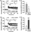
Excessive activation of N-methyl-D-aspartate receptor (NMDAR) contributes to the impairment of long-term potentiation (LTP) in knockout (KO) mice. (a) Time course of the field excitatory postsynaptic potential (fEPSP) evoked by stimulation at thalamo-lateral amygdala (LA) pathway of wild-type (WT) mice in the absence (n=11 slices from 6 mice) or presence (n=8 slices from 4 mice) of 1 μM D-2-amino-5-phosphonovaleric acid (D-APV), an NMDAR antagonist. (Inset) Schematic representation of fEPSP recorded in individual slices before (1) and 60 min after (2) the high-frequency stimulation (HFS) (five trains at 100 Hz for 1 s with 90 s interval between trains) in the absence (left) or presence (right) of D-APV. (b) The histogram showed the level of LTP 60 min after HFS at thalamo-LA pathway in WT mice in the absence or presence of 1 μM D-APV. *P<0.05 vs WT. (c) Time course of the fEPSP evoked by stimulation at thalamo-LA pathway in aquaporin-4 (AQP4) KO mice in the absence (n=13 slices from 7 animals) or presence (n=8 slices from 4 animals) of 1 μM D-APV. (Inset) Schematic representation of fEPSP recorded in individual slices before (1) and 60 min after (2) the HFS in the absence (left) or presence (right) of D-APV. (d) The histogram showed the level of LTP 60 min after HFS at thalamo-LA pathway in AQP4 KO mice in the absence or presence of 1 μM D-APV. Each point was the normalized mean±SEM of slices. ##P<0.01 vs KO.
Figure 6
Figure 6
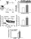
Ceftriaxone (Cef) treatment (intraperitoneally (i.p.), 200 mg/kg/day for 5 days) reverses the impairment of long-term potentiation (LTP) and fear memory in aquaporin-4 knockout (AQP4 KO) mice. (a) Experimental design of electrophysiological studies and fear conditioning tasks. (b) The expression of glutamate transporter-1 (GLT-1) in amygdala tissues of saline- and Cef-treated KO mice. (Above) Representative images of immunoblots using antibodies against GLT-1 and β-actin. (Below) Quantitative analyses of GLT-1 by densitometry are summarized. Data were expressed as percentage of value of KO. Cef treatment (i.p. 200 mg/kg/day for 5 days) increased GLT-1 expression in KO mice (n=6, ##P<0.01 vs KO). (c) Time course of the field excitatory postsynaptic potential (fEPSP) evoked by stimulation at thalamo-lateral amygdala (LA) pathway in saline- and Cef-treated KO mice. (Inset) Schematic representation of fEPSP recorded in individual slices before (1) and 60 min after (2) the LTP-inducing stimulation in saline- (left) and Cef-treated (right) KO mice. Cef treatment reversed the impairment of LTP in KO mice. (d) The histogram showed the level of LTP 60 min after high-frequency stimulation (HFS) in saline- (n=9 slices from 5 mice) and Cef-treated KO mice (n=10 slices from 5 mice). Each point was the normalized mean±SEM of slices. ##P<0.01 vs saline-treated KO mice. (e) Total percentage of freezing in saline (n=8) and Cef-treated KO mice (n=8) when tested before conditioned stimulus (pre-CS) and 24 h after conditioning training (test), respectively. Data are expressed by mean±SEM. #P<0.05 vs saline-treated KO mice.

Similar articles

Cited by

References

    1. Abel T, Lattal KM. Molecular mechanisms of memory acquisition, consolidation and retrieval. Curr Opin Neurobiol. 2001;11:180–187. - PubMed
    1. Amara SG, Fontana AC. Excitatory amino acid transporters: keeping up with glutamate. Neurochem Int. 2002;41:313–318. - PubMed
    1. Amiry-Moghaddam M, Ottersen OP. The molecular basis of water transport in the brain. Nat Rev Neurosci. 2003;4:991–1001. - PubMed
    1. Bao F, Chen M, Zhang Y, Zhao Z. Hypoalgesia in mice lacking aquaporin-4 water channels. Brain Res Bull. 2010;83:298–303. - PubMed
    1. Barad M, Gean PW, Lutz B. The role of the amygdala in the extinction of conditioned fear. Biol Psychiatry. 2006;60:322–328. - PubMed

Publication types

MeSH terms