. 2012 Jul 1;21(13):2923-35.
doi: 10.1093/hmg/dds118. Epub 2012 Mar 30.

Clustered burst firing in FMR1 premutation hippocampal neurons: amelioration with allopregnanolone

Affiliations

Clustered burst firing in FMR1 premutation hippocampal neurons: amelioration with allopregnanolone

Zhengyu Cao et al. Hum Mol Genet. .

Abstract

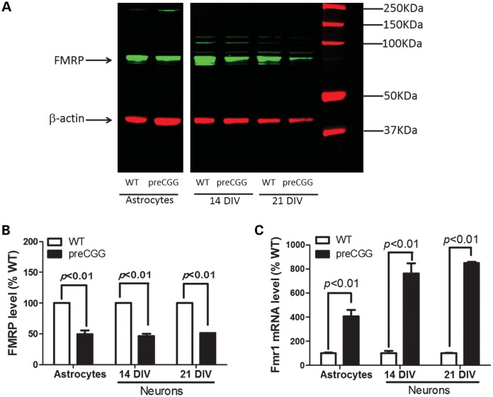
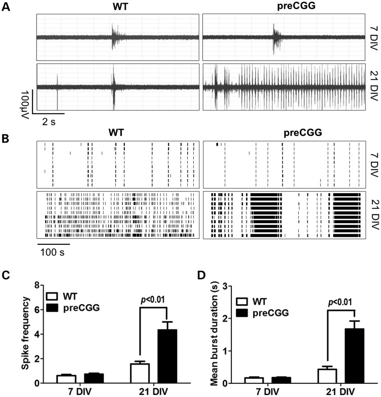
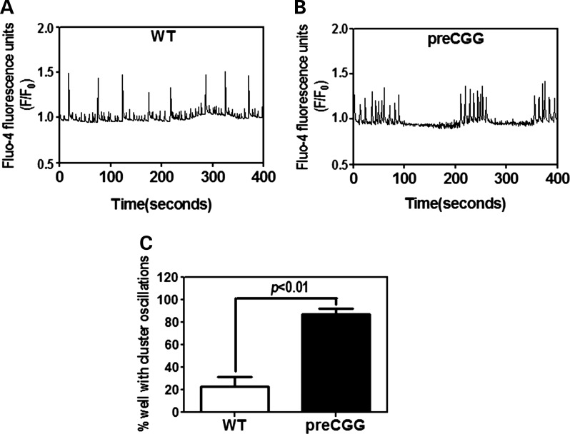
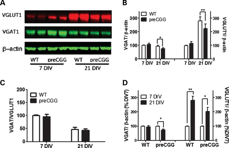
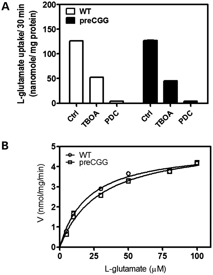
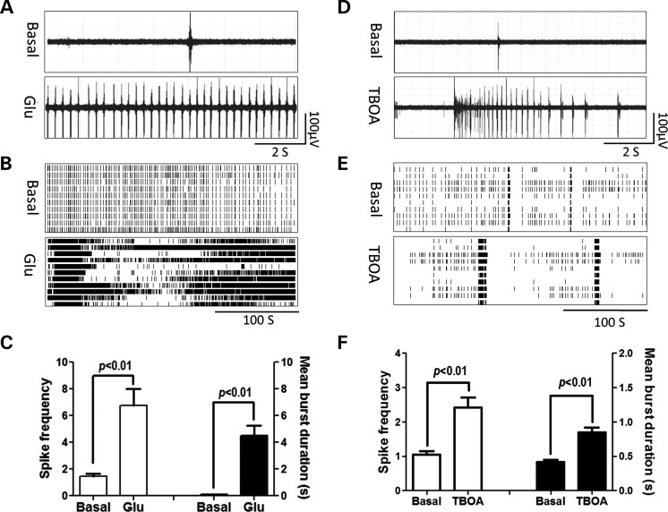
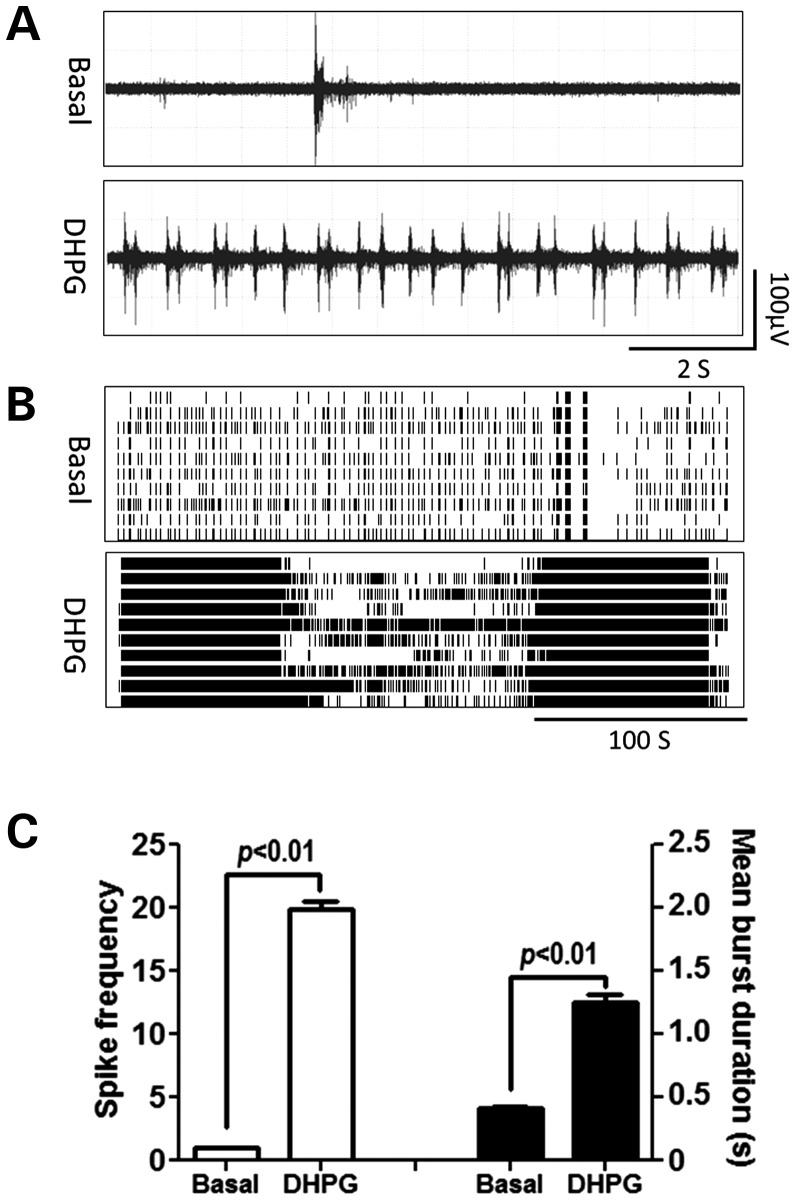
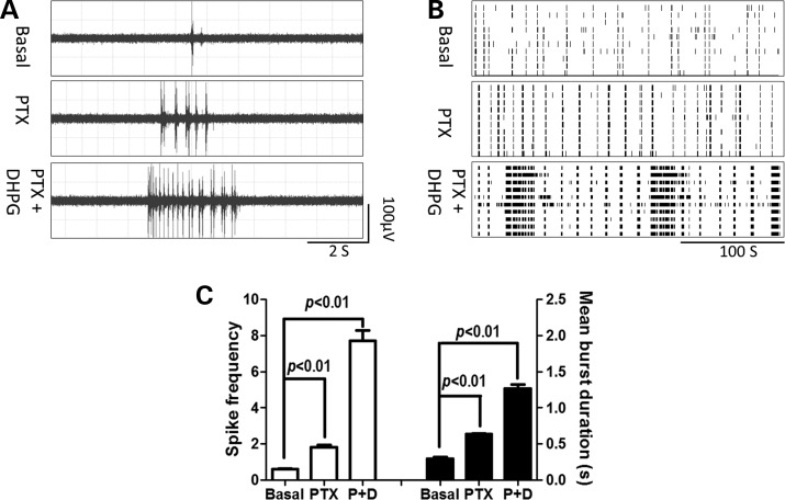
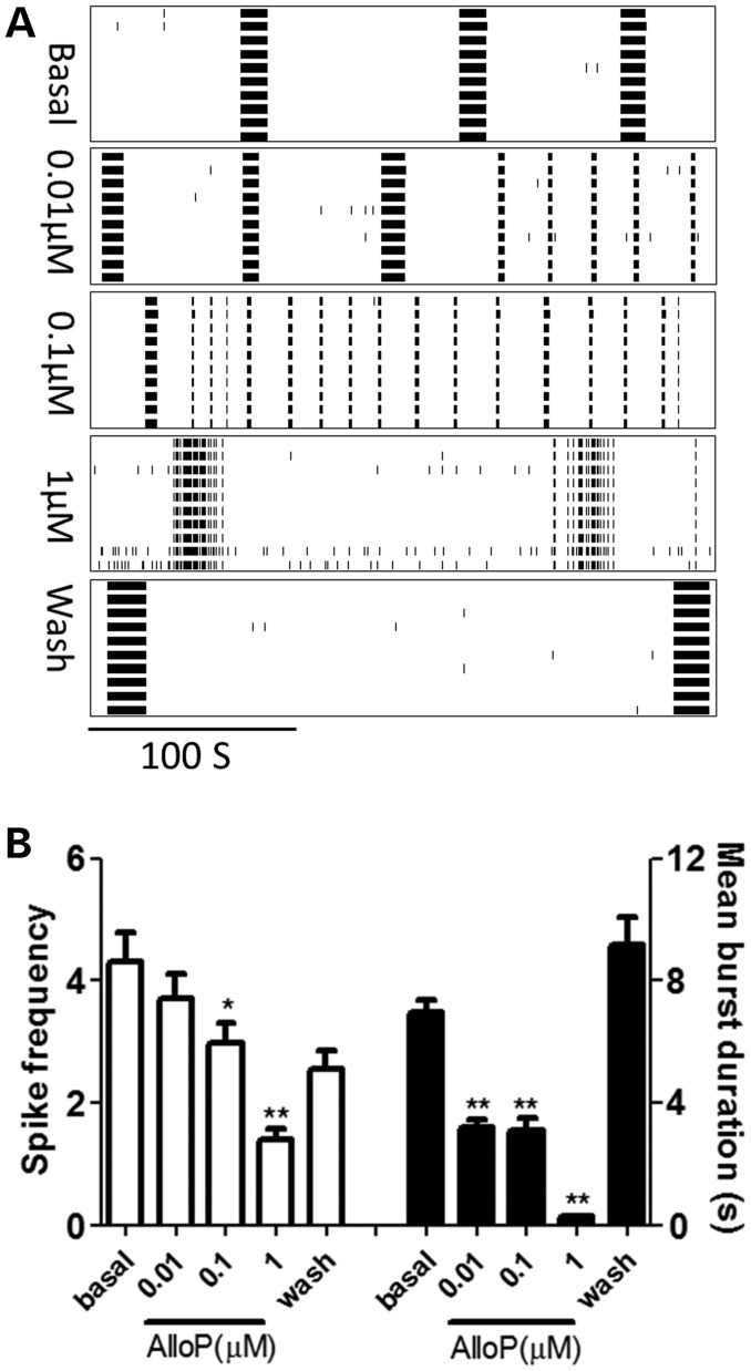
Premutation CGG repeat expansions (55-200 CGG repeats; preCGG) within the fragile X mental retardation 1 (FMR1) gene cause fragile X-associated tremor/ataxia syndrome (FXTAS). Defects in neuronal morphology and migration have been described in a preCGG mouse model. Mouse preCGG hippocampal neurons (170 CGG repeats) grown in vitro develop abnormal networks of clustered burst (CB) firing, as assessed by multielectrode array recordings and clustered patterns of spontaneous Ca(2+) oscillations, neither typical of wild-type (WT) neurons. PreCGG neurons have reduced expression of vesicular GABA and glutamate (Glu) transporters (VGAT and VGLUT1, respectively), and preCGG hippocampal astrocytes display a rightward shift on Glu uptake kinetics, compared with WT. These alterations in preCGG astrocytes and neurons are associated with 4- to 8-fold elevated Fmr1 mRNA and occur despite consistent expression of fragile X mental retardation protein levels at ∼50% of WT levels. Abnormal patterns of activity observed in preCGG neurons are pharmacologically mimicked in WT neurons by addition of Glu or the mGluR1/5 agonist, dihydroxyphenylglycine, to the medium, or by inhibition of astrocytic Glu uptake with dl-threo-β-benzyloxyaspartic acid, but not by the ionotropic Glu receptor agonists, α-2-amino-3-(5-methyl-3-oxo-1,2-oxazol-4-yl) propanoic acid or N-methyl-d-aspartic acid. The mGluR1 (7-(hydroxyimino)cyclopropa [b]chromen-1a-carboxylate ethyl ester) or mGluR5 (2-methyl-6-(phenylethynyl)pyridine hydrochloride) antagonists reversed CB firing. Importantly, the acute addition of the neurosteroid allopregnanolone mitigated functional impairments observed in preCGG neurons in a reversible manner. These results demonstrate abnormal mGluR1/5 signaling in preCGG neurons, which is ameliorated by mGluR1/5 antagonists or augmentation of GABA(A) receptor signaling, and identify allopregnanolone as a candidate therapeutic lead.


Figures

Figure 1.
Figure 1.
Premutation cultures express higher levels of Fmr1 mRNAs with decreased FMRP proteins compared with WT paired cultures. (A) Representative western blot in paired cultures of WT and preCGG hippocampal astrocytes as well as neurons. The band with molecular weight around 72 kDa is FMRP. (B) Quantification of FMRP expression levels relative to β-actin in paired WT and preCGG cultures of hippocampal astrocytes, and 14 and 21 DIV neuronal cultures. Data were pooled from two independent cultures. (C) Fmr 1 mRNA comparison between WT and preCGG paired cultures of hippocampal astrocytes and 14 as well as 21 DIV neurons. Data were pooled from two independent cultures, each performed in duplicate.
Figure 2.
Figure 2.
Hippocampal neurons with CGG expansion predominantly display a pattern of spontaneous field potential activity having CB firing. (A) Representative traces of firing for WT and preCGG hippocampal neurons at 7 and 21 DIV in a 10s epoch. (B) Representative raster plots for the neuronal firing of WT and preCGG hippocampal neurons at 7 and 21 DIV. (C and D) Quantification of the spike frequency and mean burst duration for WT and preCGG hippocampal neurons at 7 and 21 DIV. Summary data were from 26 MEAs for WT and 37 MEAs for preCGG hippocampal neurons.
Figure 3.
Figure 3.
Hippocampal neurons with CGG expansion predominately display clustered Ca2+ oscillations. Representative intracellular Ca2+ oscillations in WT (A) and preCGG (B) hippocampal neurons measured at 14 DIV. (C) Quantification of the percentage of wells displaying clustered Ca2+ oscillations. These data were summarized from five independent cultures with a total number of wells of 71 and 93 for the WT and preCGG hippocampal neurons, respectively.
Figure 4.
Figure 4.
21 DIV hippocampal neurons with CGG expansion display decreased expression levels of VGAT and VGLUT1. (A) Representative western blots for VGAT and VGLUT1. (B) Quantification of the expression levels for VGAT and VGLUT1. These data were normalized to the expression levels of VGAT or VGLUT1 on 7 DIV WT neurons. (C) Quantification of the ratio of VGAT over the VGLUT1 expression level. (D) Quantification of the developmental change of VGAT and VGLUT1. These data were normalized to the expression levels of respective 7 DIV neurons for each genotype. n = 4 from three separate culture days.
Figure 5.
Figure 5.
Glu uptake in pure hippocampal astrocyte cultures. (A) Non-selective Glu transporter inhibitors, DL-TBOA (1 mm) and l-trans-PDC (1 mm), blocked Glu uptake in both WT and preCGG hippocampal astrocytes. These data were repeated in two cultures, in triplicate, with similar results. (B) Kinetic analysis for Glu uptake in WT and preCGG hippocampal astrocytes. The experiments were repeated in four independent cultures performed in triplicate with similar results. Uptake as a function of Glu concentration was fitted by the Michaelis–Menten equation. The Vmax values for Glu uptake do not show a statistically significant difference between WT and preCGG hippocampal astrocytes. However, the Km value was shifted from 20.0 ± 2.8 µm for WT to 28.4 ± 2.2 µm for preCGG (n = 4, P< 0.01).
Figure 6.
Figure 6.
Glu and TBOA induce CB firing pattern in WT hippocampal neurons. (A and D) Representative trace for firing activity before and after WT hippocampal neurons exposed to Glu (100 µm) or TBOA (100 µm), respectively. (B and E) Representative raster plots for the neuronal firing before and after WT hippocampal neurons exposed to Glu or TBOA, respectively. (C and F) Quantification of the spike frequency (left Y axis) and mean burst duration (right Y axis) for WT hippocampal neurons before and after Glu or TBOA exposure, respectively. Data were repeated twice in duplicate with similar results.
Figure 7.
Figure 7.
DHPG produces CB firing pattern in WT hippocampal neurons. (A) Representative trace for firing activity before and after WT hippocampal neurons exposed to 10 µm DHPG. (B) Representative raster plots for the neuronal firing before and after DHPG exposure. (C) Quantification of the spike frequency (left Y axis) and mean burst duration (right Y axis) for WT hippocampal neurons before and after DHPG exposure. Data were repeated twice in duplicate with similar results.
Figure 8.
Figure 8.
Picrotoxin, together with DHPG, induces CB firing pattern in WT hippocampal neurons. (A) Representative firing traces before and after WT hippocampal neurons were exposed to picrotoxin (100 µm) or a combination of picrotoxin (100 µm) and DHPG (10 µm), respectively. (B) Representative raster plots of the neuronal firing before and after picrotoxin or picrotoxin and DHPG exposure. (C) Quantification of the spike frequency (left Y axis) and mean burst duration (right Y axis) before and after exposure to picrotoxin or picrotoxin and DHPG exposure. These data were repeated twice in two independent cultures with similar results.
Figure 9.
Figure 9.
MPEP and CPCCOEt reversed the CB firing pattern in preCGG hippocampal neurons in a concentration-dependent manner. (A and C) Representative raster plots for the neuronal firing before and after preCGG hippocampal neurons were exposed to MPEP or CPCCOEt, respectively. (B and D) Quantification of the spike frequency (left Y axis) and mean burst duration (right Y axis) for preCGG hippocampal neurons before and during exposure to different concentrations of MPEP and CPCCOEt, respectively. These data were repeated twice in duplicate with similar results.
Figure 10.
Figure 10.
Allopregnanolone reversibly reduced the spike frequency and burst duration of the preCGG hippocampal neurons. (A) Representative raster plots for the neuronal firing of preCGG hippocampal neurons before and during exposure to as well as washout of allopregnanolone. (B) Quantification of the spike frequency (left Y axis) and mean burst duration (right Y axis) for preCGG hippocampal neurons before and during exposure to as well as after washout of allopregnanolone, respectively. Data were repeated two times with independent cultures.

Similar articles

Cited by

  • Emerging topics in FXTAS.
    Hall DA, Birch RC, Anheim M, Jønch AE, Pintado E, O'Keefe J, Trollor JN, Stebbins GT, Hagerman RJ, Fahn S, Berry-Kravis E, Leehey MA. Hall DA, et al. J Neurodev Disord. 2014;6(1):31. doi: 10.1186/1866-1955-6-31. Epub 2014 Jul 30. J Neurodev Disord. 2014. PMID: 25642984 Free PMC article. Review.
  • Clinical Potential of Neurosteroids for CNS Disorders.
    Reddy DS, Estes WA. Reddy DS, et al. Trends Pharmacol Sci. 2016 Jul;37(7):543-561. doi: 10.1016/j.tips.2016.04.003. Epub 2016 May 5. Trends Pharmacol Sci. 2016. PMID: 27156439 Free PMC article. Review.
  • Insight and Recommendations for Fragile X-Premutation-Associated Conditions from the Fifth International Conference on FMR1 Premutation.
    Tassone F, Protic D, Allen EG, Archibald AD, Baud A, Brown TW, Budimirovic DB, Cohen J, Dufour B, Eiges R, Elvassore N, Gabis LV, Grudzien SJ, Hall DA, Hessl D, Hogan A, Hunter JE, Jin P, Jiraanont P, Klusek J, Kooy RF, Kraan CM, Laterza C, Lee A, Lipworth K, Losh M, Loesch D, Lozano R, Mailick MR, Manolopoulos A, Martinez-Cerdeno V, McLennan Y, Miller RM, Montanaro FAM, Mosconi MW, Potter SN, Raspa M, Rivera SM, Shelly K, Todd PK, Tutak K, Wang JY, Wheeler A, Winarni TI, Zafarullah M, Hagerman RJ. Tassone F, et al. Cells. 2023 Sep 21;12(18):2330. doi: 10.3390/cells12182330. Cells. 2023. PMID: 37759552 Free PMC article. Review.
  • Associated features in females with an FMR1 premutation.
    Wheeler AC, Bailey DB Jr, Berry-Kravis E, Greenberg J, Losh M, Mailick M, Milà M, Olichney JM, Rodriguez-Revenga L, Sherman S, Smith L, Summers S, Yang JC, Hagerman R. Wheeler AC, et al. J Neurodev Disord. 2014;6(1):30. doi: 10.1186/1866-1955-6-30. Epub 2014 Jul 30. J Neurodev Disord. 2014. PMID: 25097672 Free PMC article. Review.
  • Open-Label Allopregnanolone Treatment of Men with Fragile X-Associated Tremor/Ataxia Syndrome.
    Wang JY, Trivedi AM, Carrillo NR, Yang J, Schneider A, Giulivi C, Adams P, Tassone F, Kim K, Rivera SM, Lubarr N, Wu CY, Irwin RW, Brinton RD, Olichney JM, Rogawski MA, Hagerman RJ. Wang JY, et al. Neurotherapeutics. 2017 Oct;14(4):1073-1083. doi: 10.1007/s13311-017-0555-6. Neurotherapeutics. 2017. PMID: 28707277 Free PMC article. Clinical Trial.

References

    1. Jacquemont S., Hagerman R.J., Hagerman P.J., Leehey M.A. Fragile-X syndrome and fragile X-associated tremor/ataxia syndrome: two faces of FMR1. Lancet Neurol. 2007;6:45–55. - PubMed
    1. Hagerman R., Au J., Hagerman P. FMR1 premutation and full mutation molecular mechanisms related to autism. J. Neurodev. Disord. 2011;3:211–224. - PMC - PubMed
    1. Oostra B.A., Willemsen R. FMR1: a gene with three faces. Biochim. Biophys. Acta. 2009;1790:467–477. - PMC - PubMed
    1. Brouwer J.R., Severijnen E., de Jong F.H., Hessl D., Hagerman R.J., Oostra B.A., Willemsen R. Altered hypothalamus-pituitary-adrenal gland axis regulation in the expanded CGG-repeat mouse model for fragile X-associated tremor/ataxia syndrome. Psychoneuroendocrinology. 2008;33:863–873. - PMC - PubMed
    1. Fu Y.H., Kuhl D.P., Pizzuti A., Pieretti M., Sutcliffe J.S., Richards S., Verkerk A.J., Holden J.J., Fenwick R.G., Jr, Warren S.T., et al. Variation of the CGG repeat at the fragile X site results in genetic instability: resolution of the Sherman paradox. Cell. 1991;67:1047–1058. - PubMed

Publication types

MeSH terms