. 2012 May;23(10):1943-54.
doi: 10.1091/mbc.E11-10-0829. Epub 2012 Mar 28.

c-Jun N-terminal kinase-mediated Rad18 phosphorylation facilitates Polη recruitment to stalled replication forks

Affiliations

c-Jun N-terminal kinase-mediated Rad18 phosphorylation facilitates Polη recruitment to stalled replication forks

Laura R Barkley et al. Mol Biol Cell. 2012 May.

Abstract

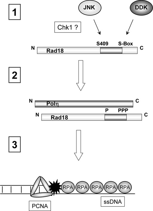
The E3 ubiquitin ligase Rad18 chaperones DNA polymerase η (Polη) to sites of UV-induced DNA damage and monoubiquitinates proliferating cell nuclear antigen (PCNA), facilitating engagement of Polη with stalled replication forks and promoting translesion synthesis (TLS). It is unclear how Rad18 activities are coordinated with other elements of the DNA damage response. We show here that Ser-409 residing in the Polη-binding motif of Rad18 is phosphorylated in a checkpoint kinase 1-dependent manner in genotoxin-treated cells. Recombinant Rad18 was phosphorylated specifically at S409 by c-Jun N-terminal kinase (JNK) in vitro. In UV-treated cells, Rad18 S409 phosphorylation was inhibited by a pharmacological JNK inhibitor. Conversely, ectopic expression of JNK and its upstream kinase mitogen-activated protein kinase kinase 4 led to DNA damage-independent Rad18 S409 phosphorylation. These results identify Rad18 as a novel JNK substrate. A Rad18 mutant harboring a Ser → Ala substitution at S409 was compromised for Polη association and did not redistribute Polη to nuclear foci or promote Polη-PCNA interaction efficiently relative to wild-type Rad18. Rad18 S409A also failed to fully complement the UV sensitivity of Rad18-depleted cells. Taken together, these results show that Rad18 phosphorylation by JNK represents a novel mechanism for promoting TLS and DNA damage tolerance.


Figures

FIGURE 1:
FIGURE 1:
Rad18 phosphorylation is genotoxin inducible and requires Chk1. (A) HA-Rad18–expressing H1299 cells were treated with 600 nM BPDE for 4 h or were left untreated for controls. CSK extracts from the cells were incubated with λ phosphatase (1 U/μl) in the presence or absence of 50 mM NaF at 37 C for 30 min. The resulting extracts were analyzed by immunoblotting with anti-HA antibodies. BPDE-induced electrophoretic mobility shifts are indicated by the arrows. (B) Control or Chk1-depleted HA-Rad18–expressing H1299 cells were treated with 600 nM BPDE for 4 h or were left untreated for controls. Anti-HA immune complexes were analyzed by SDS–PAGE and immunoblotting sequentially with anti–phosphoserine/phosphothreonine and anti-HA antibodies. Whole-cell extracts were analyzed for Chk1 expression, and PCNA was used as a loading control. (C) AdCon or AdHA-Rad18–infected H1299 cells were treated with 600 nM BPDE for 4 h or were left untreated for controls. Chromatin and soluble fractions from the cells were immunoprecipitated with anti-HA antibodies, and the resulting immunoprecipitates were analyzed by SDS–PAGE and immunoblotting with anti–phosphoserine/phosphothreonine and anti-HA antibodies. (D) AdCon or AdHA-Rad18–infected H1299 cells were transferred to phosphate-free DMEM and incubated for 4 h in the presence of 0.2 mCi/ml of 32P-orthophosphate. The 32P-orthophosphate–labeled cells were treated with 600 nM BPDE for 4 h or were left untreated for controls. Whole-cell extracts from the cells were immunoprecipitated with anti-HA antibodies, and the resulting immunoprecipitates were resolved by SDS–PAGE. The resulting gels were washed extensively in 40% methanol/10% acetic acid. The fixed gel was dried, and radioactive proteins were detected by autoradiography.
FIGURE 2:
FIGURE 2:
Rad18 S409 is phosphorylated by JNK. (A) Amino acid sequence alignment showing conservation of S409 among different species. (B) Control or Chk1-depleted HA-Rad18–expressing H1299 cells were treated with UV (20 J/m2) or left untreated for controls. After 4 h, anti-HA immune complexes were prepared from chromatin fractions and analyzed by SDS–PAGE and immunoblotting with anti–phospho-Ser-409 and anti-HA antibodies. Soluble extracts were analyzed for Chk1 expression to confirm efficient siRNA-mediated knockdown. Immunoblotting with an anti-actin antibody was used to verify equivalent protein loading between lanes. (C) Comparison of sequences flanking Rad18 S409 (underlined) with JNK1 consensus phosphorylation sites. (D) Recombinant Rad18–Rad6 complex was subject to in vitro phosphorylation by JNK. The reaction contained 0.4 μg (4 pmol) of Rad18–Rad6, and half (0.2 μg; 2 pmol) was resolved on SDS–PAGE. The phosphorylated species contained 4000 cpm (determined by scintillation counting of the excised gel slice), which is equivalent to 2 pmol of incorporated ATP incorporation. (E) Recombinant GST-Rad18 398-495 (WT), GST-Rad18 S409A, and GST (control) were tested as JNK substrates using in vitro kinase assays. Reaction products were resolved on SDS–PAGE. The resulting gel was fixed, stained, and dried. Top, an autoradiogram; bottom, the Coomassie-stained proteins. (F) Exponentially growing H1299 cells were irradiated with UVC (20 J/m2) or sham irradiated for controls. Cells were harvested 2 or 4 h after UVC treatment and lysates were analyzed for pJNK and PCNA by SDS–PAGE and immunoblotting. (G) Exponentially growing H1299 cells expressing HA-Rad18 were treated for 1 h with 1 μM SP600125 or left untreated for controls. Control and SP600125-treated cells were treated with UVC (20 J/m2) or sham irradiated. After 2 h, Rad18 was immunoprecipitated from cell extracts. Immune complexes were resolved by SDS–PAGE and probed sequentially with antibodies against Rad18 pS409 and HA. (H) H1299 cells were cotransfected with expression vectors encoding HA-Rad18 (WT), HA-Rad18 (S409A), FLAG-MKK4, and FLAG-JNK1a1 or with “empty” vector control plasmid as indicated. At 48 h posttransfection, cells were lysed and normalized for protein content. Lysates were immunoprecipitated with anti-HA, and the resulting immune complexes were resolved by SDS–PAGE, transferred to nitrocellulose, and probed sequentially with anti–Rad18 pS409 and anti-HA antibodies. Total cell lysates were also resolved and transferred to nitrocellulose and then probed with anti-FLAG to validate MKK4 and JNK expression.
FIGURE 3:
FIGURE 3:
Rad18 S409 phosphorylation promotes association with Polη. (A) RAD18/ cells were coinfected with adenovirus vectors encoding HA-Rad18 WT or HA-Rad18 S409A or with an “empty” adenovirus vector (AdCon). Cells were treated with UV (20 J/m2) and harvested after 6 h. Chromatin fractions from the cells were solubilized and immunoprecipitated with anti-HA antibodies. The resulting immune complexes were resolved by SDS–PAGE and analyzed by immunoblotting with antibodies against Rad18 S409, HA, and Polη. Total chromatin input fractions were also analyzed using anti-Polη. (B) RAD18/cells were infected with adenovirus vectors encoding HA-Rad18 WT, HA-Rad18 S409A, HA-Rad18 S409E, or with an “empty” adenovirus vector (AdCon). Cells were UV irradiated, and chromatin fractions were prepared and analyzed by SDS–PAGE and immunoblotting using antibodies against Rad18 and Polη as described in A.
FIGURE 4:
FIGURE 4:
Rad18 S409 is dispensable for Rad18–Rad6 interactions, PCNA-directed E3 ligase activity, and binding to RPA-coated ssDNA. (A) HA-Rad18–expressing H1299 cells were treated with UV (20 J/m2) or left untreated for controls. Anti-HA immune complexes were prepared from chromatin fractions and analyzed by SDS–PAGE and immunoblotting with anti-HA and anti-Rad6 antibodies. (B) H1299 cells were infected with different doses of AdCon, AdRad18WT, and AdRad18 S409A. Twenty-fours hours later, chromatin and soluble fractions were prepared and analyzed by SDS–PAGE and immunoblotting with HA and PCNA antibodies. (C) RAD18/ cells were infected with AdCon, AdRad18 WT, or Rad18 S409A. After 24 h, soluble extracts were prepared from the resulting cells. RPA/ssDNA-interacting proteins were captured as described in Materials and Methods. Proteins bound to the ssDNA-coated beads were analyzed by SDS–PAGE and immunoblotting. Top, the RPA/ssDNA-dependency of Rad18 binding to streptavidin-coated beads. Bottom, the comparison of WT Rad18 and Rad18 S409A binding to RPA/ssDNA-beads (and ssDNA and RPA were present in all pull-down samples).
FIGURE 5:
FIGURE 5:
Rad18 S409A does not promote efficient recruitment of Polη to PCNA and fails to complement the UV sensitivity of Rad18-depleted cells. (A) Triplicate cultures of H1299 cells were depleted of endogenous Rad18 and complemented with siRNA-resistant HA-Rad18 (WT) and HA-Rad18 S409A. The Rad18 dependence of UV-induced YFP-Polη and GFP-Polκ redistribution to nuclear foci was determined as described in Materials and Methods. For each experimental condition (performed in triplicate) 60 cells were scored as nuclei containing >20 TLS polymerase foci. Representative images of cells displaying YFP-Polη foci are shown in A. For each treatment, number of cells positive for nuclear foci was expressed as a percentage of YFP/GFP-polymerase–positive cells (B). For GFP-Polκ– and YFP-Polη–expressing cells we performed analysis of variation (ANOVA) followed by Tukey's test to correct for experiment-wise error rates between multiple comparisons. For UV-irradiated cells coexpressing YFP-Polη and Rad18 WT or YFP-Polη and Rad18 S409A, the difference in number of foci was significant (p < 0.05). For GFP-Polκ–expressing cells under these experimental conditions there were no statistically significant differences between groups (p > 0.05). (C) We performed immunoblotting (with anti-GFP antibodies that recognize both GFP and YFP) to confirm that expression levels of GFP-Polκ and YFP-Polη were similar under these experimental conditions. (D) H1299 cells were infected with AdCon, AdRad18 WT, or AdRad18 S409A and treated with UV (20 J/m2) or left untreated for controls. PCNA was immunoprecipitated from the resulting cells, and immunoprecipitates (as well as appropriate input fractions) were analyzed by SDS–PAGE and immunoblotting. Relative levels of PCNA-associated Polη in the adjacent lanes were calculated after densitometric analysis of the Polη immunoblot. (E) H1299 cells were transfected with siRNA directed against the 3′ untranslated region of endogenous Rad18 mRNA or with a nontargeting control siRNA, siCon. The resulting cells were transfected with CMV-driven mammalian expression plasmids encoding HA-Rad18 (WT), HA-Rad18 S409A, or “empty” pAC.CMV vector for control. Control and HA-Rad18–complemented cells were trypsinized and replated in replicate (five replicates per experimental condition) and then treated with varying doses of UV and analyzed for clonogenic survival as described in Materials and Methods. For each siRNA/complementation, the number of surviving colonies from UV-treated cultures was expressed as a percentage of colony number from unirradiated cells. On the survival curves, each data point represents the mean of five replicate determinations, and error bars represent the range. For each dose of UV, we performed ANOVA between groups followed by Tukey's multiple comparison of means test. For cells that received 6 or 10 J/m2 of UV, in ANOVA the p value was <0.0001, which is significant. Results of the Tukey test were as follows: siCon vs. siRad18, p < 0.001 (indicating reduced UV tolerance); siCon vs. siRad18 + WT Rad18, p > 0.05 (indicating no significant difference and therefore full complementation by WT Rad18); siRad18 + Rad18 WT vs. siRad18 + Rad18 S409A, p < 0.001 (indicating significant difference in phenotype between Rad18 WT and Rad18 S409A, and indicated by asterisk and double asterisk for 6 and 10 J/m2, respectively).
FIGURE 6:
FIGURE 6:
Hypothetical model showing roles of DDK and JNK in UV-inducible Rad18 phosphorylation and DNA damage tolerance. UV induces JNK-mediated phosphorylation of S409 and DDK-mediated phosphorylation of the S-box serine cluster. UV-induced phosphorylation of both S409 and the S-box is Chk1 dependent (step 1). S409 and the S-box reside in the Polη-binding region of Rad18 (residues 401–445). S409 and S-box phosphorylations promote Rad18–Polη interactions (step 2), thereby facilitating Rad18-dependent chaperoning of Polη to sites of replication stalling, where the Rad18–Polη complex associates with RPA-coated ssDNA (step 3).

Similar articles

Cited by

References

    1. Barkley LR, Ohmori H, Vaziri C. Integrating S-phase checkpoint signaling with trans-lesion synthesis of bulky DNA adducts. Cell Biochem Biophys. 2007;47:392–408. - PMC - PubMed
    1. Bi X, Barkley LR, Slater DM, Tateishi S, Yamaizumi M, Ohmori H, Vaziri C. Rad18 regulates DNA polymerase {kappa} and is required for recovery from S-phase checkpoint-mediated arrest. Mol Cell Biol. 2006;26:3527–3540. - PMC - PubMed
    1. Bi X, Slater DM, Ohmori H, Vaziri C. DNA polymerase kappa is specifically required for recovery from the benzo[a]pyrene-dihydrodiol epoxide (BPDE)-induced S-phase checkpoint. J Biol Chem. 2005;280:22343–22355. - PubMed
    1. Bienko M, et al. Ubiquitin-binding domains in Y-family polymerases regulate translesion synthesis. Science. 2005;310:1821–1824. - PubMed
    1. Bomgarden RD, Lupardus PJ, Soni DV, Yee MC, Ford JM, Cimprich KA. Opposing effects of the UV lesion repair protein XPA and UV bypass polymerase eta on ATR checkpoint signaling. EMBO J. 2006;25:2605–2614. - PMC - PubMed

Publication types

MeSH terms