. 2012 May;23(10):1874-88.
doi: 10.1091/mbc.E11-10-0881. Epub 2012 Mar 28.

cPLA2α and EHD1 interact and regulate the vesiculation of cholesterol-rich, GPI-anchored, protein-containing endosomes

Affiliations

cPLA2α and EHD1 interact and regulate the vesiculation of cholesterol-rich, GPI-anchored, protein-containing endosomes

Bishuang Cai et al. Mol Biol Cell. 2012 May.

Abstract

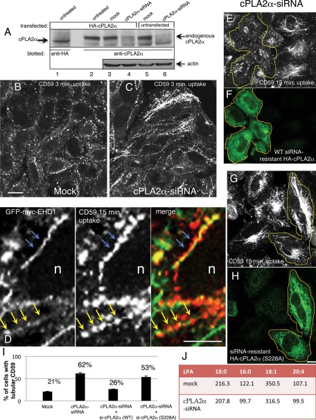
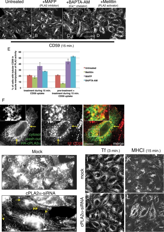
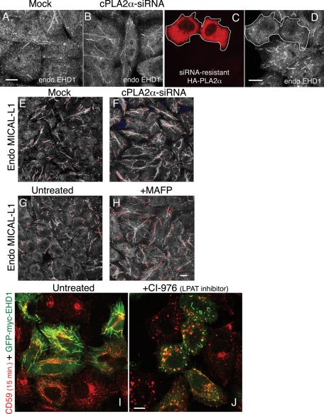
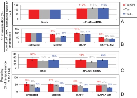
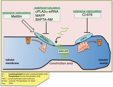
The lipid modifier phospholipase A2 catalyzes the hydrolysis of phospholipids to inverted-cone-shaped lysophospholipids that contribute to membrane curvature and/or tubulation. Conflicting findings exist regarding the function of cytosolic phospholipase A2 (cPLA2) and its role in membrane regulation at the Golgi and early endosomes. However, no studies addressed the role of cPLA2 in the regulation of cholesterol-rich membranes that contain glycosylphosphatidylinositol-anchored proteins (GPI-APs). Our studies support a role for cPLA2α in the vesiculation of GPI-AP-containing membranes, using endogenous CD59 as a model for GPI-APs. On cPLA2α depletion, CD59-containing endosomes became hypertubular. Moreover, accumulation of lysophospholipids induced by a lysophospholipid acyltransferase inhibitor extensively vesiculated CD59-containing endosomes. However, overexpression of cPLA2α did not increase the endosomal vesiculation, implying a requirement for additional factors. Indeed, depletion of the "pinchase" EHD1, a C-terminal Eps15 homology domain (EHD) ATPase, also induced hypertubulation of CD59-containing endosomes. Furthermore, EHD1 and cPLA2α demonstrated in situ proximity (<40 nm) and interacted in vivo. The results presented here provide evidence that the lipid modifier cPLA2α and EHD1 are involved in the vesiculation of CD59-containing endosomes. We speculate that cPLA2α induces membrane curvature and allows EHD1, possibly in the context of a complex, to sever the curved membranes into vesicles.


Figures

FIGURE 1:
FIGURE 1:
Depletion of cPLA2α induces hypertubulation of CD59-containing endosomes. (A) Untransfected (lanes 5 and 6) or HA-cPLA2α–overexpressing HeLa cells (lanes 1–4) were mock treated (lanes 3 and 5) or treated with cPLA2α-siRNA for 2 d (lanes 4 and 6), harvested, and lysed. Lysates were separated by 8% SDS–PAGE, transferred to nitrocellulose filters, and immunoblotted with either mouse anti-HA antibody (lane 1, to identify the band corresponding to the particular α isoform of cPLA2) and anti-cPLA2α antibody (lanes 2–6, to detect endogenous and overexpressed cPLA2α). Actin was probed as a protein loading control (lanes 3–6). Note that a band corresponding to both overexpressed and endogenous cPLA2α is greatly reduced by the siRNA treatment (lanes 4 and 6). (B, C) HeLa cells growing on coverslips were mock treated (B) or treated with cPLA2α–siRNA (C). After 48 h, cells were incubated with mouse anti-CD59 antibody for 3 min at 37°C, acid stripped, and fixed. Internalized CD59 was detected with Alexa 568–conjugated anti-mouse antibody. (D) High magnification of tubular interconnected “beads-on-a-string” endosome. HeLa cells transfected with GFP-myc-EHD1 were allowed to internalize anti-CD59 for 15 min at 37°C, then acid stripped, fixed, and stained with Alexa 568 goat anti-mouse secondary antibody. Blue arrows depict continuous CD59 and EHD1 tubules, and yellow arrows point to the postfixation commonly seen CD59 “beads” within the continuous EHD1-decorated tubular membrane. (E–H) Either siRNA-resistant wild-type HA-cPLA2α (E, F) or active-site mutant (S228A) (G, H) was transfected into cPLA2α-siRNA–treated cells. After 48 h, cells were pulsed with anti-CD59 antibody for 15 min, acid stripped, and fixed. Cells were then stained with rabbit anti-HA antibody to identify cPLA2α-expressing cells, denoted with yellow lines, followed by Alexa 568–conjugated anti-mouse and Alexa 488–conjugated anti-rabbit antibody. (I) Quantification of the percentage of cells with tubular CD59 for mock-treated, cPLA2α-siRNA–treated, and rescue-treated cells by transfecting cells with either siRNA-resistant wild-type HA-cPLA2α or S228A mutant. This experiment was repeated three times, and SE is shown. (J) Cells were either mock treated or treated with cPLA2α-siRNA for 48 h and then scraped and spun down. A small sample of each cell pellet was sonicated and subjected to total protein measurement, whereas the rest of the cell pellet was extracted with acidified 1-butanol (see Materials and Methods). Saturated (18:0, 16:0) and unsaturated (18:1, 20:4) LPA species were analyzed by liquid chromatography–tandem mass spectrometry. Data presented represents the average of two independent experiments, and the concentration of each LPA species is given in ng LPA/mg protein. Bar, 10 μm.
FIGURE 2:
FIGURE 2:
Acute manipulation of cPLA2α affects vesiculation of CD59-containing endosomes. (A–D) HeLa cells growing on coverslips were either untreated (A) or pretreated with either PLA2 inhibitor MAFP (65 μM; B) or calcium chelator BAPTA-AM (50 μM; C) for 1 h or PLA2 activator melittin (2 μM; D) for 30 min. The pretreated cells were then incubated with anti-CD59 antibody for 15 min in the presence of the corresponding pharmacological agents, followed by acid stripping, fixation, and incubation with Alexa 568–conjugated anti-mouse antibody. (E) Quantification of the percentage of cells with tubular CD59 upon the treatment of pharmacological agents was done from three experiments: Cells were pulsed with anti-CD59 antibody in the presence of pharmacological agents for 15 min either with no pretreatment or pretreatment with melittin for 10 min and with MAFP or BAPTA-AM for 20 min. SE is shown. (F) Cells growing on coverslips were transfected with HA-cPLA2α. After 16 h, cells were pulsed with mouse anti-CD59 antibody for 15 min at 37°C and acid stripped, followed by cytosol washout in PBS containing 0.05% saponin and 0.1% BSA for 30 s. After fixation, cells were stained with rabbit anti-HA antibody for 1 h, followed by Alexa 568–conjugated anti-mouse and Alexa 488–conjugated anti-rabbit antibodies. Yellow arrows in F show the colocalization of HA-cPLA2α with internalized CD59. cPLA2α affects cholesterol-containing endosomes. (G–L) Cells were mock treated (G, I, K) or treated with cPLA2α-siRNA (H, J, L). For cholesterol staining (G, H), fixed cells were quenched in 50 mM NH4Cl for 10 min and incubated with 1 mg/ml filipin for 30 min. Yellow arrows in H indicate hypertubulation detected by filipin. For transferrin uptake (I, J), HeLa cells were starved in serum-free DMEM media (containing 0.5% BSA) for 30 min and then incubated with Alexa 568–conjugated transferrin for 3 min, followed by fixation. (K–L) Cells were incubated with anti-MHC I antibody for 15 min at 37°C, acid stripped, and fixed. Internalized MHC I was detected with Alexa 568–conjugated anti-mouse antibody. Bar, 10 μm.
FIGURE 3:
FIGURE 3:
Depletion of cPLA2α induces hypertubulation of EHD1- and MICAL-L1–decorated membranes, whereas inhibition of LPAT induces vesiculation of EHD1-containing membranes. (A–F) HeLa cells growing on coverslips were mock treated (A, E) or treated with cPLA2α–siRNA (B– D, F) for 48 h and fixed. Cells were then stained with anti-EHD1 antibody, followed by Alexa 568–conjugated anti-rabbit antibody (A, B). For rescue experiments, siRNA-resistant HA-cPLA2α was transfected into cPLA2α-depleted cells (C, D) and costained with anti-HA and anti-EHD1 antibodies. The white border denotes transfected cells. (E, F) Cells were stained with mouse anti-MICAL-L1 antibody, followed by Alexa 568–conjugated anti-mouse antibody. (G, H) Cells were either left untreated (G) or pretreated with 65 μM MAFP (H) for 1 h. Cells were then fixed and stained with anti–MICAL-L1 antibodies. Saturated signal captured in images E–H are shown in pseudo red. (I, J) Cells were transfected with GFP-myc-EHD1. After 16 h, cells were either untreated (I) or pretreated with 80 μM CI-976 (J) for 1 h. Both untreated and pretreated cells were then pulsed with anti-CD59 antibody for 15 min in the absence (I) or presence of CI-976 (J), followed by acid stripping, fixation, and incubation with Alexa 568–conjugated anti-mouse antibody. Bar, 10 μm.
FIGURE 4:
FIGURE 4:
cPLA2α activity modestly affects cargoes trafficking through different endocytic pathways. (A–D) The effect of cPLA2α-siRNA and pharmacological agents on the trafficking of Tac-GPI, Tac, and Tac-LL was studied. (A) For the effect of cPLA2α-siRNA on the internalization of cargoes, Tac-GPI, Tac, and Tac-LL were transfected respectively into either mock-treated or cPLA2α-siRNA–treated cells. Cells were then pulsed with mouse anti-Tac antibody for 1 h. After acid stripping and fixation, cells were incubated with Alexa 647 goat anti–mouse F(ab)2 antibody in the presence of saponin. (B) For the effect of pharmacological agents on the internalization of cargoes, cells were untreated or pretreated for 30 min with 2 μM melittin or for 1 h with 65 μM MAFP or 50 μM BAPTA-AM. Cells were then pulsed with anti-Tac antibody for 1 h in the presence of corresponding agents, followed by acid stripping. After fixation, cells were stained with Alexa 647 goat anti–mouse F(ab)2 antibody in the presence of saponin. (C) For the effect of cPLA2α-siRNA on the recycling of cargoes to the PM, after either mock or siRNA treatment, cells were pulsed with anti-Tac antibody for 1 h, stripped, and chased in complete media for 2 h, followed by fixation. (D) For the effect of pharmacological agents on the recycling of cargoes to the PM, cells were pulsed with anti-Tac for 1 h and stripped. Cells were then chased in complete media with different pharmacological agents for 2 h, followed by fixation. Internalized Tac was measured with Alexa 647–conjugated anti–mouse F(ab)2 antibody in the presence of saponin, whereas detection of Tac reappearing at the surface was assessed in the absence of saponin. Percentage of recycling was calculated as a ratio of the reappearing Tac/internalized Tac. Measurements were carried out by flow cytometry analyses of three independent experiments for each plot. Error bars indicate SE.
FIGURE 5:
FIGURE 5:
EHD1, but not dynamin II, participates in the vesiculation of CD59 tubular endosomes. (A–D) HeLa cells growing on coverslips were transfected with the GFP-dynamin II dominant-negative mutant (K44A) for 16 h. Transfected cells are denoted by a white border in B and D. Cells were either incubated with Alexa 568–conjugated transferrin for 10 min and fixed (A, B) or pulsed with anti-CD59 antibody for 15 min at 37°C (C, D). After acid stripping (C, D), cells were fixed and stained with Alexa 568–conjugated anti-mouse antibody. (E) HeLa cells growing in 35-mm dishes were either mock treated or treated with EHD1-siRNA for 48 h. Cells were lysed, and proteins were separated by SDS–PAGE and immunoblotted with antibodies against EHD1 (top) and actin (bottom; loading control). (F, G) HeLa cells growing on coverslips were mock treated (F) or treated with EHD1-siRNA (G). After 48 h, cells were pulsed with anti-CD59 antibody for 15 min at 37°C, stripped, fixed, and then stained with Alexa 568–conjugated anti-mouse antibody. Red boxed areas are shown in higher magnification. Asterisks denote the endocytic recycling compartment. Bar, 10 μm.
FIGURE 6:
FIGURE 6:
cPLA2α and EHD1 are proximally localized and interact in vivo. (A–F) HeLa cells growing on coverslips were cotransfected with GFP-myc-EHD1 and HA-cPLA2α (A, D), HA-MICAL-L1 (positive control; B, E), or HA-JNK1 (negative control; C, F) for 16 h. Coexpression for each pair was evaluated in A–C by staining one set of cells with mouse anti-HA antibody, followed by appropriate secondary antibody. A double stain (mouse anti-HA and rabbit anti-myc antibody) was performed for the proximity assay (D–F; see Materials and Methods). Each red dot detected is a single proximity event between anti-myc and anti-HA antibodies (transfected cells, identified with GFP, are marked with green lines; D–F). (G) Quantification of the number of red dots per transfected cell was done using data from three independent experiments. (H) HeLa cells were grown in 100-mm dishes and transfected with either HA-cPLA2α or HA-JNK1 (negative control). After 20 h, cells were lysed for 1 h in buffer containing 50 mM Tris, pH 7.4, 150 mM NaCl, 0.5% Brij 98, and protease inhibitors. For pull-down, cell lysates were incubated with rabbit anti-EHD1 antibody overnight. Rabbit IP Matrix beads were added to the mixture of cell lysate and EHD1 antibody for 3 h at 4°C. Beads were washed in buffer containing 50 mM Tris, pH 7.4, 150 mM NaCl, and 0.1% Brij 98. Proteins were eluted by adding SDS loading buffer. Samples were subjected to 8% SDS–PAGE, followed by blotting with anti-HA antibody. Red arrow in H indicates HA-cPLA2α pulled down by EHD1. Red star denotes spillover of the IgG heavy chain. (I) The S. cerevisiae yeast strain AH109 was cotransformed with the indicated GAL4-binding domain (GAL4bd) fusion constructs (including GAL4bd-cPLA2α and GAL4bd-MICAL-L1) and GAL4bd-p53 (control), together with the indicated GAL4 transcription activation (GAL4ad) fusion constructs GAL4ad-EHD1 and GAL4ad-SV40 large T-antigen (control). The cotransformants were assayed for their growth on nonselective (+HIS) and selective (–HIS) media. Bar, 10 μm.
FIGURE 7:
FIGURE 7:
The EHD1(∆EH) mutant vesiculates both CD59- and MICAL-L1–containing tubular endosomes in the absence of cPLA2α. (A–D) cPLA2α-siRNA–treated cells were transfected with the mutant GFP-myc-EHD1(∆EH). (A, B) After 16 h, cells were incubated with anti-CD59 antibody for 15 min at 37°C, acid stripped, fixed, and stained with Alexa 568–conjugated anti-mouse antibody. (C, D) After transfection, cells were fixed and stained with anti–MICAL-L1 antibody for 1 h, followed by Alexa 568–conjugated anti-mouse antibody. Yellow borders indicate the transfected cells. White boxed areas are shown in higher magnification. Bar, 10 μm.
FIGURE 8:
FIGURE 8:
A speculative model describing cPLA2α and EHD1 function in vesiculation. cPLA2α is recruited onto the outer membrane leaflet of cholesterol-enriched endosomes containing cargo coming in from the clathrin-independent pathway. As a result, the level of LPL (mostly lysophosphatidic acid) locally increases, generating curvature and giving rise to a constricted region. Membrane-bound cPLA2α engages EHD1 via its interaction (likely in the context of a complex), and vesiculation proceeds, possibly through oligomerization of EHD1 and ATP hydrolysis. Local accumulation of LPL, either by activating cPLA2α with melittin or by inhibiting LPAT activity with CI-976, accelerates fission and vesiculation, whereas interfering with cPLA2α activity by knockdown or inhibition (with the inhibitor MAFP or the chelator BAPTA-AM) slows down vesiculation, and tubular structures are stabilized.

Similar articles

Cited by

References

    1. Bate C, Ingham V, Williams A. Inhibition of phospholipase A(2) increased the removal of the prion derived peptide PrP82-146 from cultured neurons. Neuropharmacology. 2011;60:365–372. - PubMed
    1. Bathena SP, Huang J, Nunn ME, Miyamoto T, Parrish LC, Lang MS, McVaney TP, Toews ML, Cerutis DR, Alnouti Y. Quantitative determination of lysophosphatidic acids (LPAs) in human saliva and gingival crevicular fluid (GCF) by LC-MS/MS. J Pharm Biomed Anal. 2011;56:402–407. - PMC - PubMed
    1. Bechler ME, Doody AM, Ha KD, Judson BL, Chen I, Brown WJ. The phospholipase A enzyme complex PAFAH Ib mediates endosomal membrane tubule formation and trafficking. Mol Biol Cell. 2011;22:2348–2359. - PMC - PubMed
    1. Brown WJ, Chambers K, Doody A. Phospholipase A2 (PLA2) enzymes in membrane trafficking: mediators of membrane shape and function. Traffic. 2003;4:214–221. - PubMed
    1. Cai B, Katafiasz D, Horejsi V, Naslavsky N. Pre-sorting endosomal transport of the GPI-anchored protein, CD59, is regulated by EHD1. Traffic. 2011;12:102–120. - PubMed

Publication types

MeSH terms