. 2012 May 1;188(9):4217-25.
doi: 10.4049/jimmunol.1102885. Epub 2012 Mar 26.

CD80 expression on B cells regulates murine T follicular helper development, germinal center B cell survival, and plasma cell generation

Affiliations

CD80 expression on B cells regulates murine T follicular helper development, germinal center B cell survival, and plasma cell generation

Kim L Good-Jacobson et al. J Immunol. .

Abstract

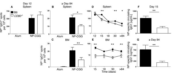
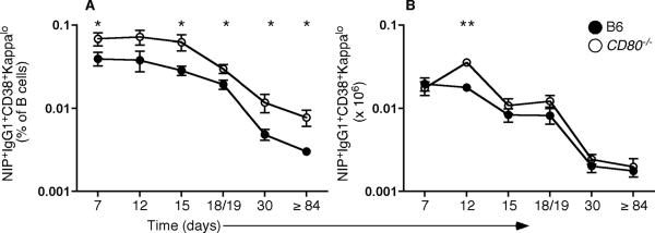
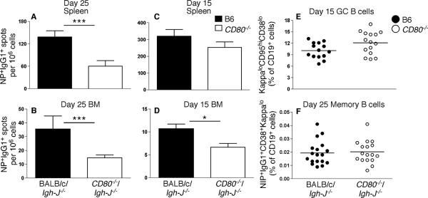
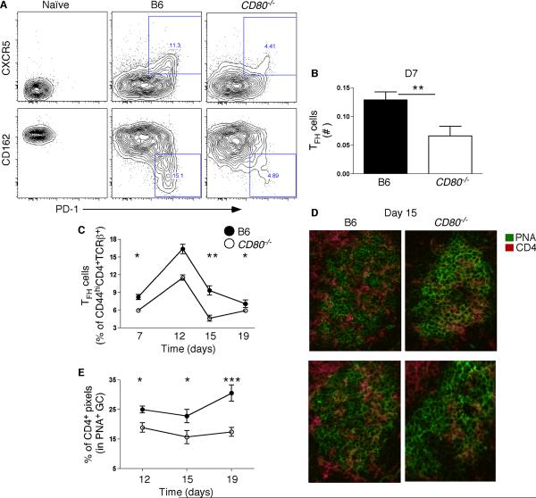
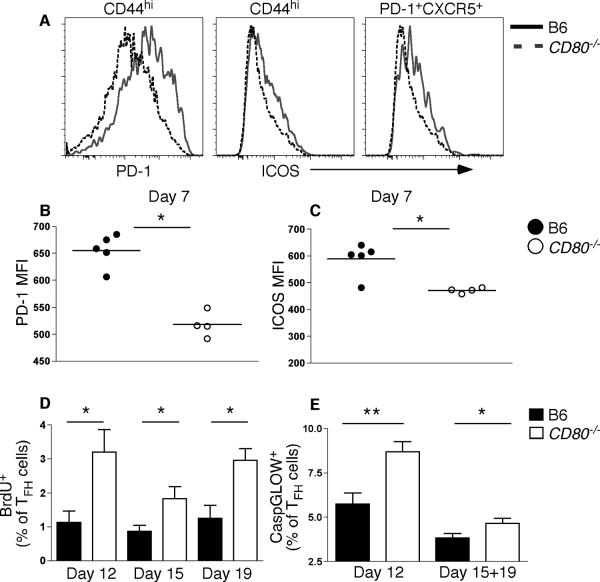
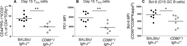
Germinal center (GC) B cells and T follicular helper (T(FH)) cells interact in the production of high-affinity long-lived plasma cells (PCs) and memory B cells, although the mechanisms regulating the formation of these long-lived populations remain unclear. Because CD80 is one of the few markers shared by human and murine memory B cells, we investigated its role in the development of GCs, memory cells, and PCs. In CD80-deficient mice, fewer long-lived PCs were generated upon immunization compared with that in B6 controls. In concert, the absence of CD80 resulted in an increase in apoptotic GC B cells during the contraction phase of the GC. CD80(-/-) mice had fewer T(FH) cells compared with that of B6, and residual T(FH) cells failed to mature, with decreased ICOS and PD-1 expression and decreased synthesis of IL-21 mRNA. Mixed bone marrow chimeras demonstrated a B cell-intrinsic requirement for CD80 expression for normal T(FH) cell and PC development. Therefore, B cell expression of CD80 plays a critical role in regulating B-T interactions in both early and late GC responses. This, in turn, results in impaired ability to produce long-lived PCs. These data provide new insights into the development of GCs and Ab-forming cells and the functions of CD80 in humoral immunity.


Figures

Figure 1
Figure 1. CD80 has a non-redundant role in PC formation
Analysis of long-lived PC responses at multiple time-points post-immunization with NP-CGG in alum. (A–C) ELISpot analysis of NP+IgG1+ AFCs in spleen (A, B) and BM (C) from CD80−/− (white bars) or B6 controls (black bars) day 12 (A; n = 4 per genotype), or ≥ day 84 (B, C; n = 18 per genotype) post-immunization with NP-CGG in alum or alum alone. Data are combined from 5 independent experiments for (B, C). (D, E) CD80−/− (open circles) or B6 (closed circles) controls were immunized with NP-CGG in alum and NP+IgG1+ AFCs in spleen (D) and BM (E) assessed at multiple time-points post-immunization. Data are combined from 2–5 independent experiments per time-point; note the log scale. ≥ day 84 data is the same as is shown in (B, C). (F, G) Circulating IgG1 in CD80−/− and B6 at day 15 (F; n = 12–14 per genotype) and ≥ day 84 (G; n = 10–11 per genotype) post-immunization, respectively. Data are combined from 3 (day 15) or 2 (≥ day 84) independent experiments per time-point. *P < 0.05, **P < 0.01 (Mann-Whitney nonparametric, two-tailed test).
Figure 2
Figure 2. Memory B cell frequency is increased in the absence of CD80
B6 (closed circles) and CD80−/− (open circles) mice were immunized with NP-CGG in alum. Live splenocytes were gated as EMACD19+NIP+IgG1+CD38+kappalo to assess memory B cell frequency (A) and number (B). *P < 0.05, **P < 0.01 (Mann-Whitney nonparametric, two-tailed test). Data shown are from 1–5 independent experiments per time-point (days 18 and 19 were combined; n = 7–17 per genotype per time point); note the log scale. The curves in both (A) and (B) were significantly different (p<0.05), as assessed by a two-way ANOVA analysis.
Figure 3
Figure 3. Increased cell death and proliferation in the GCs of CD80−/− mice
B6 and CD80−/− mice were immunized with NP-CGG in alum, and the GC response assessed at multiple time-points post-immunization. (A, B) Percentages (A) and numbers (B) of splenic B cells (CD19+EMA) with a GC phenotype (CD95hiCD38lokappalo; n = 4–10 per genotype) in CD80−/− mice (open circles) compared to B6 mice (closed circles) were assessed by flow cytometry. (C–D) B6 (closed circles) and CD80−/− (open circles) mice immunized with NP-CGG in alum, were pulsed with 3 mg/mouse of BrdU 2hr before analysis. Splenic GC B cells undergoing cell death were revealed by detection of activated caspases using CaspGLOW immediately after cell harvest at the following time points: days 12, 15 and 19 post-immunization. Summary of frequencies derived from flow cytometry analysis of splenic GC B cells that are BrdU+ (C; data are from 1 experiment per time-point; n = 4–7 per genotype) and CaspGLOW+ (D; data are combined from two experiments per time-point; n = 7–10 per genotype) *P < 0.05 as determined by a two-tailed Student's t test.
Figure 4
Figure 4. B cell intrinsic role for regulation of B cell differentiation
Mixed BM chimeras were made by mixing Igh-J−/− BM with CD80−/− (open bars or open circles) or BALB/c (black bars or closed circles) BM at an 80:20 ratio, and injecting into lethally irradiated BALB/c recipients. FACS analysis of LPS-activated splenic dendritic cells indicated that the degree of chimerization approximated the above ratio (data not shown). Mice were assessed at either days 15 or 25 post-immunization. (A–D) ELISpot analysis of mixed BM chimeras in the spleen (A, C) and BM (B, D). Data are combined from two independent experiments (n = 17–18 per genotype). (E, F) Flow cytometry analyses of GC B cell frequency at day 15 (E) and memory B cell frequency (F) of CD19+ cells. *P < 0.05, ***P < 0.001 (Mann-Whitney nonparametric, two-tailed test).
Figure 5
Figure 5. Reduced frequency of cells of a TFH phenotype in the absence of CD80
(A) Representative flow cytometry analysis of TFH cells from B6 and CD80−/− mice 12 days post-immunization. CD44hiCD4+TCRβ+ cells were gated and PD-1+CXCR5+ or PD-1+CD162lo cells were identified as TFH. (B) Numbers of TFH cells day 7 post-immunization. (C) Flow cytometry analysis of TFH frequency (gated on PD-1+CXCR5+ or PD-1+CD162lo) of CD44hiCD4+TCRβ+ cells in CD80−/− and B6 mice at days 7, 12, 15 and 19 post-immunization with NP-CGG in alum (n = 4–5 for days 7, 12, 15; n = 9–11 for day 19 per genotype). (D, E) CD80−/− mice and their B6 controls were immunized and at multiple time-points post-immunization spleens were frozen. Frozen spleen sections were stained with antibodies to CD4 and PNA. Shown in (D) are representative GCs from two different mice per genotype from day 15 post-immunization. (E) Measurement of % of CD4+ pixels in a GC as defined by PNA staining. 4–5 GCs per spleen were assessed, with two or three spleens per genotype shown. *P < 0.05, **P < 0.01, ***P < 0.001 (Mann-Whitney nonparametric, two-tailed test).
Figure 6
Figure 6. Alterations in TFH phenotype, proliferation and survival in the absence of CD80
(A) Representative flow cytometry analysis of PD-1 and ICOS expression on CD44hiCD4+TCRβ+EMA cells in B6 and CD80−/− mice 7 days post-immunization with NP-CGG in alum. Also shown is ICOS expression on P D-1+CXCR5+ cells. (B–C) CD44hiCD4+TCRβ+EMA cells were analyzed for the mean fluorescence intensity (MFI) of PD-1 (B) and ICOS (C) 7 days post-immunization. Data are representative of multiple independent experiments at days 12, 15 and/or 19. (D, E) BrdU (D) and CaspGLOW (E) analysis of CD44hiPD-1+CCR7lo CD4+ TFH cells in CD80−/− and B6 mice at days 12 (BrdU: n = 4; CaspGLOW: n = 7–8 per genotype), 15 (BrdU: n = 6–7 per genotype) and 19 (BrdU: n = 4–5; CaspGLOW days 15+19: n = 11–14 per genotype) post-immunization. CaspGLOW data are combined from at least 2 independent experiments per time point shown. *P < 0.05, **P < 0.01 (Mann-Whitney nonparametric, two-tailed test).
Figure 7
Figure 7. CD80 regulates T cell phenotype, frequency and function
Mixed BM chimeras with CD80-deficient B cells as described in Methods. (A) Frequency of splenic CD4+ T cells (CD4+TCRβ+EMA) with a TFH phenotype (CD44hiICOS+PD-1+) on day 15 in CD80−/−/ Igh-J−/− (open circles) and Balb/c/Igh-J−/− BM chimeric mice (closed circles), as assessed by flow cytometry. (B) PD-1 MFI of TFH cells at day 15 post-immunization in CD80−/−/ Igh-J−/− (open bars) and Balb/c/Igh-J−/− (black bars) BM chimeric mice. *P < 0.05, **P < 0.01 (Mann-Whitney nonparametric, two-tailed test). Data are representative of 2 independent experiments. (C) Flow cytometry analysis of Bcl-6 protein in GC B cells in CD80−/−/ Igh-J−/− and Balb/c/Igh-J−/− BM chimeric mice. Data are representative of three independent experiments at multiple time-points during the GC response in either BM chimeric or CD80−/− and B6 mice. *P < 0.05, **P < 0.01, ***P < 0.001 (Mann-Whitney nonparametric, two-tailed test).

Similar articles

Cited by

References

    1. Ahmed R, Gray D. Immunological memory and protective immunity: understanding their relation. Science. 1996;272:54–60. - PubMed
    1. Jacob J, Przylepa J, Miller C, Kelsoe G. In situ studies of the primary immune response to (4-hydroxy-3-nitrophenyl)acetyl. III. The kinetics of V region mutation and selection in germinal center B cells. J Exp Med. 1993;178:1293–1307. - PMC - PubMed
    1. Takahashi Y, Cerasoli DM, Dal Porto JM, Shimoda M, Freund R, Fang W, Telander DG, Malvey EN, Mueller DL, Behrens TW, Kelsoe G. Relaxed negative selection in germinal centers and impaired affinity maturation in bcl-xL transgenic mice. J Exp Med. 1999;190:399–410. - PMC - PubMed
    1. Takahashi Y, Dutta PR, Cerasoli DM, Kelsoe G. In situ studies of the primary immune response to (4-hydroxy-3-nitrophenyl)acetyl. V. Affinity maturation develops in two stages of clonal selection. J Exp Med. 1998;187:885–895. - PMC - PubMed
    1. Jacob J, Miller C, Kelsoe G. In situ studies of the antigen-driven somatic hypermutation of immunoglobulin genes. Immunol Cell Biol. 1992;70(Pt 2):145–152. - PubMed

Publication types

MeSH terms