. 2012;7(3):e33657.
doi: 10.1371/journal.pone.0033657. Epub 2012 Mar 16.

HIV-1 Vpr triggers mitochondrial destruction by impairing Mfn2-mediated ER-mitochondria interaction

Affiliations

HIV-1 Vpr triggers mitochondrial destruction by impairing Mfn2-mediated ER-mitochondria interaction

Chih-Yang Huang et al. PLoS One. 2012.

Abstract

Human immunodeficiency virus 1 (HIV-1) viral protein R (Vpr) has been shown to induce host cell death by increasing the permeability of mitochondrial outer membrane (MOM). The mechanism underlying the damage to the mitochondria by Vpr, however, is not clearly illustrated. In this study, Vpr that is introduced, via transient transfection or lentivirus infection, into the human embryonic kidney cell line HEK293, human CD4(+) T lymphoblast cell line SupT1, or human primary CD4(+) T cells serves as the model system to study the molecular mechanism of Vpr-mediated HIV-1 pathogenesis. The results show that Vpr injures MOM and causes a loss in membrane potential (MMP) by posttranscriptionally reducing the expression of mitofusin 2 (Mfn2) via VprBP-DDB1-CUL4A ubiquitin ligase complex, gradually weakening MOM, and increasing mitochondrial deformation. Vpr also markedly decreases cytoplasmic levels of dynamin-related protein 1 (DRP1) and increases bulging in mitochondria-associated membranes (MAM), the specific regions of endoplasmic reticulum (ER) which form physical contacts with the mitochondria. Overexpression of Mfn2 and DRP1 significantly decreased the loss of MMP and apoptotic cell death caused by Vpr. Furthermore, by employing time-lapse confocal fluorescence microscopy, we identify the transport of Vpr protein from the ER, via MAM to the mitochondria. Taken together, our results suggest that Vpr-mediated cellular damage may occur on an alternative protein transport pathway from the ER, via MAM to the mitochondria, which are modulated by Mfn2 and DRP1.


Conflict of interest statement

Competing Interests: The authors have declared that no competing interests exist.

Figures

Figure 1
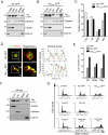
Figure 1. The C-terminal TMD is crucial to the integration of Vpr into MOM.
A, The C-terminal TMD is required for the subcellular distribution of Vpr to mitochondria. B, Using confocal immunofluorescent microscopy, Vpr-GFP was localized on the mitochondria. Mitochondria were labeled using plasmid encoding mitochondria-targeted Discosoma red fluorescent protein (DsRed-Mito). Yellow fluorescence indicates the overlapping regions between the Vpr and mitochondria (white arrow). Bar: 10 µm. C, Treatment with 100 mM sodium carbonate (pH11.5) showed that Vpr and COX IV, as integral proteins of the mitochondrial inner membrane, were resistant to sodium carbonate, while the peripherally associated matrix protein mtHsp70 was separated in the soluble fraction. All experiments were independently repeated three times. D, Mitochondria isolated from HA-Vpr expressing cells were treated with proteinase K. The level of HA-Vpr was decreased after proteinase K treatment, indicating that the N-terminal HA was sensitive to protease. E, The C-terminal GFP fragment of the Vpr-GFP was resistant to proteinase K, indicating that the C-terminus of Vpr was protected. F, Resistance of the C-terminal GFP fragment of Vpr52–96-GFP to proteinase K treatment indicated that C-terminal GFP fragments were oriented toward the mitochondria. G, Following silencing of Tom22 or Tom40, cells were transfected with Vpr-GFP for 48 hours. Although expression of Tom22 or Tom40 was significantly reduced in gene silencing cells, expression of Vpr-GFP was not affected. H, The mitochondrial level of Vpr was not affected either in Tom22KD or Tom40KD cells as shown by Western blotting. I, The relative expression of mitochondrial Vpr-GFP in Tom22KD and Tom40KD cells was similar to that in control cells, although the mitochondrial protein COX IV was markedly reduced. The results are shown as the means ± S.D. of three independent experiments. ***The difference of relative expression is statistically significant (p<0.001) between the control and Tom22KD cells or that and Tom40KD cells.
Figure 2
Figure 2. Transport of Vpr to the ER.
A, Confocal immunofluorescent microscopy localized Vpr at the ER. ER was labeled using plasmid encoding ER-targeted Discosoma red fluorescent protein (DsRed-ER). Yellow fluorescence indicates the overlapped regions between Vpr and ER (white arrow). Bar: 10 µm. B, Treatment with 100 mM sodium carbonate confirmed that Vpr and the ER integral protein Calnexin were present in the alkaline-resistant fraction. C, Isolated microsomal fractions treated with trypsin indicate that Vpr inserted into the ER by the C-terminal TMD. D, Using immunogold electron microscopy, Vpr particles (15 nm) were shown to be located on the membrane of mitochondria (arrowhead) and ER (arrow). These results are representative of three independent experiments.
Figure 3
Figure 3. Vpr is presented in the MAM.
A, Using Percoll self-generating gradient fractionation, Vpr-GFP was localized in the ER, MAM, and mitochondria. Cyto: cytosolic fraction, LM: microsomal fraction, Mito: mitochondrial fraction. B, Likewise, Vpr52–96-GFP was located in the ER, MAM, and mitochondria. C, Relative expression levels of Vpr-GFP and Vpr52–96-GFP in different subcellular fractions are similar. D, Immunofluorescent confocal microscopy showed that Vpr was co-localized with the MAM marker, PSS-1 protein. Bulged MAM became evident in Vpr52–96-GFP expressing cells. Yellow fluorescence (white arrow) indicates an overlap between Vpr (green fluorescence) and MAM (red fluorescence). Bar: 10 µm. E, Colocalization of Vpr52–96-GFP or Vpr-GFP with the marker of ER, MAM and mitochondria was assessed based on confocal images, and 100–120 cells were scored in three independent experiments. The results showed that both proteins were present in the three organelles. F, HEK293 cells were transfected with the plasmid encoding HA-Vpr for 48 hours. Cells were harvested and fractionated on a self-generated Percoll gradient. HA-Vpr was localized in the ER, MAM, and mitochondria. Cyto: cytosolic fraction, LM: microsomal fraction, Mito: mitochondrial fraction. G, HEK293 cells were either transfected with the plasmid encoding Vpr-GFP, Vpr52–96-GFP, or HA-Vpr for 48 hours, or infected with Lenti-Vpr (48 and 72 hours). Cells were harvested and cell cycle was analyzed by flow cytometry. Vpr-induced G2 arrest was observed in Vpr-expressing cells.
Figure 4
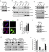
Figure 4. Vpr influences the expression level of GRP78 and Mfn2 proteins.
A, At 48 hours post-transfection, expression of Vpr-GFP or Vpr52–96-GFP up-regulated GRP78 level, but reduced Mfn2 level in HEK293 cells. B, Vpr-related decrease in the expression of Mfn2 was time-dependent after Lenti-Vpr infection. C, Vpr-related loss of mitochondrial membrane potential (MMP) was also time-dependent, indicating that the effect of Vpr on MMP change was gradual, not immediate. D, Infection of HEK293 cells with lentivirus carrying siRNA to Mfn2 markedly reduced protein levels 48 hours after viral treatment. E, Similar to that mediated by Vpr, the Mfn2 silencing-induced loss of MMP was time-dependent. Results are the means ± S.D. of three independent experiments. For panels C and E, * (p<0.05) and *** (p<0.001) indicate significantly different from the control. F, HEK293 cells were transfected with the plasmid encoding HA-Vpr for 48 hours. Cells were harvested and analyzed by Western blotting. The expression of Mfn2 was reduced in HA-Vpr expressing cells.
Figure 5
Figure 5. Vpr protein influences the morphology of mitochondria and integrity of MOM.
A, Electron microscopic analysis of HEK293 cells showed that mitochondria were intact with a double-layer membrane and regular arrangement of cristae. Bar: 200 nm. B, Similar to wild-type cells, expression of GFP did not influence the morphology of the mitochondria. Bar: 200 nm. C, Following Vpr-GFP expression, marked changes in the architecture of the cristae was observed. Vpr led to swollen cristae, condensed matrix (arrowhead) and gradually disappearance of outer membrane (arrow). Bar: 200 nm. D, The same phenomenon was observed with Vpr52–96-GFP expressing cells. Mitochondria were condensed with swollen cristae. Bar: 200 nm. E, Based on TEM images, Vpr induced mitochondrial fragmentation and the length of mitochondria was evidently shorter. Twenty five cells were scored in three independent experiments with means ± S.D. F, Vpr also resulted in the condensation of mitochondria. For panels E and F, 25 cells were scored in three independent experiments with means ± S.D. *** indicates statistically significant difference (p<0.001) between the control and Vpr-expressing cells.
Figure 6
Figure 6. Knockdown of DRP1 (DRP1KD) expression induces the accumulation of Vpr in the MAM. Ectopic expression of Vpr, on the other hand, induces nuclear translocation of DRP1.
A, Treatment with siRNA for 48 hours reduced the expression of DRP1 by approximately 80%. B, In DRP1KD cells, protein levels of Vpr, Calnexin and GRP78 were increased in the MAM, while those of Vpr were decreased in the ER and mitochondria, indicating that the knockdown of DRP1 expression induced the accumulation of Vpr in the MAM. The results are representative of two independent experiments. C, The presence of Vpr or Vpr52–96 increased the levels of DRP1, in particular phosphorylated DRP1 (pDRP1), in the nucleus, as determined by Western blotting. These results are representative of two independent experiments. D, As shown by confocal immunofluorescence microscopy, ectopic expression of Vpr52–96 increased nuclear levels of Vpr and DRP1 (arrows with white margin). In some cells, both Vpr and DRP1 were co-localized in the nucleolus (white arrows). Bar: 10 mm. E, The increase of nuclear DRP1 in Vpr- or Vpr52–96-expressing cells. Fifty cells were scored in three independent experiments with means ± S.D. * (p<0.05) and ** (p<0.01) indicate significantly different from the control. F, HA-Vpr increased nuclear levels of DRP1. G, and H, The effect of gene knockdown of Mfn2 (Mfn2KD) or ATAD3A (ATAD3AKD) on the organelle distribution of Vpr was determined by Western blotting. Vpr level was increased in the cytosolic fractions, but it was reduced in the mitochondrial fractions in both Mfn2KD and ATAD3AKD cells. Band intensities were calculated using Image J. Relative intensities are shown at the bottom of each panel.
Figure 7
Figure 7. Transportation of Vpr protein from the ER/MAM to the mitochondria as determined by the time-lapse confocal fluorescence microscopy.
A, Confocal fluorescence time-lapse images of Vpr52–96-GFP and CFP-ER co-expressed HEK293 cells show the formation of Vpr-containing transport vesicles (green fluorescence) from the ER/MAM (red fluorescence; yellow fluorescence indicates the merge of the green and the red fluorescence. Arrows indicate the region (ER/MAM, yellow fluorescence) where the Vpr52–96-GFP emerges. Intensity of yellow fluorescence in the ER/MAM decreases following the appearance of Vpr-containing transport vesicles (green fluorescence, T = 14 s–28 s). B, Fusion of the Vpr-containing transport vesicles (green fluorescence, Vpr52–96-GFP) into the mitochondria (red fluorescence, MitoTracker-Red) in real time. As shown in time-lapse series, Vpr-containing transport vesicle (green fluorescence) approached a mitochondrion (red fluorescence, T = 28 s), and then fused into the mitochondrion (T = 56 s). Arrows indicate Vpr52–96-GFP molecules that merge into the mitochondrion, and the fusion spot appears as yellow fluorescence (arrow). Results of the confocal fluorescence microscopy demonstrate that Vpr is budding off from the ER/MAM and forms within a spherical vesicle, which moves to the vicinity of the mitochondrion and then fuse into the organelle.
Figure 8
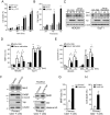
Figure 8. Vpr-mediated mitochondrial damage causes cell death in HEK293 and CD4+ T cells under normal growth condition or serum starvation.
A, HEK293 cells were transfected with GFP vector the plasmid encoding Vpr-GFP or Vpr52–96-GFP, and harvested at different time (hours) post-transfection for PI staining. The percentage of dead cells among GFP-expressing cells was determined by flow cytometry. *** (p<0.001) indicates significantly different from the GFP vector control. B, HEK293 cells were infected with lenti-vector (control) or Lenti-Vpr and harvested at different time (hours) post-infection for PI staining. The percentage of dead cells was determined by flow cytometry. *** (p<0.001) indicates significantly different from the control. C, HEK293 and SupT1 cells were grown in 10% FBS or starved for 24 hours, and infected with Vpr-expressing lentivirus for 48 and 72 hours. The expression of Mfn2 was decreased in serum-starved HEK293 or SupT1 cells. The quantitative expression of Mfn2 was measured by Image J and normalized with the expression of β-actin. C indicates Vpr negative lentiviral control. D, MMP loss was determined after Vpr-expressing lentivirus infection. Vpr significantly impaired MMP in serum-starved HEK293 and SupT1 cells. E, Vpr expression led to cell death in serum-starved HEK293 and SupT1 cells. For panels D and E, results are the means ± S.D. of three independent experiments. * (p<0.05), ** (p<0.01) and ** (p<0.001) indicate significantly higher than the Vpr negative lentiviral control (Con.). † (p<0.05) and †† (p<0.01) indicate significantly different between 10% FBS and serum starvation. F, Human primary CD4+ T cells were isolated from peripheral blood mononuclear cell (PBMC) and infected with Vpr-expressing lentivirus for 72 hours. The expression of Mfn2 was decreased and the expression of nuclear DRP1 was increased in human primary CD4+ cells. The relative expression levels of Mfn2 and DRP1 were measured by Image J and normalized with the expression of β-actin. G, MMP loss was determined after Vpr-expressing lentivirus infection. Vpr led to a significant MMP loss in human primary CD4+ T cells. H, Vpr expression led to cell death in human primary CD4+ T cells. For panels G and H, results are the means ± S.D. of three independent experiments. ** (p<0.01) and *** (p<0.001) indicate significantly higher than control human primary CD4+ T cells. Band intensities were calculated using Image J. Relative intensities are shown at the bottom of each panel.
Figure 9
Figure 9. Enforced expression of Mfn2 and DRP1 relieves Vpr-induced apoptosis.
After transient transfection of Mfn2, or DRP1 expression plasmid for 24 hours, cells were infected with Vpr-expressing lentivirus for 72 hours. A, In contrast to Lenti-Vpr negative (-) HEK293 cells (medium control and vector control), the 89 kDa cleavage fragment of PARP appeared exclusively in Lenti-Vpr positive (+) HEK293 cells. PARP cleavage was reduced in Mfn2-, DRP1- and Mfn2/DRP1-overexpressed Lenti-Vpr (+) HEK293 cells. B, PARP cleavage occurred only in Lenti-Vpr (+) SupT1 cells. The cleavage of PARP was reduced in Mfn2-, DRP1- and Mfn2/DRP1-expressed Lenti-Vpr (+) SupT1 cells. C, Overexpression of Mfn2, DRP1, and Mfn2/DRP1 decreased the percentage of MMP loss in Lenti-Vpr (+) HEK293 and SupT1 cells. D, Overexpression of Mfn2, DRP1, and Mfn2/DRP1 reduced Vpr-induced apoptosis. Results are the means ± S.D. of three independent experiments. For panels C and D, † (p<0.05) and †† (p<0.01) indicate significantly lower than Lenti-Vpr (+) cells pretreated with mock (medium control) or vector transfection.
Figure 10
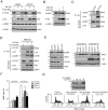
Figure 10. Vpr downregulated Mfn2 expression via VprBP-DDB1-CUL4A ubiquitin ligase complex.
A, HEK293 cells were transfected respectively with plasmids encoding HA-Vpr and Flag-Mfn2 for 32 hours and treated with proteasome inhibitor MG132 (5 µM) for 16 hours. The expression of Mfn2 was recovered after MG132 treatment. B, HEK293 cells were infected with Lenti-Vpr for 56 hours and treated with proteasome inhibitor MG132 (5 µM) for 16 hours. The expression of Mfn2 was not decreased after MG132 treatment. C, HEK293 cells were transfected with the plasmid encoding HA-Vpr and harvested after 48 hrs. Cell lysates were immunoprecipitated with anti-HA antibody, and detected by Western blotting. D, HEK293 cells were transfected respectively with plasmids encoding HA-Vpr and Flag-Mfn2 for 32 hours and treated with MG132 (5 µM) for 16 hours. Cell lysates were immunoprecipitated with anti-HA antibodies, and the coimmunoprecipitated proteins were detected by Western blotting. * indicates the ubiquitinated Mfn2. E, HEK293 cells were silenced by shRNA against DDB1, VprBP or CUL4A, and infected with lentivirus carrying Vpr for 72 hours. The ubiquitinated Mfn2 was detected in VprBPKD, DDB1KD, CUL4AKD cells. Moreover, Vpr infection did not decrease Mfn2 expression in VprBPKD, DDB1KD, CUL4AKD cells. * indicates the ubiquitinated Mfn2. F, The MMP loss was determined by flow cytometry after Lenti-Vpr infection. Knockdown of DDB1, VprBP and CUL4A reduced the percentage of MMP loss after Lenti-Vpr infection. G, HEK293 cells were infected with Lenti-Vpr for 24 hours and transfected with the plasmid encoding FLAG-Mfn2 for 48 hours. Cells were harvested and analyzed by Western blotting and flow cytometry. Band intensities were calculated using Image J. Relative intensities are shown at the bottom of each panel.

Similar articles

Cited by

References

    1. Emerman M, Malim MH. HIV-1 regulatory/accessory genes: keys to unraveling viral and host cell biology. Science. 1998;280:1880–1884. - PubMed
    1. Bukrinsky M, Adzhubei A. Viral protein R of HIV-1. Rev Med Virol. 1999;9:39–49. - PubMed
    1. Muthumani K, Choo AY, Hwang DS, Chattergoon MA, Dayes NN, et al. Mechanism of HIV-1 viral protein R-induced apoptosis. Biochem Biophys Res Commun. 2003;304:583–592. - PubMed
    1. Andersen JL, Planelles V. The role of Vpr in HIV-1 pathogenesis. Curr HIV Res. 2005;3:43–51. - PubMed
    1. Muthumani K, Choo AY, Premkumar A, Hwang DS, Thieu KP, et al. Human immunodeficiency virus type 1 (HIV-1) Vpr-regulated cell death: insights into mechanism. Cell Death Differ. 2005;12(Suppl 1):962–970. - PubMed

Publication types

MeSH terms

Substances