. 2012;8(3):e1002564.
doi: 10.1371/journal.pgen.1002564. Epub 2012 Mar 15.

Autoregulation of the Drosophila Noncoding roX1 RNA Gene

Affiliations

Autoregulation of the Drosophila Noncoding roX1 RNA Gene

Chiat Koo Lim et al. PLoS Genet. 2012.

Abstract

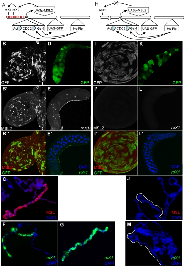
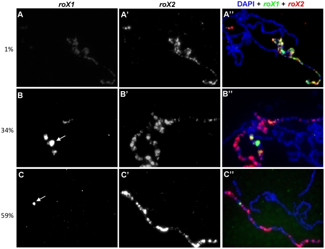
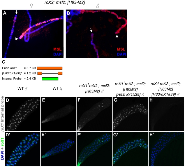
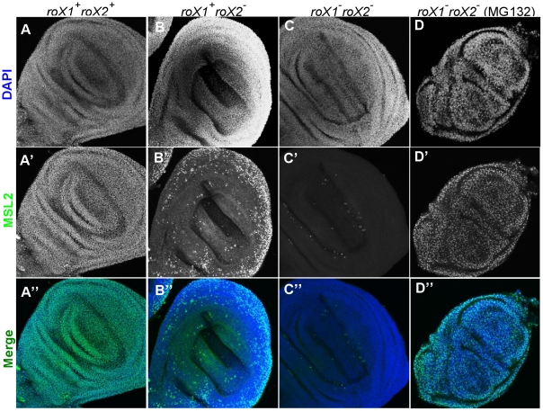
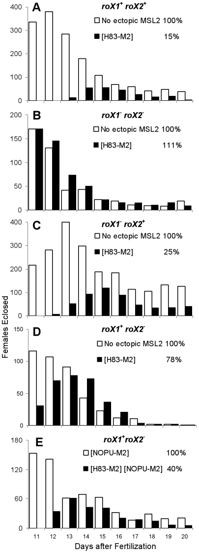
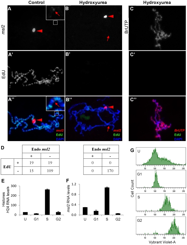
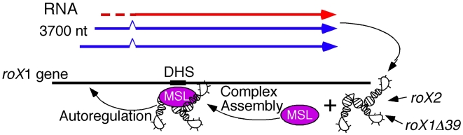
Most genes along the male single X chromosome in Drosophila are hypertranscribed about two-fold relative to each of the two female X chromosomes. This is accomplished by the MSL (male-specific lethal) complex that acetylates histone H4 at lysine 16. The MSL complex contains two large noncoding RNAs, roX1 (RNA on X) and roX2, that help target chromatin modifying enzymes to the X. The roX RNAs are functionally redundant but differ in size, sequence, and transcriptional control. We wanted to find out how roX1 production is regulated. Ectopic DC can be induced in wild-type (roX1(+) roX2(+)) females if we provide a heterologous source of MSL2. However, in the absence of roX2, we found that roX1 expression failed to come on reliably. Using an in situ hybridization probe that is specific only to endogenous roX1, we found that expression was restored if we introduced either roX2 or a truncated but functional version of roX1. This shows that pre-existing roX RNA is required to positively autoregulate roX1 expression. We also observed massive cis spreading of the MSL complex from the site of roX1 transcription at its endogenous location on the X chromosome. We propose that retention of newly assembled MSL complex around the roX gene is needed to drive sustained transcription and that spreading into flanking chromatin contributes to the X chromosome targeting specificity. Finally, we found that the gene encoding the key male-limited protein subunit, msl2, is transcribed predominantly during DNA replication. This suggests that new MSL complex is made as the chromatin template doubles. We offer a model describing how the production of roX1 and msl2, two key components of the MSL complex, are coordinated to meet the dosage compensation demands of the male cell.


Conflict of interest statement

The authors have declared that no competing interests exist.

Figures

Figure 1
Figure 1. MSL proteins alone cannot drive roX1 expression late in development.
A) 4 day old larvae were heatshocked to induce expression of Flp, resulting in the removal of the blocking sequence from GAL4 and subsequent expression of both MSL2 and GFP. MSL2 is expected to initiate roX transcription and MSL complex assembly. (B) GFP+ clones mark imaginal disc cells that have successfully removed the blocking sequences from GAL4 (B′–B″). Induction of MSL2 results in punctate subnuclear foci in imaginal disc cells. (C) MSL2 immunostaining of polytene chromosome shows late MSL2 paints the entire X chromosome. (D) Whole salivary gland showing GFP induced in some cells. (E–E′) roX1 FISH of whole mount of similar GFP+ salivary glands or (F) polytene squashes shows successful induction of roX1 expression in a subset of cells. (G) roX1 FISH of wildtype males (H) The same experiment was repeated in roX1+roX2 larvae. However, in the absence of roX2, MSL2 fails to drive roX1 expression. (I) Despite the presence of GFP+ (late MSL2 expressing) cells, MSL2 is not detectable over the X in (I′–I″) imaginal disc cells or (J) polytene chromosomes. (K) Whole salivary gland showing successful GFP expression in roX1+roX2 larvae. (L–L′) Expression of roX1 is never observed painting the X or as nascent transcripts at band 3F in separately processed GFP+ glands or on (M) polytene squashes.
Figure 2
Figure 2. Late induction of roX1 expression requires roX2 RNA.
In nuclei where dosage compensation was successfully turned on after late msl2 induction, extensive roX2 was observed painting the entire X chromosome. (A) However, only 1% of the chromosomes showed extensive roX1 painting. 34% and 59% of chromosomes showed roX1 expression confined to several Mbp around (B) or just at the endogenous roX1 locus (C), respectively. The remaining chromosomes (6%) had no roX1 expression despite the presence of roX2 (data not shown). roX1 and roX2 were detected by biotin (green, A–C) and digoxigenin (red, A′–C′) labeled antisense riboprobes, respectively. The merged figure is shown in A″–C″. White arrows denote the endogenous roX1 locus at band 3F.
Figure 3
Figure 3. roX1 RNA is needed to sustain endogenous roX1 transcription in males.
X chromosomes from neighboring cells display a mosaic pattern in which the MSL complex either succeeded (arrowhead) or failed (arrow) to paint the X from roX2; msl2; H83M2 female (A) and male (B) salivary glands. (C) Endogenous roX1 and H83roX1Δ39 transcripts (Orange) and antisense riboprobe recognizing only full length roX1 (green). Whole mount roX1 FISH using the internal probe on salivary glands from (D) wild type male, (E) wild type female, (F) roX1+ roX2/Y; msl2; H83M2 mosaic male, (G) roX1+roX2/Y; msl2; H83M2 H83-roX1Δ39/+ male, (H) roX1 roX2 /Y; msl2; H83M2 H83-roX1Δ39/+ male. The X chromosomes in G are fully painted in all cells with MSL complex relying upon roX1-Δ39 RNA (Figure S1D), but the truncated roX1 RNA is not recognized by the internal probe.
Figure 4
Figure 4. Dosage compensation fails in many diploid cells relying exclusively on roX1 and H83M2.
(A–A″) Males and females relying entirely on H83M2 showed subnuclear MSL2 staining in all wing imaginal disc cells when both roX1 and roX2 RNAs were present. (B–B″) Many cells lacked dosage compensation if roX1 + was the only source of roX RNA. (C–C″) MSL2 did not accumulate over the X when both roX1 and roX2 were absent. (D–D″) Nuclear staining of MSL2 was easily detected in the absence of roX RNA after treatment with MG132, a proteasome inhibitor.
Figure 5
Figure 5. roX2 mutant females escape the toxic effects of H83M2.
Adult females eclosing each day were counted from a population of eggs laid on day one. White bars show non-transgenic females without ectopic MSL2 (except E), and black bars show females carrying the H83M2 transgene. Each experiment varied the roX genotype. (A) roX1+ roX2+, (B) no roX, (C) roX2+ only, (D) roX1+ only, (E) roX1+ only. Cumulative viability of H83M2 females is shown as a percentage of their non-transgenic sisters.
Figure 6
Figure 6. Transcription of msl2 is correlated with cell cycle.
Nascent msl2 transcripts were detected with antisense FISH riboprobe in salivary glands. (A) Transgenic H83M2 expression (bright signal indicated by red triangle) was observed at the 87A insertion site in all nuclei. Hybridization to the endogenous msl2 locus at 23F (faint band indicated by red arrow in inset) was seen in only a minority of nuclei. Because of the difference in signal intensities between the two msl2 loci, the inset is enhanced to show the weaker signal. (A′) EdU incorporation shows that this is one of the few nuclei undergoing endoreplication. (B) After treatment with 1 mM of HU (hydroxyurea), no cell transcribed the endogenous msl2 gene (red arrow) but the transcription of H83M2 continued (red triangle). (B′) HU blocked EdU incorporation from any cell. (C) Simultaneous treatment of salivary glands with HU, BrUTP, and EdU showed that blocking replication did not inhibit bulk transcription in these cells. (D) Many nuclei without (left table, N = 162) or with (right table, N = 170) HU treatment were scored for expression of endogenous msl2 and EdU incorporation. Nascent transcripts were detected at the H83M2 transgene in 100% of the nuclei (data not represented in the table). After sorting growing S2 cells into their respective phase of cell cycle via FACS, qPCR was done to quantify H2A (E) and msl2 (F) transcripts levels normalized to PKA. (G) The FACS profile of unsorted (U) and sorted S2 cells (G1, S and G2 cell cycle). The sorted cells have a slightly higher content of Vybrant Violet-A dye because the cells are collected into tubes containing Vybrant Violet-A. The Y-axes are drawn on different scales.
Figure 7
Figure 7. Autoregulation model.
The earliest roX1 transcripts (red) made at blastoderm originate from an uncharacterized MSL-independent promoter. This RNA may assemble with MSL protein subunits to produce the first functional MSL complexes needed to bind the internal DHS enhancer that drives sustained transcription (blue) from the male-specific promoters. When present, roX2 RNA can also drive roX1 transcription. Components of the replication pre-initiation complex also bind the DHS sequence in male cells (Figure S8A). The msl2 transcripts are made predominantly during replication and new MSL2 protein is needed to assemble and stabilize newly made roX1 RNA.

Similar articles

Cited by

References

    1. Khalil AM, Guttman M, Huarte M, Garber M, Raj A, et al. Many human large intergenic noncoding RNAs associate with chromatin-modifying complexes and affect gene expression. Proc Natl Acad Sci U S A. 2009;106:11667–11672. - PMC - PubMed
    1. Orom UA, Derrien T, Beringer M, Gumireddy K, Gardini A, et al. Long noncoding RNAs with enhancer-like function in human cells. Cell. 2010;143:46–58. - PMC - PubMed
    1. Deng X, Meller VH. Non-coding RNA in fly dosage compensation. Trends Biochem Sci. 2006;31:526–532. - PubMed
    1. Gelbart M, Kuroda M. Drosophila dosage compensation: a complex voyage to the X chromosome. Development. 2009;136:1399–1410. - PMC - PubMed
    1. Hilfiker A, Hilfiker-Kleiner D, Pannuti A, Lucchesi JC. mof, a putative acetyl transferase gene related to the Tip60 and MOZ human genes and to the SAS genes of yeast, is required for dosage compensation in Drosophila. EMBO J. 1997;16:2054–2060. - PMC - PubMed

Publication types

MeSH terms

LinkOut - more resources