

Brain region differences in regulation of Akt and GSK3 by chronic stimulant administration in mice
- PMID: 22434044
- PMCID: PMC3335955
- DOI: 10.1016/j.cellsig.2012.03.001
Brain region differences in regulation of Akt and GSK3 by chronic stimulant administration in mice
Abstract
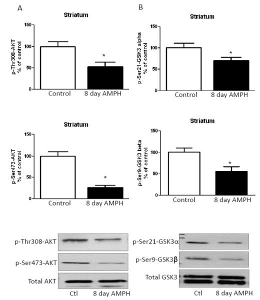
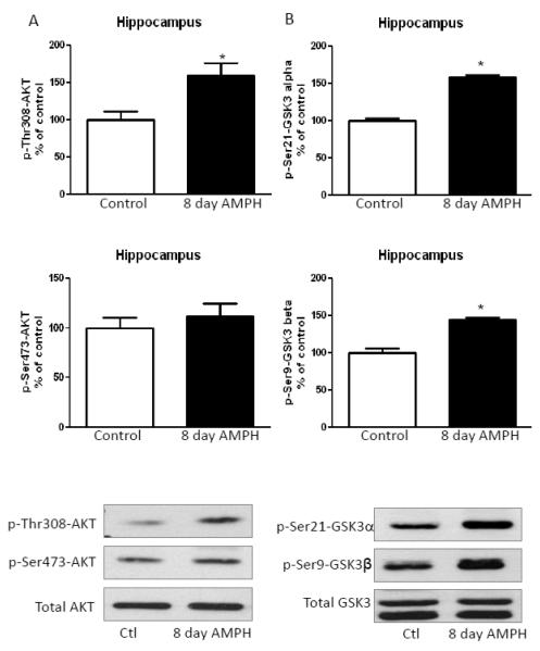
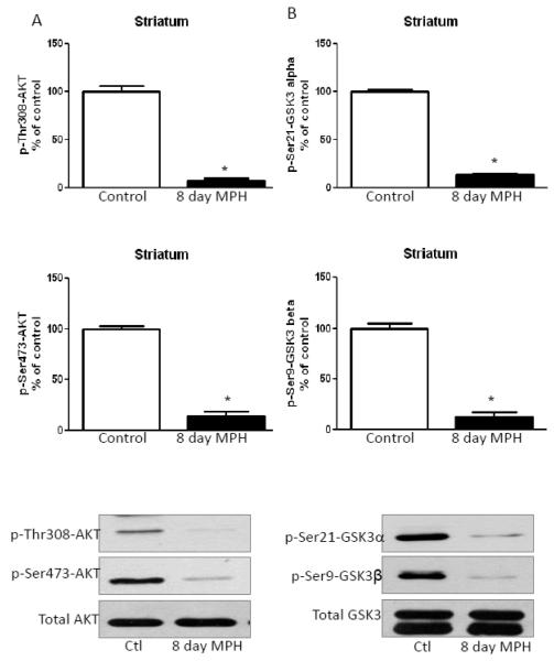
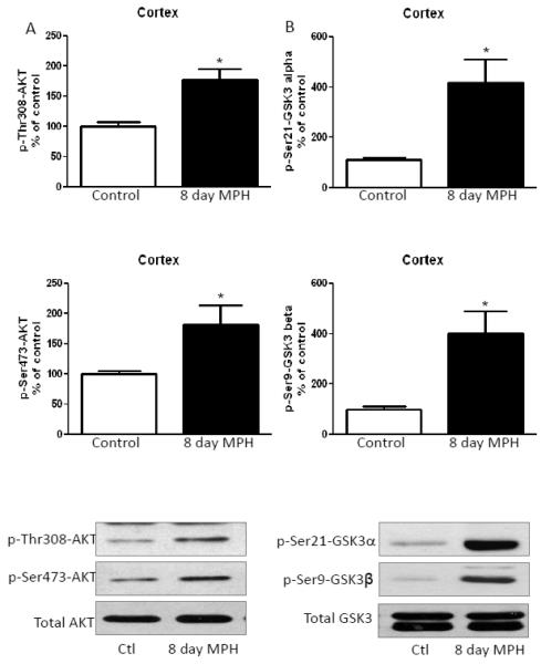
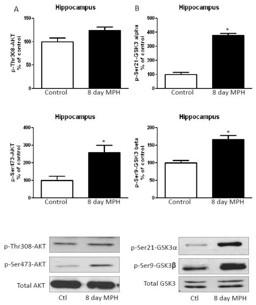
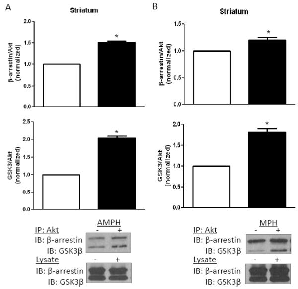
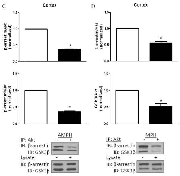
Acute amphetamine administration activates glycogen synthase kinase-3 (GSK3) by reducing its inhibitory serine-phosphorylation in mouse striatum and cerebral cortex. This results from Akt inactivation and is required for certain behavioral effects of amphetamine, such as increased locomotor activity. Here we tested if regulation of Akt and GSK3 was similarly affected by longer-term administration of amphetamine, as well as of methylphenidate, since each of these is administered chronically in patients with attention deficit hyperactivity disorder (ADHD). Akt is activated by post-translational phosphorylation on Thr308, and modulated by Ser473 phosphorylation, whereas phosphorylation on Ser21/9 inhibits the two GSK3 isoforms, GSK3α and GSK3β. After eight days of amphetamine or methylphenidate treatment, striatal Akt and GSK3 were dephosphorylated similar to reported changes after acute amphetamine treatment. Oppositely, in the cerebral cortex and hippocampus Akt and GSK3 phosphorylation increased after eight days of amphetamine or methylphenidate treatment. These opposite brain region changes in Akt and GSK3 phosphorylation matched opposite changes in the association of Akt with β-arrestin and GSK3, which after eight days of amphetamine treatment were increased in the striatum and decreased in the cerebral cortex. Thus, whereas the acute dephosphorylating effect of stimulants on Akt and GSK3 in the striatum was maintained, the response switched in the cerebral cortex after eight days of amphetamine or methylphenidate treatment to cause increased phosphorylation of Akt and GSK3. These results demonstrate that prolonged administration of stimulants causes brain region-selective differences in the regulation of Akt and GSK3.
Copyright © 2012 Elsevier Inc. All rights reserved.
Figures

Similar articles
-
Examination of methylphenidate-mediated behavior regulation by glycogen synthase kinase-3 in mice.Eur J Pharmacol. 2013 Jan 5;698(1-3):252-8. doi: 10.1016/j.ejphar.2012.10.018. Epub 2012 Oct 23. Eur J Pharmacol. 2013. PMID: 23099259 Free PMC article.
-
Striatal Akt/GSK3 signaling pathway in the development of L-Dopa-induced dyskinesias in MPTP monkeys.Prog Neuropsychopharmacol Biol Psychiatry. 2010 Apr 16;34(3):446-54. doi: 10.1016/j.pnpbp.2009.12.011. Epub 2009 Dec 16. Prog Neuropsychopharmacol Biol Psychiatry. 2010. PMID: 20026151
-
Glycogen synthase kinase-3 levels and phosphorylation undergo large fluctuations in mouse brain during development.Bipolar Disord. 2012 Dec;14(8):822-30. doi: 10.1111/bdi.12023. Bipolar Disord. 2012. PMID: 23167932 Free PMC article.
-
An update on the pharmacokinetic considerations in the treatment of ADHD with long-acting methylphenidate and amphetamine formulations.Expert Opin Drug Metab Toxicol. 2019 Nov;15(11):937-974. doi: 10.1080/17425255.2019.1675636. Epub 2019 Nov 8. Expert Opin Drug Metab Toxicol. 2019. PMID: 31581854 Review.
-
Glycogen synthase kinase 3 (GSK3) in the heart: a point of integration in hypertrophic signalling and a therapeutic target? A critical analysis.Br J Pharmacol. 2008 Mar;153 Suppl 1(Suppl 1):S137-53. doi: 10.1038/sj.bjp.0707659. Epub 2008 Jan 21. Br J Pharmacol. 2008. PMID: 18204489 Free PMC article. Review.
Cited by
-
Transcriptional signatures of participant-derived neural progenitor cells and neurons implicate altered Wnt signaling in Phelan-McDermid syndrome and autism.Mol Autism. 2020 Jun 19;11(1):53. doi: 10.1186/s13229-020-00355-0. Mol Autism. 2020. PMID: 32560742 Free PMC article.
-
Targeted Treatments for Fragile X Syndrome.Adv Neurobiol. 2023;30:225-253. doi: 10.1007/978-3-031-21054-9_10. Adv Neurobiol. 2023. PMID: 36928853
-
Overlapping Molecular Pathways Leading to Autism Spectrum Disorders, Fragile X Syndrome, and Targeted Treatments.Neurotherapeutics. 2021 Jan;18(1):265-283. doi: 10.1007/s13311-020-00968-6. Epub 2020 Nov 19. Neurotherapeutics. 2021. PMID: 33215285 Free PMC article. Review.
-
Deletion of GSK3β in D2R-expressing neurons reveals distinct roles for β-arrestin signaling in antipsychotic and lithium action.Proc Natl Acad Sci U S A. 2012 Dec 11;109(50):20732-7. doi: 10.1073/pnas.1215489109. Epub 2012 Nov 27. Proc Natl Acad Sci U S A. 2012. PMID: 23188793 Free PMC article.
-
Glycogen synthase kinase-3β inhibition in the medial prefrontal cortex mediates paradoxical amphetamine action in a mouse model of ADHD.Front Behav Neurosci. 2015 Mar 20;9:67. doi: 10.3389/fnbeh.2015.00067. eCollection 2015. Front Behav Neurosci. 2015. PMID: 25852508 Free PMC article.
References
Publication types
MeSH terms
Substances
Grants and funding
LinkOut - more resources
Full Text Sources
Molecular Biology Databases
