Imiquimod attenuates the growth of UVB-induced SCC in mice through Th1/Th17 cells
- PMID: 22431065
- PMCID: PMC3397281
- DOI: 10.1002/mc.21901
Imiquimod attenuates the growth of UVB-induced SCC in mice through Th1/Th17 cells
Abstract
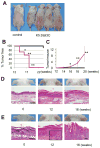
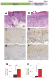
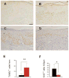
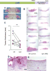
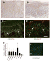
Imiquimod (IMQ), a Toll-like receptor (TLR) 7/8 agonist, has been used to treat various skin neoplasms, including genital warts, actinic keratoses, and superficial basal cell carcinomas. Although IMQ has been recognized to activate both innate and adaptive immunity, the underlying mechanism(s) by which IMQ exerts its anti-tumor activity in vivo remains largely unknown. In this study, we took advantage of skin cancer-prone mice to characterize the effects of IMQ on ultraviolet irradiation (UV)-induced de novo carcinogenesis. Transgenic mice with keratinocytes expressing constitutively activated Stat3 (K5.Stat3C mice) developed squamous cell carcinomas (SCC in situ) as early as after 14 wk of UVB irradiation, while wild-type mice required much higher doses of UVB with more than 25 wk of UVB irradiation to produce SCC. Topical treatment of K5.Stat3C mice with IMQ attenuated UVB-induced epidermal dysplasia (SCC in situ). In addition, SCC growth due to increased total irradiation doses was significantly attenuated by IMQ treatment. Topical IMQ treatment induced T cell and plasmacytoid dendritic cell infiltrates at the tumor sites, where levels of IL-12/23p40, IL-12p35, IL-23p19, IL-17A, and IFN-γ mRNAs were up-regulated. Immunohistochemistry revealed T cell infiltrates consisting of T1, Th17, and CD8(+) T cells. We speculate that topical IMQ treatment attenuates the de novo growth of UVB-induced SCC through activation of Th17/Th1 cells and cytotoxic T lymphocytes.
Keywords: TLR agonist; Th1/Th17; UVB-induced skin cancer.
© 2012 Wiley Periodicals, Inc.
Conflict of interest statement
The authors state no conflict of interest.
Figures
Similar articles
-
Effects of Topical Application of Betamethasone on Imiquimod-induced Psoriasis-like Skin Inflammation in Mice.Kobe J Med Sci. 2016 Sep 9;62(4):E79-E88. Kobe J Med Sci. 2016. PMID: 28239073 Free PMC article.
-
Constitutive activation and targeted disruption of signal transducer and activator of transcription 3 (Stat3) in mouse epidermis reveal its critical role in UVB-induced skin carcinogenesis.Oncogene. 2009 Feb 19;28(7):950-60. doi: 10.1038/onc.2008.453. Epub 2009 Jan 12. Oncogene. 2009. PMID: 19137019 Free PMC article.
-
IL-27 activates Th1-mediated responses in imiquimod-induced psoriasis-like skin lesions.J Invest Dermatol. 2013 Feb;133(2):479-88. doi: 10.1038/jid.2012.313. Epub 2012 Sep 6. J Invest Dermatol. 2013. PMID: 22951728
-
Topical imiquimod or fluorouracil therapy for basal and squamous cell carcinoma: a systematic review.Arch Dermatol. 2009 Dec;145(12):1431-8. doi: 10.1001/archdermatol.2009.291. Arch Dermatol. 2009. PMID: 20026854 Review.
-
UV-Induced Molecular Signaling Differences in Melanoma and Non-melanoma Skin Cancer.Adv Exp Med Biol. 2017;996:27-40. doi: 10.1007/978-3-319-56017-5_3. Adv Exp Med Biol. 2017. PMID: 29124688 Review.
Cited by
-
Imiquimod regulating Th1 and Th2 cell-related chemokines to inhibit scar hyperplasia.Int Wound J. 2019 Dec;16(6):1281-1288. doi: 10.1111/iwj.13183. Epub 2019 Sep 2. Int Wound J. 2019. PMID: 31475447 Free PMC article.
-
Combination Treatment of Topical Imiquimod Plus Anti-PD-1 Antibody Exerts Significantly Potent Antitumor Effect.Cancers (Basel). 2021 Aug 5;13(16):3948. doi: 10.3390/cancers13163948. Cancers (Basel). 2021. PMID: 34439104 Free PMC article.
-
Imiquimod induces apoptosis of squamous cell carcinoma (SCC) cells via regulation of A20.PLoS One. 2014 Apr 17;9(4):e95337. doi: 10.1371/journal.pone.0095337. eCollection 2014. PLoS One. 2014. PMID: 24743316 Free PMC article.
-
Expression of IL-23/Th17-related cytokines in basal cell carcinoma and in the response to medical treatments.PLoS One. 2017 Aug 22;12(8):e0183415. doi: 10.1371/journal.pone.0183415. eCollection 2017. PLoS One. 2017. PMID: 28829805 Free PMC article.
-
Targeting PRPK and TOPK for skin cancer prevention and therapy.Oncogene. 2018 Oct;37(42):5633-5647. doi: 10.1038/s41388-018-0350-9. Epub 2018 Jun 14. Oncogene. 2018. PMID: 29904102 Free PMC article.
References
-
- Stockfleth E, Meyer T, Benninghoff B, Christophers E. Successful treatment of actinic keratosis with imiquimod cream 5%: a report of six cases. Br J Dermatol. 2001;144:1050–1053. - PubMed
-
- Persaud AN, Shamuelova E, Sherer D, et al. Clinical effect of imiquimod 5% cream in the treatment of actinic keratosis. J Am Acad Dermatol. 2002;47:553–556. - PubMed
-
- Szeimies RM, Gerritsen MJ, Gupta G, et al. Imiquimod 5% cream for the treatment of actinic keratosis: results from a phase III, randomized, double-blind, vehicle-controlled, clinical trial with histology. J Am Acad Dermatol. 2004;51:547–555. - PubMed
-
- Marks R, Gebauer K, Shumack S, et al. Imiquimod 5% cream in the treatment of superficial basal cell carcinoma: results of a multicenter 6-week dose-response trial. J Am Acad Dermatol. 2001;44:807–813. - PubMed
-
- Sterry W, Ruzicka T, Herrera E, et al. Imiquimod 5% cream for the treatment of superficial and nodular basal cell carcinoma: randomized studies comparing low-frequency dosing with and without occlusion. Br J Dermatol. 2002;147:1227–1236. - PubMed
Publication types
MeSH terms
Substances
Grants and funding
LinkOut - more resources
Full Text Sources
Medical
Research Materials
Miscellaneous

