. 2012;7(3):e33041.
doi: 10.1371/journal.pone.0033041. Epub 2012 Mar 12.

Keratinocyte growth factor induces gene expression signature associated with suppression of malignant phenotype of cutaneous squamous carcinoma cells

Affiliations

Keratinocyte growth factor induces gene expression signature associated with suppression of malignant phenotype of cutaneous squamous carcinoma cells

Mervi Toriseva et al. PLoS One. 2012.

Abstract

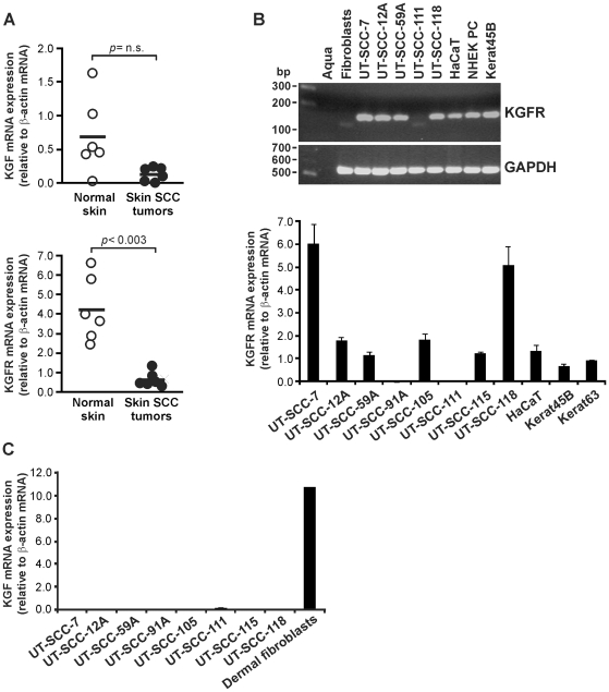
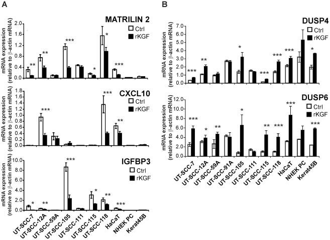
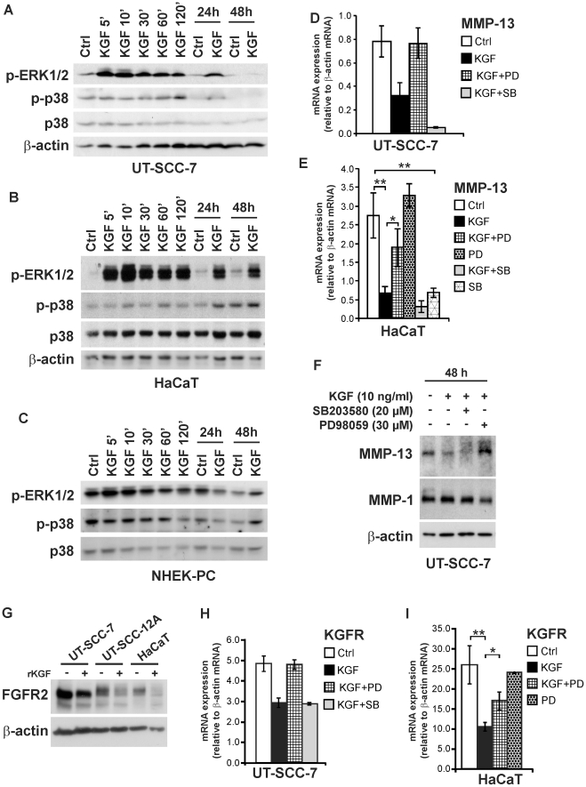
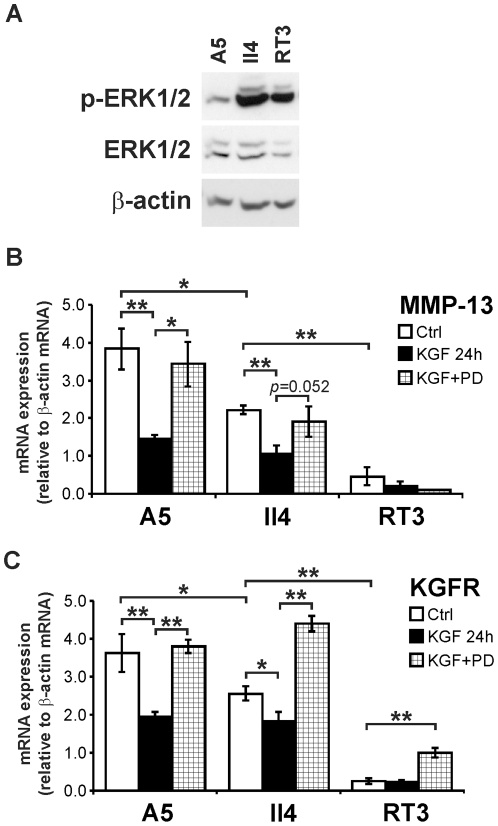
Keratinocyte growth factor (KGF, fibroblast growth factor-7) is a fibroblast-derived mitogen, which stimulates proliferation of epithelial cells. The expression of KGF by dermal fibroblasts is induced following injury and it promotes wound repair. However, the role of KGF in cutaneous carcinogenesis and cancer progression is not known. We have examined the role of KGF in progression of squamous cell carcinoma (SCC) of the skin. The expression of KGF receptor (KGFR) mRNA was lower in cutaneous SCCs (n = 6) than in normal skin samples (n = 6). Expression of KGFR mRNA was detected in 6 out of 8 cutaneous SCC cell lines and the levels were downregulated by 24-h treatment with KGF. KGF did not stimulate SCC cell proliferation, but it reduced invasion of SCC cells through collagen. Gene expression profiling of three cutaneous SCC cell lines treated with KGF for 24 h revealed a specific gene expression signature characterized by upregulation of a set of genes specifically downregulated in SCC cells compared to normal epidermal keratinocytes, including genes with tumor suppressing properties (SPRY4, DUSP4, DUSP6, LRIG1, PHLDA1). KGF also induced downregulation of a set of genes specifically upregulated in SCC cells compared to normal keratinocytes, including genes associated with tumor progression (MMP13, MATN2, CXCL10, and IGFBP3). Downregulation of MMP-13 and KGFR expression in SCC cells and HaCaT cells was mediated via ERK1/2. Activation of ERK1/2 in HaCaT cells and tumorigenic Ha-ras-transformed HaCaT cells resulted in downregulation of MMP-13 and KGFR expression. These results provide evidence, that KGF does not promote progression of cutaneous SCC, but rather suppresses the malignant phenotype of cutaneous SCC cells by regulating the expression of several genes differentially expressed in SCC cells, as compared to normal keratinocytes.


Conflict of interest statement

Competing Interests: The authors have declared that no competing interests exist.

Figures

Figure 1
Figure 1. Expression of KGF and KGFR mRNA in cutaneous SCC tumors and cell lines.
(A) Cutaneous SCC tumor tissue samples (n = 6) and normal skin samples (n = 6) were analyzed for KGF (upper panel) and KGFR (lower panel) mRNA levels by qPCR. The results were normalized for β-actin mRNA levels in each sample. A dot represents the mean of duplicate analysis of one sample. Statistical analysis with independent samples T-test (n.s., not significant). (B, upper panel) Human skin SCC cell lines (UT-SCC-7, -12A, -59A, -111, -118), HaCaT cells and normal epidermal keratinocytes (NHEK PC, Kerat45B) were analyzed for the expression of KGFR mRNA using RT-PCR. RNA from normal human skin fibroblasts served as a negative control. Amplification of a fragment of housekeeping gene GAPDH transcript was used as a loading control. (B, lower panel) KGFR mRNA expression was quantified in human skin SCC cell lines (UT-SCC-7, -12A, -59A, -91A, -105, -111, -115, and -118), HaCaT cells and normal epidermal keratinocytes (Kerat45B, Kerat63) with qPCR (n = 3–4). (C) KGF mRNA expression was quantified in human skin SCC cell lines (UT-SCC-7, -12A, -59A, -91A, -105, -111, -115, and -118), and normal human dermal fibroblasts with qPCR (n = 3–4).
Figure 2
Figure 2. Lack of mitogenic response to KGF in skin SCC cells.
(A) Growth supplement-starved normal primary keratinocytes (Kerat63), serum-starved HaCaT cells and primary (UT-SCC-12A) and metastatic (UT-SCC-7) cutaneous SCC cells were treated with recombinant KGF (rKGF; 10 ng/ml) for 2 h followed by addition of BrdU. DNA-synthesis was determined as incorporation of BrdU into DNA after 18 h incubation. *p<0.02; **p<0.005, with Mann-Whitney U-test, n = 5–6. (B) Keratinocytes (Kerat63) (n = 8), HaCaT cells (n = 12), UT-SCC-12A (n = 12) and UT-SCC-7 (n = 12) cells were treated as in (A). The relative amount of viable cells was determined using colorimetric assay of WST-1 reagent metabolism. *p<0.02; **p<0.000001, with Mann-Whitney U-test. (C) Serum-starved primary human skin fibroblasts were infected with recombinant adenovirus (RAdKGF) coding for human KGF, or with empty control adenovirus (RAdpCA3) (MOI 200) overnight and incubated in DMEM containing 0.5% FCS for 72 h. The conditioned medium (c.m.) was analyzed for the presence of KGF by western blotting, and KGF concentration was quantified with ELISA. ProMMP-1 was visualized as control in c.m. by western blotting. (D–F) Serum-starved HaCaT cells (D), UT-SCC-12A cells (E) and UT-SCC-7 (F) cells were incubated with c.m. in indicated concentrations or with rKGF for 18 h. BrdU was added and DNA-synthesis was analyzed after 8 h incubation. *a p<0.05, **a p<0.01, *p<0.05 compared to 0% c.m., **p<0.02 compared to 0% c.m. with Mann-Whitney U-test, n = 4–5.
Figure 3
Figure 3. Dynamic molecular network induced by KGF in skin SCC cells.
KGF-treatment of skin SCC cells induces up- and downregulation of variety of ERK1/2-regulated genes including upregulation of DUSP6, ETV5, SPRY1, and SPRY4 genes. KGF also induces downregulation of MMP7, MMP13 and CXCL10. The data presented show KGF-induced changes as an average in three SCC cell lines. The image was generated using Ingenuity Pathways Analysis (IPA). The symbols represent gene function, as indicated in legend on the right. Red color indicates gene upregulation and green color downregulation by KGF. The genes with fold change >1.5 are indicated with the strongest red (up) and green (down) colors. The rainbow color indicates a group of genes with up- or downregulation. Some of the groups (FGF, MMP, MKP1/3/4) are “opened” to show the regulation of individual genes. The arrows and lines indicate direct (solid line) and indirect (dashed line) physical and functional interactions. The arrows show the direction of regulation. The relations found in this study are highlighted with yellow arrows.
Figure 4
Figure 4. The expression of matrilin 2, CXCL10, IGFBP3, DUSP4 and DUSP6 is regulated by KGF in cutaneous SCC cells.
Cutaneous SCC cell lines (UT-SCC-7, -12A, -59A, -91A, -105, -111, -115, and -118), HaCaT cells and normal keratinocytes (NHEK PC, Kerat45B) were serum starved, treated with recombinant KGF (rKGF; 10 ng/ml) for 24 h and analyzed for (A) matrilin 2, CXCL10 and IGFBP3 mRNA and (B) DUSP4 and DUSP6 mRNA expression with qPCR. The results were normalized for β-actin mRNA levels in each sample. Note that the cell lines UT-SCC-91A and UT-SCC-111 do not express KGFR mRNA. *p<0.05, **p<0.01, ***p<0.001, with independent samples T-test, n = 3–4.
Figure 5
Figure 5. KGF downregulates the expression of MMP-13 and MMP-7 and suppresses invasion of cutaneous SCC cells.
(A, C) Cutaneous SCC cell lines (UT-SCC-7, - 12A, -59A, -105, -111, and -118) and HaCaT cells were serum starved and treated with recombinant KGF (rKGF; 10 ng/ml). After 24-h treatment, total RNA was extracted and analyzed for the expression of MMP-13 (A) and MMP-7 (C) mRNA by qPCR. The results were normalized for β-actin mRNA levels in the same samples. *p<0.05, **p<0.01, ***p<0.0001, with independent samples T-test, n = 3–4. (B) HaCaT cells and cutaneous SCC cells (UT-SCC-7, - 12A, and -118) were incubated with recombinant KGF (rKGF) for 72 h, and the medium samples were analyzed for MMP-13 and MMP-2 protein using western immunoblotting. Each pair of control and KGF-treated sample contain equal amount of total protein. (D) Cutaneous SCC cells (UT-SCC-7, -12A) (3×10⁵cells) treated with KGF (10 ng/ml) for 24 h were applied in collagen gel coated invasion chamber and incubated for 48 h in the presence of KGF. The cells that had invaded through collagen matrix were stained, photographed and counted. Statistical analysis with independent samples T-test, n = 4–6.
Figure 6
Figure 6. KGF-elicited downregulation of MMP-13 and KGFR expression in cutaneous SCC and HaCaT cells is mediated via ERK1/2.
Serum-starved (A) skin SCC cells (UT-SCC-7), (B) HaCaT cells, and (C) human epidermal keratinocytes (NHEK-PC) were incubated with KGF (10 ng/ml) for different periods of time, as indicated. Cell lysates were analyzed for phosphorylated forms of ERK1/2 and p38 MAP-kinases (p-ERK1/2 and p-p38, respectively) and for total p38 by western immunoblotting. β-actin was determined as loading control. (D, E) Serum-starved (D) UT-SCC-7 cells (n = 2) and (E) HaCaT cells (n = 3) were pretreated with MEK1 inhibitor PD98059 (30 µM) or with p38 inhibitor SB203580 (20 µM) for 1 h and KGF (10 ng/ml) was added, as indicated. After 24 h, total RNA was harvested and analyzed for MMP-13 mRNA by qPCR. The results were normalized for β-actin mRNA levels in each sample. (F) UT-SCC-7 cells were treated as in (D) and incubated for 48 h. Equal aliquots of conditioned media were analyzed for MMP-13 and MMP-1 by western immunoblotting. The level of β-actin in corresponding cell lysstes was determined as loading control. (G) Serum-starved skin SCC cells (UT-SCC-7, -12A) and HaCaT cells were treated with recombinant KGF (10 ng/ml) for 24 h and equal amounts of total cell lysates were analyzed for FGFR2 by western immunoblotting. The expression level of β-actin was visualized for estimation of equal loading. (H, I) UT-SCC-7 and HaCaT cells were treated as in (D, E). After 24 h, total RNA was harvested and analyzed for KGFR mRNA by qPCR. The results were normalized for β-actin mRNA levels in each sample. *p<0.05, **p<0.01, with independent samples T-test, n = 3.
Figure 7
Figure 7. KGF downregulates MMP-13 and KGFR expression in Ha-ras-transformed HaCaT cells via ERK1/2.
Three Ha-ras-transformed HaCaT cell-derived lines (A5, II4, and RT3) were used. A5 is benign tumorigenic cell line, II4 forms invasive malignant tumors, and RT3 cells form metastatic SCCs in vivo. (A) A5, II4, and RT3 cultures were serum-starved overnight and the cell lysates were analyzed for phosphorylated ERK1/2 (p-ERK1/2) and total ERK1/2 by western blotting. β-actin was determined as loading control. (B, C) Serum-starved A5, II4, and RT3 cells were pre-treated with PD98059 (30 µM) for 1 h and KGF (10 ng/ml) was added. After 24 h, total RNA was extracted and analyzed for MMP-13 mRNA (B) and KGFR mRNA (C) using qPCR. The amplification results were normalized for β-actin mRNA levels in each sample. *p<0.05, **p<0.005, with independent samples T-test, n = 3.
Figure 8
Figure 8. Activation of ERK1/2 downregulates MMP-13 and KGFR expression in HaCaT cells.
Serum-starved HaCaT cells were infected with control adenovirus RAdLacZ or with adenovirus RAdMEK1ca harboring constitutively active MEK1 at MOI 500. (A) Cell lysates were harvested 18 h after infection and analyzed for phosphorylated ERK1/2 (p-ERK1/2) and total ERK1/2 by western blotting. β-actin was determined as loading control. (B) Total RNA was extracted 48 h after infection and analyzed for MMP-13 mRNA by qPCR. The amplification results were normalized for GAPDH mRNA levels in each sample. (C) Total RNA was extracted 24 h after infection and analyzed for KGFR by qPCR, as described in (B).

Similar articles

Cited by

References

    1. Finch PW, Rubin JS, Miki T, Ron D, Aaronson SA. Human KGF is FGF-related with properties of a paracrine effector of epithelial cell growth. Science. 1989;245:752–755. - PubMed
    1. Jameson J, Ugarte K, Chen N, Yachi P, Fuchs E, et al. A role for skin γδT cells in wound repair. Science. 2002;296:747–749. - PubMed
    1. Miki T, Bottaro DP, Fleming TP, Smith CL, Burgess WH, et al. Determination of ligand-binding specificity by alternative splicing: two distinct growth factor receptors encoded by a single gene. Proc Natl Acad Sci U S A. 1992;89:246–250. - PMC - PubMed
    1. Andreadis ST, Hamoen KE, Yarmush ML, Morgan JR. Keratinocyte growth factor induces hyperproliferation and delays differentiation in a skin equivalent model system. FASEB J. 2001;15:898–906. - PubMed
    1. Grose R, Fantl V, Werner S, Chioni AM, Jarosz M, et al. The role of fibroblast growth factor receptor 2b in skin homeostasis and cancer development. EMBO J. 2007;26:1268–1278. - PMC - PubMed

Publication types

MeSH terms