

Syndecan-4 regulates early neutrophil migration and pulmonary inflammation in response to lipopolysaccharide
- PMID: 22427536
- PMCID: PMC3423465
- DOI: 10.1165/rcmb.2011-0294OC
Syndecan-4 regulates early neutrophil migration and pulmonary inflammation in response to lipopolysaccharide
Abstract
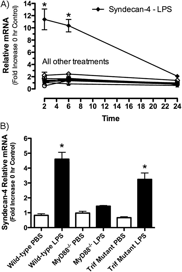
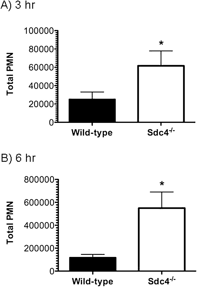
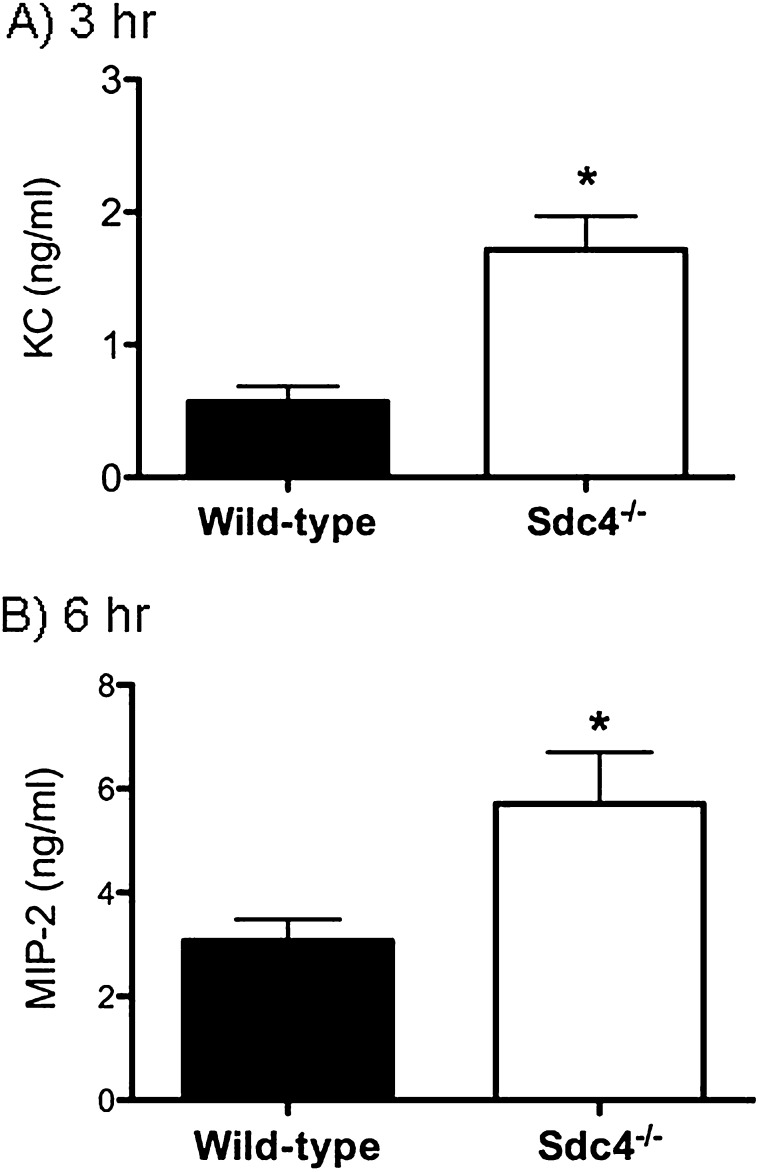
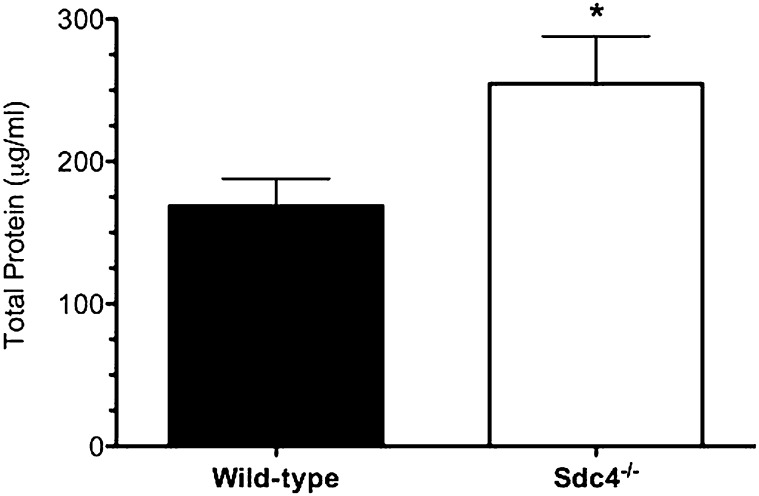
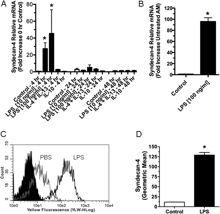
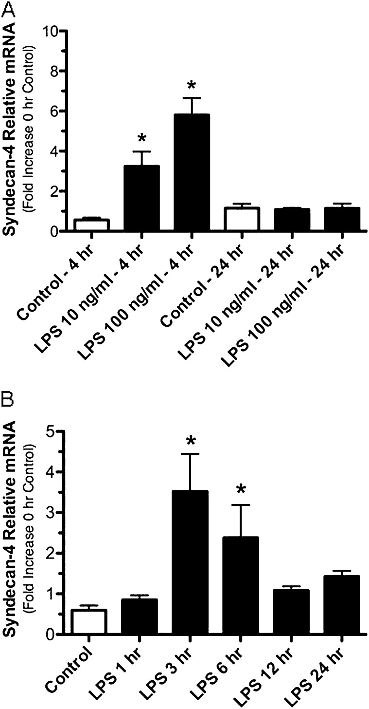
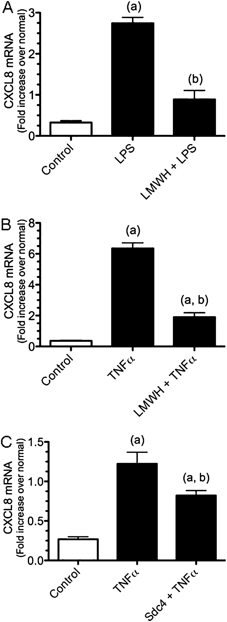
Proteoglycans (PGs) and their associated glycosaminoglycan side chains are effectors of inflammation, but little is known about changes to the composition of PGs in response to lung infection or injury. The goals of this study were to identify changes to heparan sulfate PGs in a mouse model of gram-negative pneumonia, to identify the Toll-like receptor adaptor molecules responsible for these changes, and to determine the role of the heparan sulfate PG in the innate immune response in the lungs. We treated mice with intratracheal LPS, a component of the cell wall of gram-negative bacteria, to model gram-negative pneumonia. Mice treated with intratracheal LPS had a rapid and selective increase in syndecan-4 mRNA that was regulated through MyD88-dependent mechanisms, whereas expression of several other PGs was not affected. To determine the role of syndecan-4 in the inflammatory response, we exposed mice deficient in syndecan-4 to LPS and found a significant increase in neutrophil numbers and amounts of CXC-chemokines and total protein in bronchoalveolar lavage fluid. In studies performed in vitro, macrophages and epithelial cells treated with LPS had increased expression of syndecan-4. Studies performed using BEAS-2B cells showed that pretreatment with heparin and syndecan-4 decreased the expression of CXCL8 mRNA in response to LPS and TNF-α. These findings indicate that the early inflammatory response to LPS involves marked up-regulation of syndecan-4, which functions to limit the extent of pulmonary inflammation and lung injury.
Figures
Similar articles
-
Innate immune signaling induces expression and shedding of the heparan sulfate proteoglycan syndecan-4 in cardiac fibroblasts and myocytes, affecting inflammation in the pressure-overloaded heart.FEBS J. 2013 May;280(10):2228-47. doi: 10.1111/febs.12161. Epub 2013 Feb 24. FEBS J. 2013. PMID: 23374111
-
Both TRIF- and MyD88-dependent signaling contribute to host defense against pulmonary Klebsiella infection.J Immunol. 2009 Nov 15;183(10):6629-38. doi: 10.4049/jimmunol.0901033. Epub 2009 Oct 21. J Immunol. 2009. PMID: 19846873 Free PMC article.
-
Effects of alpha 1-antitrypsin on endotoxin-induced lung inflammation in vivo.Inflamm Res. 2010 Jul;59(7):571-8. doi: 10.1007/s00011-010-0164-x. Epub 2010 Mar 18. Inflamm Res. 2010. PMID: 20238140
-
Neutrophil-Dependent Immunity During Pulmonary Infections and Inflammations.Front Immunol. 2021 Oct 19;12:689866. doi: 10.3389/fimmu.2021.689866. eCollection 2021. Front Immunol. 2021. PMID: 34737734 Free PMC article. Review.
-
Role of syndecan-4 in breast cancer pathophysiology.Am J Physiol Cell Physiol. 2022 Nov 1;323(5):C1345-C1354. doi: 10.1152/ajpcell.00152.2022. Epub 2022 Sep 12. Am J Physiol Cell Physiol. 2022. PMID: 36094435 Review.
Cited by
-
Syndecan-4 is a major syndecan in primary human endothelial cells in vitro, modulated by inflammatory stimuli and involved in wound healing.J Histochem Cytochem. 2015 Apr;63(4):280-92. doi: 10.1369/0022155415568995. Epub 2015 Jan 9. J Histochem Cytochem. 2015. PMID: 25575567 Free PMC article.
-
Proteomic analysis reveals exercise training induced remodelling of hepatokine secretion and uncovers syndecan-4 as a regulator of hepatic lipid metabolism.Mol Metab. 2022 Jun;60:101491. doi: 10.1016/j.molmet.2022.101491. Epub 2022 Apr 2. Mol Metab. 2022. PMID: 35381388 Free PMC article.
-
Targeting of CCL2-CCR2-Glycosaminoglycan Axis Using a CCL2 Decoy Protein Attenuates Metastasis through Inhibition of Tumor Cell Seeding.Neoplasia. 2016 Jan;18(1):49-59. doi: 10.1016/j.neo.2015.11.013. Neoplasia. 2016. PMID: 26806351 Free PMC article.
-
Syndecan-4 as a biomarker to predict clinical outcome for glioblastoma multiforme treated with WT1 peptide vaccine.Future Sci OA. 2016 Oct 3;2(4):FSO96. doi: 10.4155/fsoa-2015-0008. eCollection 2016 Dec. Future Sci OA. 2016. PMID: 28116121 Free PMC article.
-
ADAMTS proteases and the tumor immune microenvironment: Lessons from substrates and pathologies.Matrix Biol Plus. 2020 Dec 30;9:100054. doi: 10.1016/j.mbplus.2020.100054. eCollection 2021 Feb. Matrix Biol Plus. 2020. PMID: 33718860 Free PMC article. Review.
References
Publication types
MeSH terms
Substances
Grants and funding
- R21 RR030249/RR/NCRR NIH HHS/United States
- HL098067/HL/NHLBI NIH HHS/United States
- RR030249/RR/NCRR NIH HHS/United States
- P01 GM037696/GM/NIGMS NIH HHS/United States
- R01 HL082658/HL/NHLBI NIH HHS/United States
- HL082658/HL/NHLBI NIH HHS/United States
- HL081764/HL/NHLBI NIH HHS/United States
- P01 HL098067/HL/NHLBI NIH HHS/United States
- P30 DK089507/DK/NIDDK NIH HHS/United States
- R01 HL081764/HL/NHLBI NIH HHS/United States
- GM37696/GM/NIGMS NIH HHS/United States
- R01 HL093022/HL/NHLBI NIH HHS/United States
- DK089507/DK/NIDDK NIH HHS/United States
LinkOut - more resources
Full Text Sources
Medical
Molecular Biology Databases
