Review
. 2012 Mar 16;148(6):1223-41.
doi: 10.1016/j.cell.2012.02.039.

CNVs: harbingers of a rare variant revolution in psychiatric genetics

Affiliations
Review

CNVs: harbingers of a rare variant revolution in psychiatric genetics

Dheeraj Malhotra et al. Cell. .

Abstract

The genetic bases of neuropsychiatric disorders are beginning to yield to scientific inquiry. Genome-wide studies of copy number variation (CNV) have given rise to a new understanding of disease etiology, bringing rare variants to the forefront. A proportion of risk for schizophrenia, bipolar disorder, and autism can be explained by rare mutations. Such alleles arise by de novo mutation in the individual or in recent ancestry. Alleles can have specific effects on behavioral and neuroanatomical traits; however, expressivity is variable, particularly for neuropsychiatric phenotypes. Knowledge from CNV studies reflects the nature of rare alleles in general and will serve as a guide as we move forward into a new era of whole-genome sequencing.


Figures

Figure 1
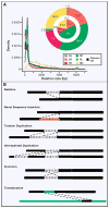
Figure 1. Types of structural variation and its evolving definition
(A) Size distribution of 22,025 deletions (release set) identified from whole genome sequencing of 179 unrelated individuals and 2 trios (i.e., child-mother-father) by the 1000 genomes project. Deletions were identified by four different types of structural variation detection approaches, which all rely on mapping sequenced reads to reference human genome sequence and subsequently identifying discordant patterns that are characteristic of deletions. Read pair (RP) methods detect clusters of “discordant” paired end reads in which mapping span and/or orientation is inconsistent with the reference genome. Read depth (RD) methods detect CNVs based on the regional depth of coverage. Split read (SR) methods detect individual reads that span the breakpoint of an SV. Assembly (AS) methods detect differences between sequence contigs assembled from the sample genome and the reference genome sequence. Paired depth (PD) refers to hybrid methods that combine RP and RD. Pie charts display the contribution of different SV detection approaches to the release set. Outer pie = based on number of SV calls; inner pie = based on total number of variable nucleotides. (B) Schematic representation of deletion, novel sequence insertion (red color bar), tandem duplication, interspersed duplication, inversion and translocation in test genome (lower black bar) compared to human reference genome sequence (upper black bar). Green colored bar represent a different chromosome from reference genome. Figure adapted from Mills et al 2010.
Figure 2
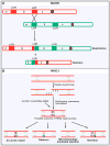
Figure 2. Four major mechanisms underlying human genomic rearrangements and CNV formation
(A) Non allelic homologous recombination (NAHR) occurs by unequal crossing over between flanking segmental duplications (SDs represented by two red and two green bars on respective homologous chromosomes), which result in reciprocal deletion and duplication of intervening sequence (b). These homologous chromosomes segregate from each other at the next cell division, thus leading to a change in copy number in both daughter cells.(B) In classical non homologous end joining (NHEJ) double-strand break repair pathway, the ends of DNA double-strand breaks are repaired through many rounds of enzymatic activity (including tethering of DNA ends by the Ku protein, followed by recruitment of DNA-dependent protein kinase, DNA-PKcs by Ku and DNA-PKcs mediated activation of the Artemis nuclease, which trims back overhangs in preparation for ligation). The different types of DNA double strand breaks fixed by NHEJ combined with other sundry alternate repair mechanisms including microhomology mediated end joining (MMEJ) leads to diverse repaired products. Although limited base pairing can guide accurate repair, deletions of variable size, and to a lesser extent insertions, are formed. (C) A simple model of FoSTeS/MMBIR is described. When a replication fork encounters a nick (striking arrowhead) in a template strand, one arm of the fork breaks off, producing a collapsed fork. At the single double-strand end, the 5′ end of the lagging strand (dashed red colored lines) is resected, giving a 3′ overhang. The 3′ single-strand end of lagging strand template (solid red colored lines) invades the sister leading strand DNA (green colored lines) guided by regions of microhomology (MH, red and green colored boxes), forming a new low processivity replication fork. The extended end dissociates repeatedly (due to migration of holiday junction or some other helicase activity) with 5′ ends resected and reforms the fork. Whether the template switch occurs in front of or behind the position of the original collapse determines whether there is a deletion or duplication respectively. The 3′ end invasion of lagging strand template can reform replication forks on different genomic templates (>100kb apart), before returning to the original sister chromatid and forming a processive replication fork that completes replication. Thus, the final product usually contains sequence from different genomic regions (not shown). Each line represents a DNA nucleotide strand. Polarity is indicated by arrows on 3′ end. New DNA synthesis is shown by dashed lines. (D) LINE-1 retrotransposition. A full-length L1 (red, green and orange bar on gray chromosome) is transcribed and translation of ORF1 (red) and ORF2(orange) protein encoded by the L1 messenger RNA (mRNA) leads to ribonucleoprotein (RNP) formation. L1 RNP is transported to the nucleus, and retrotransposition occurs by target-site primed reverse transcription (TPRT). During TPRT, the L1 endonuclease (EN) activity of ORF2 nicks target genomic DNA (black lines), exposing a free 3′-OH that serves as a primer for reverse transcription of the L1 RNA. The mechanistic details of target site second-strand cleavage, second-strand complementary DNA (cDNA) synthesis, and completion of L1 integration require further elucidation. TPRT results in the insertion of a new, often 5′-truncated L1 copy at a new genomic location that generally is flanked by target-site duplications. Alu, SINE-R/VNTR/Alu (SVA), and cellular mRNAs can also hijack the L1-encoded protein(s) in the cytoplasm to mediate their transmobilization. ORF: open reading frame; FoSTeS: fork stalling and template switching; MMBIR: microhomology mediated break induced replication.
Figure 3
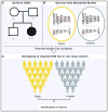
Figure 3. Key Plays from the CNV Playbook
(A) Family based studies of de novo mutation, (B) and Case-control studies of genome wide CNV burden, with CNV positions denoted by a red star (C) followed by Single marker tests for association in large cohorts.

Similar articles

Cited by

References

    1. Alkan C, Coe BP, Eichler EE. Genome structural variation discovery and genotyping. Nat Rev Genet. 2011;12:363–376. - PMC - PubMed
    1. Alkuraya FS. Homozygosity mapping: one more tool in the clinical geneticist’s toolbox. Genet Med. 2010;12:236–239. - PubMed
    1. Bailey A, Le Couteur A, Gottesman I, Bolton P, Simonoff E, Yuzda E, Rutter M. Autism as a strongly genetic disorder: evidence from a British twin study. Psychol Med. 1995;25:63–77. - PubMed
    1. Bakkaloglu B, O’Roak BJ, Louvi A, Gupta AR, Abelson JF, Morgan TM, Chawarska K, Klin A, Ercan-Sencicek AG, Stillman AA, et al. Molecular cytogenetic analysis and resequencing of contactin associated protein-like 2 in autism spectrum disorders. Am J Hum Genet. 2008;82:165–173. - PMC - PubMed
    1. Beck CR, Garcia-Perez JL, Badge RM, Moran JV. LINE-1 elements in structural variation and disease. Annu Rev Genomics Hum Genet. 2011;12:187–215. - PMC - PubMed

Publication types