. 2012 Mar 13;22(3):597-609.
doi: 10.1016/j.devcel.2011.12.024.

Sox9 directs hypertrophic maturation and blocks osteoblast differentiation of growth plate chondrocytes

Affiliations

Sox9 directs hypertrophic maturation and blocks osteoblast differentiation of growth plate chondrocytes

Peter Dy et al. Dev Cell. .

Abstract

The transcription factor Sox9 is necessary for early chondrogenesis, but its subsequent roles in the cartilage growth plate, a highly specialized structure that drives skeletal growth and endochondral ossification, remain unclear. Using a doxycycline-inducible Cre transgene and Sox9 conditional null alleles in the mouse, we show that Sox9 is required to maintain chondrocyte columnar proliferation and generate cell hypertrophy, two key features of functional growth plates. Sox9 keeps Runx2 expression and β-catenin signaling in check and thereby inhibits not only progression from proliferation to prehypertrophy, but also subsequent acquisition of an osteoblastic phenotype. Sox9 protein outlives Sox9 RNA in upper hypertrophic chondrocytes, where it contributes with Mef2c to directly activate the major marker of these cells, Col10a1. These findings thus reveal that Sox9 remains a central determinant of the lineage fate and multistep differentiation program of growth plate chondrocytes and thereby illuminate our understanding of key molecular mechanisms underlying skeletogenesis.


Figures

Figure 1
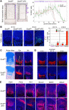
Figure 1. Sox9 is required to maintain functional growth plates
A. Histological aspect and Sox9 expression of proximal tibia growth plates from Sox9fl/fl and Sox9fl/flATC fetuses treated with Dox from E15.5 and collected at E16.5, E17.5, and E18.5. Sections were stained with Alcian blue (which binds aggrecan) and nuclear fast red, or hybridized with a Sox9 RNA probe (red) and DNA-binding DAPI dye (blue). EP, epiphyses; CZ, columnar zone; HZ, hypertrophic zone. B. Histological aspect of the entire tibia of Sox9fl/fl, Sox9fl/+ATC and Sox9fl/flATC littermates treated as described in panel A. EB, endochondral bone. Dotted line, middle of the bone. C. High-magnification pictures of hypertrophic zones at E17.5. See also Figure S1.
Figure 2
Figure 2. Sox9 is required for chondrocyte proliferation, survival, and hypertrophy, but delays prehypertrophy
A. BrdU incorporation assay in tibia proximal growth plates of Sox9fl/fl and Sox9fl/flATC littermates treated with Dox from E15.5 and harvested at E17.5. Left, representative pictures of BrdU antibody staining. The box shows 35 segments in the epiphysis (EP) and columnar zone (CZ), in which BrdU-positive cells (dark brown nuclei) were counted. Right, graph showing the percentage of positive cells in control and mutant growth plates in each segment. Data are presented as the average with standard deviation of measurements made in 3 non-adjacent sections from each of 3 embryos per genotype. B. TUNEL assay in embryos similar to those shown in panel A, but collected at E17.5 and E18.5. Left, representative pictures of data. Dying cells are seen as green dots and cell nuclei are blue. Right, quantification of the percentage of TUNEL positive cells in the columnar (CZ) and prehypertrophic/hypertrophic (PH) zones. Data are shown as the averages with standard deviation obtained for three non-adjacent sections in each of 3 embryos per genotype. C. Ppr and Ihh RNA in situ hybridization of sections similar to those in panel A. The double arrow shows the length of the columnar zone. D. Col10a1 RNA in situ hybridization of sections from Sox9fl/fl and Sox9fl/flATC littermates treated with Dox from E15.5 and collected at E16.5, E17.5, and E18.5. E. RNA in situ hybridization of sections similar to those in panel A. Probes are indicated. See also Figure S2.
Figure 3
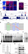
Figure 3. Sox9 protein outlives its RNA in upper hypertrophic chondrocytes and binds next to Mef2c on the Col10a1 promoter
A. Sox9 RNA in situ hybridization and protein immunostaining in tibia sections from E17.5 Sox9fl/fl and Sox9fl/flATC littermates treated with Dox from E15.5. Dotted line, middle of the prehypertrophic zone. B. High-magnification picture of the growth plate region boxed in yellow in panel A. C. Top, mammalian conservation plot for the mouse Col10a1 locus from −5.0 to +1.5 kb relative to the transcription start site (+1). Peaks denoting high conservation are seen at −4.15kb, and in the promoter, exon 1 and exon 2. Bottom left, enlargement of the −4.15kb region. Double arrows designate the enhancers described by Zheng et al. (2009) and used in this study in Col10a1 reporters (Dy). Bottom right, enlargement of the −380/+646bp region used in this study in Col10a1 reporters. D. Alignment of the −207/−154bp Col10a1 upper-strand wild-type sequence (wt) with sequences (1 to 9) harboring 4 consecutive nucleotide changes. Mef2c and Sox9 binding sites and mutant nucleotides are highlighted. Arrows show Sox-like sites oriented head to head. Double-stranded oligonucleotides corresponding to these sequences were used in EMSA. E. EMSA with extracts from Cos1 cells forced to express no exogenous protein (none), SOX9, or Mef2c. Probes are indicated on top of the lanes. Arrowheads, SOX9/DNA (blue) and Mef2c/DNA (green) complexes. Free, unbound probes. F. EMSA performed as in panel E with the wild-type probe and combinations of empty, SOX9, and Mef2c extracts. Gels were run longer than in panel E to separate SOX9/DNA from Mef2c/DNA complexes. G. Real-time RT-PCR quantification of Col10a1 RNA present in primary chondrocytes treated with no drug, okadaic acid (OKA), or forskolin for 24h. Each bar corresponds to the average with standard deviation of measurements made in triplicate cultures in a representative experiment. H. Chromatin immunoprecipitation in primary chondrocytes transfected with FLAG-SOX9 and Myc-Mef2c expression plasmids and treated without (−) or with OKA. Antibodies were non-immune (n.i.), or directed against RNA polymerase 2, FLAG or Myc. PCR products for Col10a1 and Gapdh promoters were obtained from input material and precipitated chromatin, and resolved by electrophoresis.
Figure 4
Figure 4. Sox9 and Mef2c act cooperatively to activate Col10a1
A. Relative activity of Col10a1 reporters transfected in primary chondrocytes along with SOX9 and Mef2c expression plasmids. Cells were treated with forskolin or okadaic acid. Dotted lines, expected values from additive effects of SOX9 and Mef2c. B. Effect of mutations (Δ) in the Sox9 and Mef2c binding sites on the relative activity of a Col10a1 reporter transfected in primary chondrocytes treated with okadaic acid. C. Histology and Col10a1 expression in tibia proximal growth plates of E18.5 Sox9/Mef2c mutants generated using Prx1Cre. Left, pictures of sections stained with Alcian blue and hybridized with a Col10a1 RNA probe. Right, quantification of the density of Col10a1 RNA level in hypertrophic zones. Data are presented as the averages with standard deviation obtained for three sections per embryo. Stars, p value <0.05 (Student's t-test). D. Histology and gene expression analysis of tibia proximal growth plates of E18.5 Sox9/Mef2c mutants generated using ATC and treated with Dox from E15.5. Arrows, distance from the articular surface to the onset of marker gene expression. See also Figure S3.
Figure 5
Figure 5. Sox9 prevents osteoblastic differentiation of chondrocytes
A. RNA in situ hybridization of tibia proximal growth plates from E17.5 Sox9fl/fl and Sox9fl/flATC littermates treated with Dox from E15.5. Arrows, distance from the articular surface to the prehypertrophic zone. Dotted lines delineate the prehypertrophic/osteoblastic zone. B. RNA in situ hybridization of tibia proximal growth plates from E18.5 Sox9fl/fl and Sox9fl/flATC littermates treated with Dox from E15.5. C. Staining with the von Kossa reagent of the same growth plates as in panel B. The region boxed in green on the left is shown at high magnification on the right. Double arrows, mineralized cartilage matrix (dark brown signal). See also Figure S4.
Figure 6
Figure 6. Sox9 inhibits β-catenin signaling, but β-catenin signaling does not inhibit Sox9 in the growth plate
Histology analysis and RNA in situ hybridization of tibia proximal growth plates from E17.5 Sox9fl/fl, Sox9fl/flATC, and Sox9fl/flCtnnbfl/flATC littermates treated with Dox from E15.5. Dotted lines delineate the prehypertrophic/osteoblastic zones. See also Figure S5.
Figure 7
Figure 7. Proposed roles of Sox9 in the growth plate
In a wild-type growth plate (left), Sox9 is expressed in the columnar, prehypertrophic and upper hypertrophic zones. It promotes (green) columnar chondrocyte proliferation and prevents (red) prehypertrophy by keeping expression of the major chondrocyte maturation regulators Runx2 and Mef2c in check. Sox9 then contributes, along with Mef2c, to activate hypertrophy. Sox9 deletion in growth plate chondrocytes (right) swiftly leads to columnar cell growth arrest, prehypertrophy, and osteoblastic differentiation, through upregulation of Runx2, Osx, and Mef2c expression and increased β-catenin signaling.

Similar articles

Cited by

References

    1. Akiyama H. Control of chondrogenesis by the transcription factor Sox9. Mod. Rheumatol. 2008;18:213–219. - PubMed
    1. Akiyama H, Chaboissier MC, Martin JF, Schedl A, de Crombrugghe B. The transcription factor Sox9 has essential roles in successive steps of the chondrocyte differentiation pathway and is required for expression of Sox5 and Sox6. Genes Dev. 2002;16:2813–2828. - PMC - PubMed
    1. Akiyama H, Lyons JP, Mori-Akiyama Y, Yang X, Zhang R, Zhang Z, Deng JM, Taketo MM, Nakamura T, Behringer RR, McCrea PD, de Crombrugghe B. Interactions between Sox9 and beta-catenin control chondrocyte differentiation. Genes Dev. 2004;18:1072–1087. - PMC - PubMed
    1. Arnold MA, Kim Y, Czubryt MP, Phan D, McAnally J, Qi X, Shelton JM, Richardson JA, Bassel-Duby R, Olson EN. MEF2C transcription factor controls chondrocyte hypertrophy and bone development. Dev. Cell. 2007;12:377–389. - PubMed
    1. Bhattaram P, Penzo-Méndez A, Sock E, Colmenares C, Kaneko KJ, Vassilev A, Depamphilis ML, Wegner M, Lefebvre V. Organogenesis relies on SoxC transcription factors for the survival of neural and mesenchymal progenitors. Nat. Commun. 2010;1:9. - PMC - PubMed

Publication types

MeSH terms