Co-expression of endothelial and neuronal nitric oxide synthases in the developing vasculatures of the human fetal eye
- PMID: 22411126
- PMCID: PMC3358532
- DOI: 10.1007/s00417-012-1969-9
Co-expression of endothelial and neuronal nitric oxide synthases in the developing vasculatures of the human fetal eye
Abstract
Background: Nitric oxide (NO) is a multifunctional gaseous molecule that regulates various physiological functions in both neuronal and non-neuronal cells. NO is synthesized by nitric oxide synthases (NOSs), of which three isoforms have been identified. Neuronal NOS (nNOS) and endothelial NOS (eNOS) constitutively produce low levels of NO as a cell-signaling molecule in response to an increase in intracellular calcium concentration. Recent data have revealed a predominant role of eNOS in both angiogenesis and vasculogenesis.
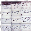
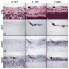
Methods: The immunohistochemical localization of nNOS and eNOS was investigated during embryonic and fetal ocular vascular development from 7 to 21 weeks gestation (WG) on sections of cryopreserved tissue.
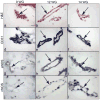
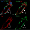
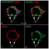
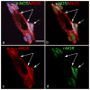
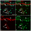
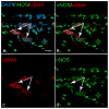
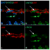
Results: eNOS was confined to endothelial cells of developing vessels at all ages studied. nNOS was prominent in nuclei of vascular endothelial and smooth muscle cells in the fetal vasculature of vitreous and choriocapillaris. nNOS was also prominent in the nuclei of CXCR4(+) progenitors in the inner retina and inner neuroblastic layer.
Conclusions: These findings demonstrate co-expression of n- and eNOS isoforms in different compartments of vasoformative cells during development. Nuclear nNOS was present in vascular and nonvascular progenitors as well as endothelial cells and pericytes. This suggests that nNOS may play a role in the transcription regulatory systems in endothelial cells and pericytes during ocular hemo-vasculogenesis, vasculogenesis, and angiogenesis.
Conflict of interest statement
Figures
Similar articles
-
Low nitric oxide synthases (NOSs) in eyes with age-related macular degeneration (AMD).Exp Eye Res. 2010 Jan;90(1):155-67. doi: 10.1016/j.exer.2009.10.004. Epub 2009 Oct 15. Exp Eye Res. 2010. PMID: 19836390 Free PMC article.
-
Development of the hyaloid, choroidal and retinal vasculatures in the fetal human eye.Prog Retin Eye Res. 2018 Jan;62:58-76. doi: 10.1016/j.preteyeres.2017.10.001. Epub 2017 Nov 2. Prog Retin Eye Res. 2018. PMID: 29081352 Free PMC article. Review.
-
Maturation of the fetal human choriocapillaris.Invest Ophthalmol Vis Sci. 2009 Jul;50(7):3503-11. doi: 10.1167/iovs.08-2614. Epub 2009 Mar 5. Invest Ophthalmol Vis Sci. 2009. PMID: 19264887 Free PMC article.
-
Normal vascular development in mice deficient in endothelial NO synthase: possible role of neuronal NO synthase.Mol Vis. 2003 Oct 8;9:549-58. Mol Vis. 2003. PMID: 14551528
-
Physiological and Pathophysiological Relevance of Nitric Oxide Synthases (NOS) in Retinal Blood Vessels.Front Biosci (Landmark Ed). 2024 May 16;29(5):190. doi: 10.31083/j.fbl2905190. Front Biosci (Landmark Ed). 2024. PMID: 38812321 Review.
Cited by
-
Role of neuronal nitric oxide synthase on cardiovascular functions in physiological and pathophysiological states.Nitric Oxide. 2020 Sep 1;102:52-73. doi: 10.1016/j.niox.2020.06.004. Epub 2020 Jun 23. Nitric Oxide. 2020. PMID: 32590118 Free PMC article. Review.
-
Neurovascular anatomy of the developing human fetal penis and clitoris.J Anat. 2024 Jul;245(1):35-49. doi: 10.1111/joa.14029. Epub 2024 Feb 28. J Anat. 2024. PMID: 38419143 Free PMC article.
-
Hemoglobin induced NO/cGMP suppression Deteriorate Microcirculation via Pericyte Phenotype Transformation after Subarachnoid Hemorrhage in Rats.Sci Rep. 2016 Feb 25;6:22070. doi: 10.1038/srep22070. Sci Rep. 2016. PMID: 26911739 Free PMC article.
-
Nitrosative Stress in Retinal Pathologies: Review.Antioxidants (Basel). 2019 Nov 11;8(11):543. doi: 10.3390/antiox8110543. Antioxidants (Basel). 2019. PMID: 31717957 Free PMC article. Review.
-
Effect of Shuangdan Mingmu capsule, a Chinese herbal formula, on oxidative stress-induced apoptosis of pericytes through PARP/GAPDH pathway.BMC Complement Med Ther. 2021 Apr 10;21(1):118. doi: 10.1186/s12906-021-03238-w. BMC Complement Med Ther. 2021. PMID: 33838689 Free PMC article.
References
-
- Moncada S, Palmer RM, Higgs EA. Nitric oxide: physiology, pathophysiology, and pharmacology. Pharmacol Rev. 1991;43:109–142. - PubMed
-
- Neufeld AH, Shareef S, Pena J. Cellular localization of neuronal nitric oxide synthase (NOS-1) in the human and rat retina. J Comp Neurol. 2000;416:269–275. - PubMed
-
- Giove TJ, Deshpande MM, Eldred WD. Identification of alternate transcripts of neuronal nitric oxide synthase in the mouse retina. J Neurosci Res. 2009;87:3134–3142. - PubMed
Publication types
MeSH terms
Substances
Grants and funding
LinkOut - more resources
Full Text Sources
Medical

