. 2012 May 4;287(19):15966-80.
doi: 10.1074/jbc.M112.346817. Epub 2012 Mar 8.

NEU1 and NEU3 sialidase activity expressed in human lung microvascular endothelia: NEU1 restrains endothelial cell migration, whereas NEU3 does not

Affiliations

NEU1 and NEU3 sialidase activity expressed in human lung microvascular endothelia: NEU1 restrains endothelial cell migration, whereas NEU3 does not

Alan S Cross et al. J Biol Chem. .

Abstract

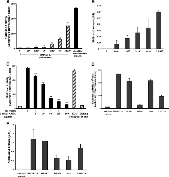
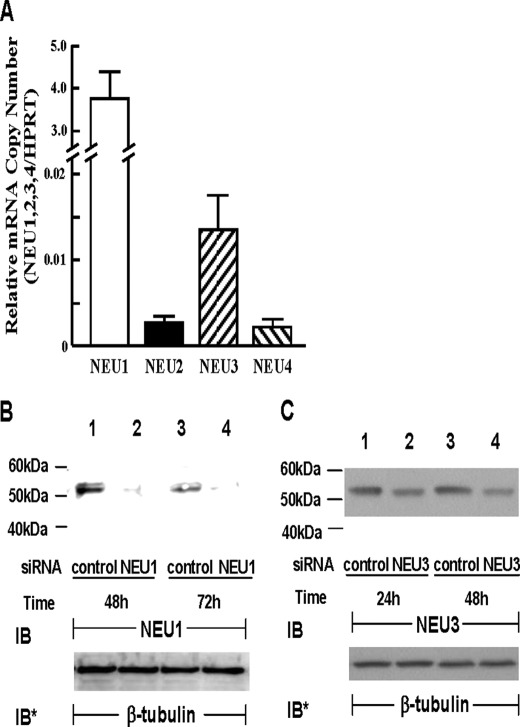
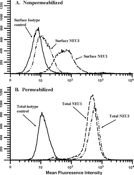
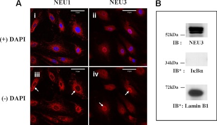
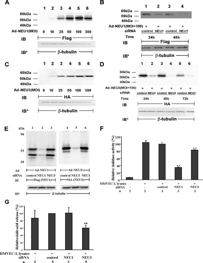
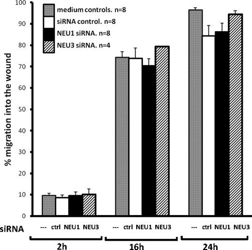
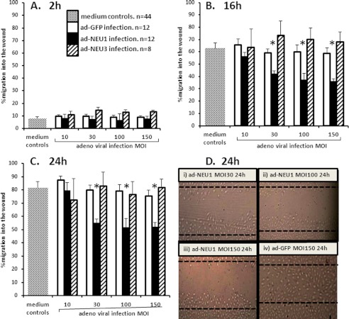
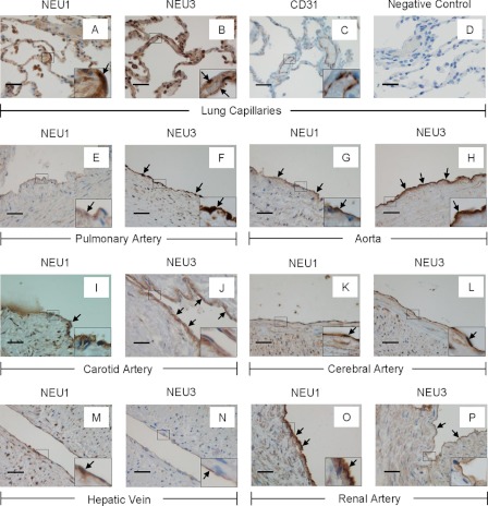
The microvascular endothelial surface expresses multiple molecules whose sialylation state regulates multiple aspects of endothelial function. To better regulate these sialoproteins, we asked whether endothelial cells (ECs) might express one or more catalytically active sialidases. Human lung microvascular EC lysates contained heat-labile sialidase activity for a fluorogenic substrate, 2'-(4-methylumbelliferyl)-α-D-N-acetylneuraminic acid (4-MU-NANA), that was dose-dependently inhibited by the competitive sialidase inhibitor, 2,3-dehydro-2-deoxy-N-acetylneuraminic acid but not its negative control. The EC lysates also contained sialidase activity for a ganglioside mixture. Using real time RT-PCR to detect mRNAs for the four known mammalian sialidases, NEU1, -2, -3, and -4, NEU1 mRNA was expressed at levels 2700-fold higher that those found for NEU2, -3, or -4. Western analyses indicated NEU1 and -3 protein expression. Using confocal microscopy and flow cytometry, NEU1 was immunolocalized to both the plasma membrane and the perinuclear region. NEU3 was detected both in the cytosol and nucleus. Prior siRNA-mediated knockdown of NEU1 and NEU3 each decreased EC sialidase activity for 4-MU-NANA by >65 and >17%, respectively, and for the ganglioside mixture by 0 and 40%, respectively. NEU1 overexpression in ECs reduced their migration into a wound by >40%, whereas NEU3 overexpression did not. Immunohistochemical studies of normal human tissues immunolocalized NEU1 and NEU3 proteins to both pulmonary and extrapulmonary vascular endothelia. These combined data indicate that human lung microvascular ECs as well as other endothelia express catalytically active NEU1 and NEU3. NEU1 restrains EC migration, whereas NEU3 does not.


Figures

FIGURE 1.
FIGURE 1.
EC sialidase activity. Increasing HMVEC-L cell numbers were assayed for sialidase activity as liberation of sialic acid from the fluorogenic substrate, 4-MU-NANA (A) or from a ganglioside mixture (B). For comparison, sialidase activity for a known concentration of purified clostridial neuraminidase is indicated (A). In C, HMVEC-Ls were assayed for sialidase activity before and after boiling and in the presence of increasing concentrations (5–500 μg/ml) of the competitive sialidase inhibitor, 2-deoxy-NANA, or its negative control, KDO (500 μg/ml). Equivalent cell numbers of HMVEC-Ls, HPAECs, HBMEs, Hrvts, and HMEC-1s were assayed for sialidase activity for the 4-MU-NANA substrate (D) and the ganglioside mixture (E). In A–E, vertical bars represent the mean (±S.E.) sialidase activity, expressed as either arbitrary fluorescence units (A, C, and D) or sialic acid release in μm (B and E). The n for each experimental and control group is ≥2. *, significantly increased compared with background sialidase activity in the cell-free control at p < 0.05. **, significantly decreased compared with sialidase activity in total HMVEC-L lysates at p < 0.05.
FIGURE 2.
FIGURE 2.
Expression of NEU1–4 in endothelia. A, total RNA isolated from HMVEC-Ls was reverse-transcribed, and the resulting cDNA was used as a template for amplification with primers corresponding to NEU1, NEU2, NEU3, and NEU4 as well as HPRT as a housekeeping gene control. The mRNAs for each sialidase was normalized to HPRT (n = 2). B, HMVEC-Ls were transfected with NEU1-targeting or control siRNAs, cultured for 48 or 72 h, and lysed, and the lysates were processed for NEU1 immunoblotting. C, HMVEC-Ls were transfected with NEU3-targeting or control siRNAs, cultured for 24 or 48 h, and lysed, and the lysates were processed for NEU3 immunoblotting. B and C, to control for protein loading and transfer, blots were stripped and reprobed for β-tubulin. IB, immunoblot; IB*, immunoblot after stripping. Molecular mass in kDa is indicated on the left. Each blot is representative of three or more independent experiments.
FIGURE 3.
FIGURE 3.
Surface and intracellular pools of NEU1 and NEU3. A, nonpermeabilized HMVEC-Ls were studied by flow cytometry for surface NEU1 and NEU3. B, permeabilized HMVEC-Ls were studied by flow cytometry for total NEU1 and NEU3. HMVEC-Ls, some of which were permeabilized with Triton X-100, were incubated with rabbit polyclonal antibodies raised against human NEU1 or NEU3 or a species-matched control antibody, washed, and incubated with FITC-conjugated goat anti-rabbit antibody. Each plot is representative of three or more independent experiments.
FIGURE 4.
FIGURE 4.
Immunolocalization of NEU1 and -3 in HMVEC-Ls. Ai, subconfluent HMVEC-Ls were fixed, permeabilized, blocked, and incubated with rabbit polyclonal antibodies raised against NEU1 (i and iii) or NEU3 (ii and iv) and counterstained with DAPI (overlay in i and ii). Arrows indicate the perinuclear regions in NEU1-stained (iii) and nuclear localization in NEU3-stained (iv) cells. In each panel, the scale bar represents 50 μm. Each photomicrograph is representative of two or more independent experiments. B, the nuclear fraction of HMVEC-Ls was probed with anti-NEU3 antibody. To verify nuclear fractionation, the blot was stripped and reprobed for the cytoplasmic marker protein, IκBα, and the nuclear marker protein, lamin B1. IB, immunoblot; IB*, immunoblot after stripping. Molecular mass in kDa is indicated on the left. Each blot is representative of two independent experiments.
FIGURE 5.
FIGURE 5.
NEU1 and NEU3 sialidase activity in HMVEC-Ls. A, HMVEC-Ls were transiently infected with increasing m.o.i. of Ad-NEU1-FLAG. At 48 h, cells were lysed, and the lysates were processed for immunoblotting with anti-FLAG antibodies. B, HMVEC-Ls were transiently infected with Ad-NEU1-FLAG at an m.o.i. of 100. After 48 h, the HMVEC-Ls overexpressing FLAG-tagged NEU1 were transfected with either NEU1-targeting or control siRNAs, cultured for 24 or 48 h, and lysed, and the lysates were processed for immunoblotting with anti-FLAG antibodies. C, HMVEC-Ls were transiently infected with increasing m.o.i. of Ad-NEU3-HA. At 48 h, cells were lysed, and the lysates were processed for immunoblotting with anti-HA antibodies. D, HMVEC-Ls were transiently infected with Ad-NEU3-HA at an m.o.i. = 100. After 48 h, the HMVEC-Ls overexpressing HA-tagged NEU3 were transfected with either NEU3-targeting or control siRNAs, cultured for 24, 48, or 72 h, and lysed, and the lysates were processed for immunoblotting with anti-HA antibodies. E, HMVEC-Ls were transiently infected with either Ad-NEU1-FLAG (lanes 1–3) or Ad-NEU3-HA (lanes 4–6), each at m.o.i. = 100. After 48 h, the cells were transfected with NEU1-targeting (lanes 2 and 6), NEU3-targeting (lanes 3 and 5), or control (lanes 1 and 4) siRNAs, cultured for 48 h, and lysed, and the lysates were processed for immunoblotting with either anti-FLAG (lanes 1–3) or anti-HA (lanes 4–6) antibodies. In A–E, to control for protein loading and transfer, blots were stripped and reprobed for β-tubulin. IB, immunoblot; IB* = immunoblot after stripping. Molecular mass in kDa is indicated on the left. Each blot is representative of three or more independent experiments. F and G, after transfection with NEU1-targeting, NEU3-targeting, and control siRNAs, HMVEC-Ls were fluorometrically assayed for sialidase activity for the 4-MU-NANA substrate (F) or the ganglioside mixture (G). In F and G, vertical bars represent mean (±S.E.) sialidase activity, expressed as either arbitrary fluorescence units (F) or sialic acid release in μm (G). The n for each experimental and control group is indicated below the bars. *, significantly increased compared with background sialidase activity in the cell-free control. **, significantly decreased compared with sialidase activity in lysates of control siRNA-transfected HMVEC-Ls at p < 0.05.
FIGURE 6.
FIGURE 6.
Effect of NEU1/NEU3 depletion on HMVEC-L migration in a wounding assay. HMVEC-Ls transfected with NEU1-targeting, NEU3-targeting, or control siRNAs were cultured to confluence, after which a single wound was made across each monolayer. At 0, 2, 16, and 24 h, images of each monolayer were captured, and cell migration into the wound after 2, 16, and 24 h was compared with that observed in the same wounded monolayer at 0 h. Vertical bars represent the mean (±S.E.) migration of cells into the wound at 2, 16, and 24 h. n, the number of independent experiments is indicated.
FIGURE 7.
FIGURE 7.
Effect of NEU1/NEU3 overexpression on HMVEC-L migration in a wounding assay. HMVEC-Ls infected with increasing m.o.i. of Ad-NEU1-FLAG, Ad-NEU3-HA, or Ad-GFP were cultured to confluence, after which a single wound was made across each monolayer. At 0, 2, 16, and 24 h, images of each monolayer were captured, and cell migration into the wound after 2, 16, and 24 h was compared with that observed in the same wounded monolayer at 0h. In A, B, and C, vertical bars represent the mean (±S.E.) migration of cells into the wound at 2 h (A), 16 h (B), and 24 h (C). n, numbers of independent experiments are indicated. *, significantly decreased compared with the Ad-GFP infected cells at p < 0.05. D, at 24 h, representative images of wounded HMVEC-L monolayers are shown, including Ad-NEU1, m.o.i. = 30 (i), Ad-NEU1, m.o.i. = 100 (ii), Ad-NEU1, m.o.i. = 150 (iii), Ad-GFP, m.o.i. = 150 (iv). Dotted lines indicate wound boundaries at 0 h.
FIGURE 8.
FIGURE 8.
NEU1 and NEU3 immunostaining in vascular endothelia in human tissues. Sections of human tissues were probed with rabbit antibodies raised against either human NEU1 or NEU3 followed by biotinylated peroxidase-conjugated goat anti-rabbit IgG antibody and developed with diaminobenzidine and counterstained with hematoxylin. NEU1 and NEU3 immunostaining appears brown and is indicated by arrows. Shown are expanded pulmonary alveoli-NEU1 (A), expanded pulmonary alveoli-NEU3 (B), CD31 immunostaining of expanded pulmonary alveoli (C), a negative control in which sections of expanded lung were incubated with nonimmune rabbit IgG in lieu of the anti-NEU1/3 antibody followed by secondary goat anti-rabbit antibody (D), pulmonary artery-NEU1 (E), pulmonary artery-NEU3 (F), aorta-NEU1 (G), aorta-NEU3 (H), carotid artery-NEU1 (I), carotid artery-NEU3 (J), cerebral artery-NEU1 (K), cerebral artery-NEU3 (L), hepatic vein-NEU1 (M), hepatic vein-NEU3 (N), renal artery-NEU1 (O), and renal artery-NEU3 (P). Each tissue section was photographed at either 400× or 1000× (see the insets). In each panel the scale bar represents 50 μm. Arrows indicate immunostaining for either NEU1 or 3.

Similar articles

Cited by

References

    1. Smith C. W. (2008) Adhesion molecules and receptors. J. Allergy Clin. Immunol. 121, S375–S379 - PubMed
    1. Stevens T., Garcia J. G., Shasby D. M., Bhattacharya J., Malik A. B. (2000) Mechanisms regulating endothelial cell barrier function. Am. J. Physiol. Lung Cell. Mol. Physiol. 279, L419–L422 - PubMed
    1. Varki A. (2008) Sialic acids in human health and disease. Trends Mol. Med. 14, 351–360 - PMC - PubMed
    1. Schauer R. (2009) Sialic acids as regulators of molecular and cellular interactions. Curr. Opin. Struct. Biol. 19, 507–514 - PMC - PubMed
    1. Born G. V., Palinski W. (1985) Unusually high concentrations of sialic acids on the surface of vascular endothelia. Br. J. Exp. Pathol. 66, 543–549 - PMC - PubMed

Publication types

MeSH terms