. 2012 Oct 15;17(8):1066-82.
doi: 10.1089/ars.2011.4288. Epub 2012 Apr 18.

Targeted deletion of nrf2 impairs lung development and oxidant injury in neonatal mice

Affiliations

Targeted deletion of nrf2 impairs lung development and oxidant injury in neonatal mice

Hye-Youn Cho et al. Antioxid Redox Signal. .

Abstract

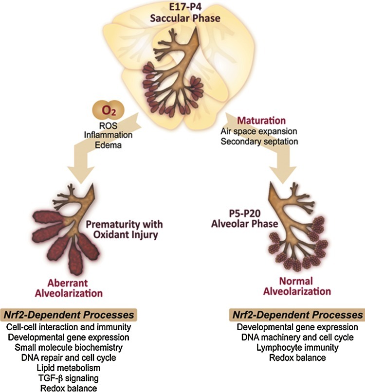
Aims: Nrf2 is an essential transcription factor for protection against oxidant disorders. However, its role in organ development and neonatal disease has received little attention. Therapeutically administered oxygen has been considered to contribute to bronchopulmonary dysplasia (BPD) in prematurity. The current study was performed to determine Nrf2-mediated molecular events during saccular-to-alveolar lung maturation, and the role of Nrf2 in the pathogenesis of hyperoxic lung injury using newborn Nrf2-deficient (Nrf2(-/-)) and wild-type (Nrf2(+/+)) mice.

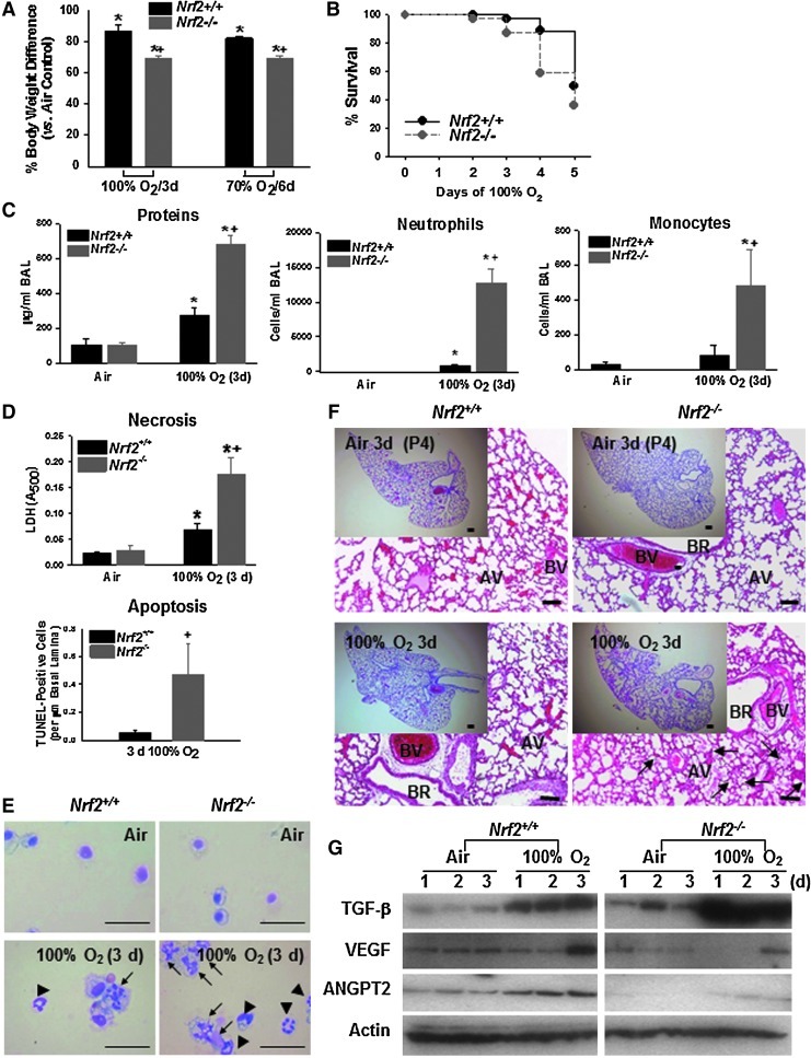
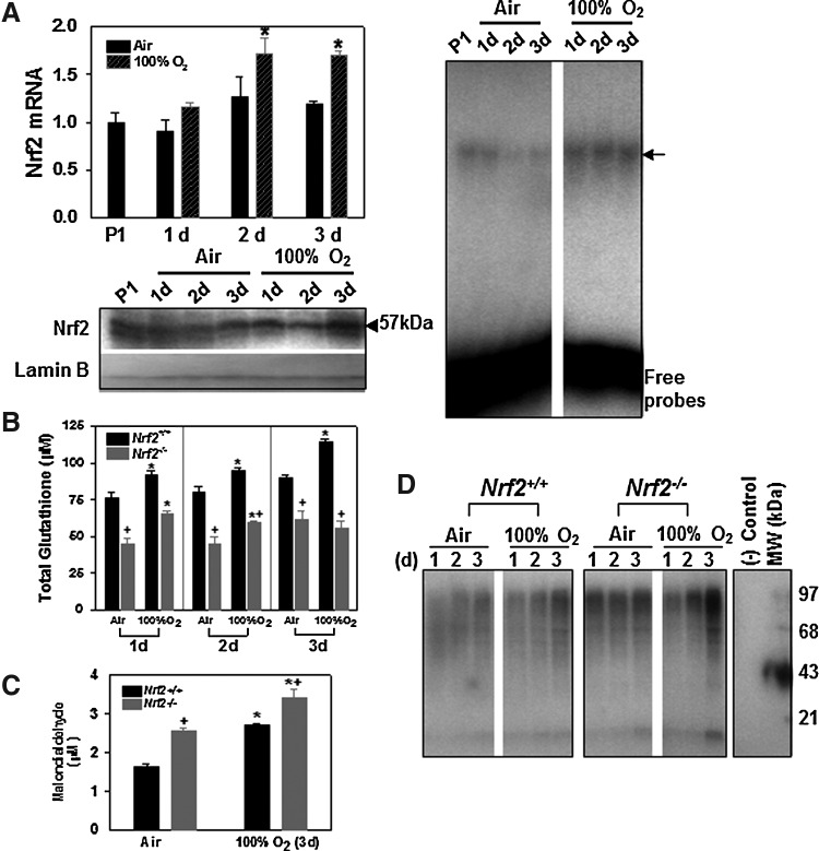
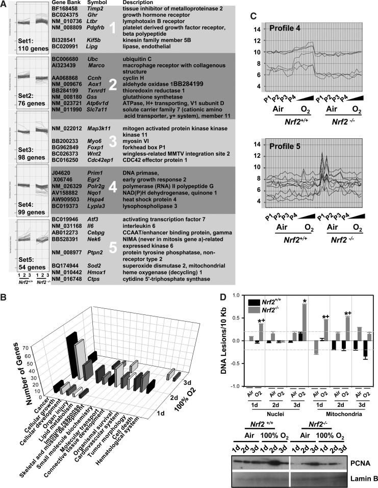
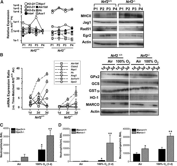
Results: Pulmonary basal expression of cell cycle, redox balance, and lipid/carbohydrate metabolism genes was lower while lymphocyte immunity genes were more highly expressed in Nrf2(-/-) neonates than in Nrf2(+/+) neonates. Hyperoxia-induced phenotypes, including mortality, arrest of saccular-to-alveolar transition, and lung edema, and inflammation accompanying DNA damage and tissue oxidation were significantly more severe in Nrf2(-/-) neonates than in Nrf2(+/+) neonates. During lung injury pathogenesis, Nrf2 orchestrated expression of lung genes involved in organ injury and morphology, cellular growth/proliferation, vasculature development, immune response, and cell-cell interaction. Bioinformatic identification of Nrf2 binding motifs and augmented hyperoxia-induced inflammation in genetically deficient neonates supported Gpx2 and Marco as Nrf2 effectors.

Innovation: This investigation used lung transcriptomics and gene targeted mice to identify novel molecular events during saccular-to-alveolar stage transition and to elucidate Nrf2 downstream mechanisms in protection from hyperoxia-induced injury in neonate mouse lungs.

Conclusion: Nrf2 deficiency augmented lung injury and arrest of alveolarization caused by hyperoxia during the newborn period. Results suggest a therapeutic potential of specific Nrf2 activators for oxidative stress-associated neonatal disorders including BPD.


Figures

FIG. 1.
FIG. 1.
Gene expression profiles during postnatal lung development. (A) Expression kinetics of ≥2-fold suppressed (n=324, blue) or upregulated (n=198, orange-red) genes at P1–P3 relative to age P4 in Nrf2+/+ mice (p<0.01). Average transcript levels of individual genes at each time were normalized to those at P4. Color bar indicates average expression intensity. (B) Differential transcript expression patterns during P1–P4 in Nrf2+/+ and Nrf2−/− mice were identified by visual data mining (Spotfire). Signal intensity of individual sample transcript (each vertical line) is indicated as log2-normalized value. (C) Selected differentially expressed genes (≥2-fold, from Supplementary Tables S1A, B) are presented. Genes in blue or in orange are ≥2-fold lower or higher, respectively, at least one time during P1–P3 than in P4. (D) Suppression of lung proliferating cell nuclear antigen (PCNA) at P1 relative to later postnatal times was confirmed by Western blotting and immunohistochemistry in Nrf2+/+ mice. Nuclear lamin B level was determined as a loading control. PCNA-positive nuclei were histologically detected in pulmonary artery and main stem bronchi, and the populations were increased throughout the lungs by P4 (P3 tissues not shown). Arrows, PCNA-stained cells. AV, alveoli; BR, bronchiole; BV, blood vessel. Representative light photomicrographs are shown (n=3/group). Bars indicate 100 μm. (To see this illustration in color the reader is referred to the web version of this article at www.liebertonline.com/ars)
FIG. 2.
FIG. 2.
Effect of Nrf2 deletion on transcriptome of late saccular phase lungs. (A) Biological functions and disorders of 9737 Nrf2-dependent transcripts (p<0.05) altered during postnatal ages P1–P4 were identified by ingenuity pathway analysis (IPA) and the number of genes in each function and disorder are depicted in a pie chart. (B) The number of transcripts significantly different (≥2-fold, p<0.01) between Nrf2+/+ and Nrf2−/− neonates at each postnatal day. Selected lung genes overexpressed (black) or suppressed (gray) in Nrf2−/− relative to Nrf2+/+ neonates (from Supplementary Tables S2a, b) are presented. (C) Visual data profiling analysis (Spotfire) classified Nrf2-dependent profile patterns during P1–P4 (gene list in Supplementary Table S3). Signal intensity of individual sample transcript (each vertical line) is indicated as log2-normalized value.
FIG. 3.
FIG. 3.
Enhanced susceptibility of Nrf2−/− neonates to hyperoxia. Significantly increased hyperoxia susceptibility of newborn Nrf2−/− mice relative to Nrf2+/+ newborns was determined by lower body weight gain after 3 days of 100% O2 or after 6 days of 70% O2 (A); more severe mortality after 1–5 days of 100% O2 exposure (B); enhanced total protein concentration, and the number of neutrophils and monocytes in bronchoalveolar lavage (BAL) at 3 days after 100% O2 (C); and heightened necrotic and apoptotic airway cell death after 3 days of 100% O2 (D). All data are presented as mean±standard error of the mean (SEM). *Significantly different from genotype-matched air controls (p<0.05). +Significantly different from exposure-matched Nrf2+/+ mice (p<0.05). n=5–17/group for mean % body weight data. n=10–12 for 2 days and n=22–39 for 3–5 days mortality data. n=6/group for BAL data. Necrotic lung cell death was quantified in aliquots of BAL using a colorimetric lactate dehydrogenase (LDH) assay (n=6/group). Airway cell apoptosis was determined by deoxynucleotidyl transferase-mediated dUTP nick-end labeling (TUNEL) and National Institutes of Health (NIH) Image analysis (n=3/group). TUNEL-stained lung cells were barely detected in air controls of either genotype. (E) Representative Giemsa-stained cytocentrifuged BAL fluid cells indicate markedly greater lung cell death (arrows) by lysis or apoptosis and airway neutrophilic infiltration (arrow heads) in Nrf2−/− than in Nrf2+/+ mice after 3 days of 100% O2. Bars=50 μm. (F) Augmented adverse lung histopathology in Nrf2−/− neonates relative to Nrf2+/+ after hyperoxia indicated by greater pulmonary epithelial thickening, perivascular-peribronchiolar edema, and protein exudates in air space (arrows) after 3 days of 100% O2. Representative light photomicrographs of hematoxylin and eosin (H&E)-stained lung tissue sections are presented (n=3–9/group). Bars=100 μm. (G) Differential protein expression of transforming growth factor beta (TGF-β), and angiogenesis factors for lung development, vascular endothelial growth factor (VEGF), and angiopoietin 2 (ANGPT2), between Nrf2+/+ and Nrf2−/− neonates basally and after O2. Representative band images of multiple applications are shown (n=3/group).
FIG. 4.
FIG. 4.
Lung Nrf2 activation and redox status after hyperoxia. (A) Hyperoxia increased Nrf2 message as determined by semi-quantitative reverse transcription–polymerase chain reaction (RT-PCR) in Nrf2+/+ mice after 2–3 days of 100% O2, while basal level of Nrf2 mRNA did not vary significantly between ages P1 and P4. Data presented as mean±SEM (n=3/group) after normalization to the level at P1. *Significantly different from time-matched air controls (p<0.05). Nuclear translocation of Nrf2 determined by Western blot analysis of lung nuclear protein aliquots (20 μg) was enhanced by 1–3 days of 100% O2 over the corresponding air controls in Nrf2+/+ mice. Nuclear lamin B level was determined as a loading control. Gel shift analysis demonstrated enhanced total antioxidant response element (ARE) binding activity of lung nuclear proteins at 1–3 days of O2 relative to time-matched air controls in Nrf2+/+ mice. Arrow indicates shifted bands for nuclear protein (5 μg) bound to ARE consensus sequence. Representative digitized bands from duplicate Western blotting and gel shift analysis are presented. (B) Reduced lung glutathione in Nrf2−/− mice compared with Nrf2+/+ mice at baseline and after hyperoxia. Mean±SEM presented (n=3/group). *Significantly different from genotype-matched air control mice (p<0.05). +Significantly lower than exposure-matched Nrf2+/+ mice (p<0.05). (C) Different levels of lung lipid peroxidation evaluated by malondialdehyde (MDA) level in BAL from Nrf2+/+ and Nrf2−/− neonates after 3 days air and O2. Mean±SEM (n=4/group) is presented. *Significantly different from genotype-matched air control mice (p<0.05). +Significantly different from O2-exposed Nrf2+/+ neonates (p<0.05). (D) Heightened oxidized proteins in Nrf2−/− neonates determined by immunoblotting analysis of carbonyl moieties detected in 30–100 kDa lung proteins after 3 days of air or O2. (−) control, nonderivatized protein samples. MW, protein molecular weight marker.
FIG. 5.
FIG. 5.
Effect of Nrf2 deletion on lung transcriptome during pathogenesis of hyperoxia-induced lung injury. (A) Nrf2-dependently changed genes during hyperoxia (n=437, p<0.01) were grouped into 5 k-means cluster profiles (GeneSpring). Transcript expression is indicated as relative log ratio after normalization to time-matched Nrf2+/+ air control. Selected genes from each cluster are listed. (B) Nrf2-dependent genes modulated by hyperoxia on each day were classified into biological functions and disorders by IPA, and plotted against the number of genes associated. (C) Visual profiling analysis (Spotfire) clustered several distinct genes showing unique Nrf2-dependent expression pattern during hyperoxia exposure. Profile 4 includes thioredoxin reductase 1 (Txnrd1) and cellular repressor of E1A-stimulated genes 1 (Creg1). Profile 5 includes major histocompatibility complex, class II genes (e.g., H2-Ea) and 5-methyltetrahydrofolate-homocysteine methyltransferase (Mtr). Signal intensity of individual sample transcript (each vertical line) is indicated as log2-normalized value. (D) Lesion frequencies in genomic (DNA polymerase β gene) and mitochondrial DNA were compared in Nrf2+/+ and Nrf2−/− mice after air or hyperoxia (100%). All data were normalized to 1 day air-exposed Nrf2+/+ mice and group mean±SEM is presented (n=3/group). Background noise level (dashed lines) is set at±0.15. *Significantly different from genotype- and time-matched air controls (p<0.05). +Significantly different from exposure- and time-matched Nrf2+/+ mice (p<0.05). PCNA level was determined as a marker for S-phase cells undergoing proliferation by western blotting in nuclear extracts of Nrf2+/+ and Nrf2−/− lungs. Nuclear lamin B level was determined as a loading control. Representative band images from replicates are shown.
FIG. 6.
FIG. 6.
Validation of microarray profiles and role for Nrf2 effectors in hyperoxia-induced lung injury pathogenesis. Selected transcripts differentially expressed between Nrf2+/+ and Nrf2−/− neonates at P1–P4 (A) and during hyperoxia exposure (B) were confirmed by quantitative (q)RT-PCR and/or Western blot analyses. qRT-PCR graphs present fold differences of each gene expression relative to P4 level in Nrf2+/+ after normalization to corresponding 18s expression (A) or depict fold differences relative to time-matched Nrf2+/+ air control of 18s-normalized data (B). Group mean±SEM is presented (n=3/group). Actin level was determined as internal control for western blotting. Representative digitized bands from multiple blot analysis are presented. H2-Q1, histocompatibility 2, Q region locus 1; H2-D1, histocompatibility 2, D region locus 1; Inta4, integrin alpha 4; Nqo1, NAD(P)H:quinone oxidoreductase 1; Akr1b8, aldo-keto reductase family 1, member B8; Hc, hemolytic complement; Aox1, aldehyde oxidase 1; Jag1, jagged 1; Rad51, RAD51 homolog; Egr2, early growth response 2; Clstn2, calsyntenin 2; Ang3, angiogenin, ribonuclease A family, member 3; Slc7a11, solute carrier family 7, member 11; Gpx2, glutathione peroxidase 2; Gclc or GCS, glutamate cysteine ligase, catalytic subunit; Gstm or GST-μ, glutathione S-transferase M; Ho1, heme oxygenase-1; Marco, macrophage receptor with collagenous structure. Functional roles of ARE-bearing Gpx2 (C) and Marco (D) in hyperoxia-induced lung inflammation were determined by BAL analysis in gene-targeted neonates after 100% of O2 (3 days). Mean±SEM (n=4/group) is presented. *Significantly different from genotype-matched air control (p<0.05). +Significantly different from exposure-matched wild-type controls (p<0.05).
FIG. 7.
FIG. 7.
Hypothetical schematics depicting proposed role for pulmonary Nrf2 in saccular-to-alveolar transition and hyperoxia-induced lung injury pathogenesis learned from mice. During early postnatal lung maturation and development, Nrf2 modulates expression of genes associated with DNA replication machinery, cell cycle regulation, development, and host defense and redox balance in mice. Abnormally high expression of genes for antigen presentation and T lymphocyte immunity are also evident in Nrf2−/− mice, indicating a role for Nrf2 in suppression of aberrant acquired immunity. Hyperoxia exposure during the late saccular phase causes oxidative injuries to lung proteins and lipids, and genomic and mitochondrial DNA damages are coupled to the tissue and protein edema, inflammation, cell death, and abnormal alveolar formation, which are similar to the bronchopulmonary dysplasia (BPD) phenotypes of prematurity. In Nrf2−/− lungs, these phenotypes are significantly augmented. Suppression of DNA repair device and redox capacity, interruption of cell cycle machinery and tissue development factors, alteration of lipid metabolism and small molecule biochemistry process, and potentiation of TGF-β signaling and fibrogenic factors in Nrf2−/− lungs in response to hyperoxia suggest functional roles for Nrf2 in the pathogenesis of BPD-like phenotypes. (To see this illustration in color the reader is referred to the web version of this article at www.liebertonline.com/ars)

Similar articles

Cited by

References

    1. Ahola T. Lapatto R. Raivio KO. Selander B. Stigson L. Jonsson B. Jonsbo F. Esberg G. Stovring S. Kjartansson S. Stiris T. Lossius K. Virkola K. Fellman V. N-acetylcysteine does not prevent bronchopulmonary dysplasia in immature infants: a randomized controlled trial. J Pediatr. 2003;143:713–719. - PubMed
    1. Arredouani M. Yang Z. Ning Y. Qin G. Soininen R. Tryggvason K. Kobzik L. The scavenger receptor MARCO is required for lung defense against pneumococcal pneumonia and inhaled particles. J Exp Med. 2004;200:267–272. - PMC - PubMed
    1. Baraldi E. Filippone M. Chronic lung disease after premature birth. N Engl J Med. 2007;357:1946–1955. - PubMed
    1. Barker GF. Manzo ND. Cotich KL. Shone RK. Waxman AB. DNA damage induced by hyperoxia: quantitation and correlation with lung injury. Am J Respir Cell Mol Biol. 2006;35:277–288. - PMC - PubMed
    1. Bauer AK. Fostel J. Degraff LM. Rondini EA. Walker C. Grissom SF. Foley J. Kleeberger SR. Transcriptomic analysis of pathways regulated by toll-like receptor 4 in a murine model of chronic pulmonary inflammation and carcinogenesis. Mol Cancer. 2009;8:107. - PMC - PubMed

Publication types