. 2012 Mar 7;14(2):R39.
doi: 10.1186/bcr3135.

Targeting of the non-mutated tumor antigen HER2/neu to mature dendritic cells induces an integrated immune response that protects against breast cancer in mice

Affiliations

Targeting of the non-mutated tumor antigen HER2/neu to mature dendritic cells induces an integrated immune response that protects against breast cancer in mice

Bei Wang et al. Breast Cancer Res. .

Abstract

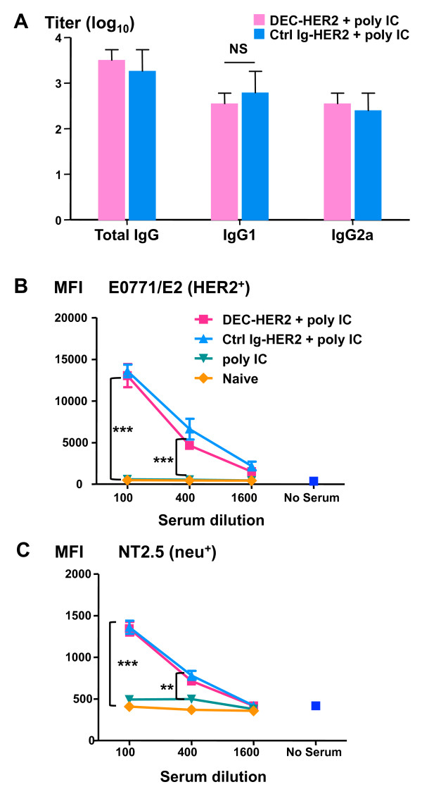
Introduction: Given their relative simplicity of manufacture and ability to be injected repeatedly, vaccines in a protein format are attractive for breast and other cancers. However, soluble human epidermal growth factor receptor (HER2)/neu protein as a vaccine has not been immunogenic. When protein is directly targeted to antigen uptake receptors, such as DEC205 (DEC), efficient processing and presentation of antigen take place. The aim of this study was to determine the immunogenicity of a HER2 protein vaccine that directly targets to DEC+ dendritic cells (DCs) in a mouse breast cancer model.

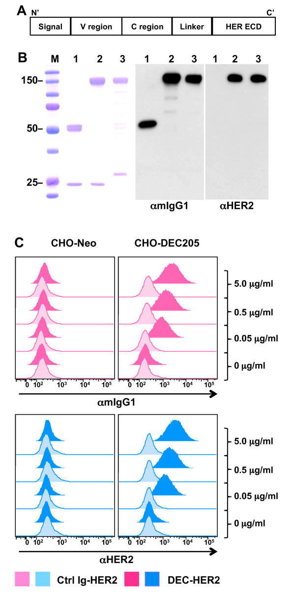
Methods: We genetically engineered the HER2 extracellular domain into a monoclonal antibody specific for DEC (DEC-HER2). Mice of various genetic backgrounds were immunized with DEC-HER2 in combination with DC maturation stimuli (poly IC ± CD40 Ab). Vaccine-induced T cell immunity was determined by analyzing the ability of CD4+/CD8+ T cell to produce interferon (IFN)-gamma and proliferate upon antigen rechallenge. Sera were assessed for the presence of antigen specific antibody (Ab). For vaccine efficacy, FVB/N mice were immunized with DEC-HER2 in combination with poly IC and protection against neu-expressing mammary tumors was assessed. Protection mechanisms and tumor-specific T cell responses were also evaluated.

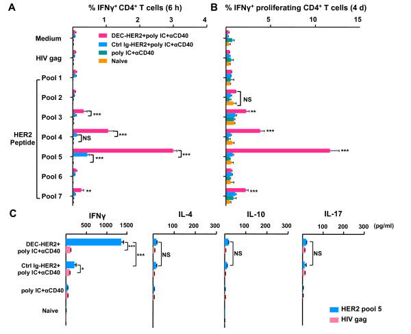
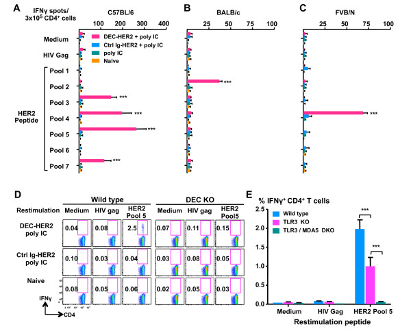
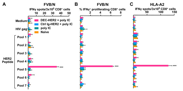
Results: We demonstrate that DEC-HER2 fusion mAb, but not Ctrl Ig-HER2, elicits strong, broad and multifunctional CD4+ T cell immunity, CD8+ T cell responses, and humoral immunity specific for HER2 antigen. Cross-reactivity to rat neu protein was also observed. Importantly, mice xeno-primed with DEC-HER2 were protected from a neu-expressing mammary tumor challenge. Both CD4+ and CD8+ T cells mediated the tumor protection. Robust anti-tumor T cell immunity was detected in tumor protected mice.

Conclusions: Immunization of mice with HER2 protein vaccine targeting DEC+ DCs in vivo induced high levels of T- and B-cell immunity. Non-targeted HER2 protein was poorly immunogenic for CD4+ and CD8+ T cells. This vaccination approach provided long-term survival benefit for mice challenged with neu-expressing tumor following as little as 2.7 μg of HER2 protein incorporated in the vaccine. Vaccine-induced CD4+ and CD8+ T cells were both essential for tumor protection. This immunization strategy demonstrates great potential towards the development of vaccines for breast cancer patients.


Figures

Figure 1
Figure 1
Characterization of HER2 fusion monoclonal antibody (mAb). (A) Structure of HER2 fusion mAb. C', carboxyl-terminus; C region, constant region; ECD, extracellular domain (amino acid 22-653); N', amino-terminus; V region, variable region. (B) Fusion mAb (DEC-HER2 or Ctrl Ig-HER2) was produced by transient transfection of 293T cells with the appropriate vector and purified on a protein G column. Imperial protein staining (left panel) and Western blotting of the fusion mAb are shown, and the anti-mouse IgG1-HRP (αmIgG) and anti-HER2-Biotin/SAv-HRP (αHER2) antibodies are indicated below the figures. Lane 1: empty DEC mAb; lane 2: DEC-HER2 mAb; lane 3: Ctrl Ig-HER2 mAb. M, molecular weight standards (in kilodaltons). (C) Fluorescence-activated cell sorting staining data show the binding capacity of graded doses (0, 0.05, 0.5, and 5 μg/mL) of the indicated fusion mAb to the DEC or neomycin (Neo) stably transfected CHO cells. Binding was detected by fluorescence-labeled secondary antibody specific for mouse IgG1 or HER2 antigen. CHO, Chinese hamster ovary; HER, human epidermal growth factor receptor; Ig, immunoglobulin.
Figure 2
Figure 2
A single dose of DEC-HER2 fusion monoclonal antibody (mAb) vaccine immunizes HER2-specific CD4+ T cells in vivo. (A) Groups of C57BL/6 mice were vaccinated intraperitoneally with 5 μg of DEC-HER2 or Ctrl Ig-HER2 in combination with poly IC (50 μg) and anti-CD40 antibody (25 μg). Additional groups received adjuvants alone or were left untreated. Two weeks after immunization, splenocytes were re-stimulated without added peptides (medium alone) or with 2 μg/mL HER2 peptide pools 1 to 7 or irrelevant HIV gag peptides. Intracellular cytokine staining was determined by fluorescence-activated cell sorting after 6 hours of in vitro stimulation. The percentage of IFNγ+CD4+ T cells is shown as mean ± standard error with three or four mice per group. The results of one of three independent experiments are shown. (B) CFSE dilution assay. Mice were immunized as in (A), and bulk splenocytes were labeled with 2.5 μM CFSE and re-stimulated with 0.2 μg of HER2 peptide pool 1-7 or HIV gag peptide mix or medium alone for 4 days. T-cell proliferation was analyzed in combination with intracellular cytokine staining of the cells re-stimulated for the last 6 hours. Percentages of proliferating (CFSElow) and IFNγ+CD4+ T cells are shown with three or four mice per group. The results of one of two independent experiments are shown. (C) Th1/Th2/Th17 cytokine enzyme-linked immunosorbent assay (ELISA). Mice were immunized as in (A). Two weeks after the boost, splenic CD4+ T cells and CD11c+ cells were isolated. CD4+ cells (3 × 10⁵) were incubated with 1 × 10⁵ CD11c+ cells in 96-well U-bottomed plates in the presence of 2 μg/mL HER2 peptide pool 5 or HIV gag peptides for 48 hours. Concentrations of IFNγ/IL-4/IL-10/IL-17 in culture supernatant were measured by ELISA. The results of one of two independent experiments (n = 4 mice per group) are shown. *P < 0.05, **P < 0.01, ***P < 0.001. CFSE, 5,6-carboxy fluorescein diacetate succinimidyl ester; HER, human epidermal growth factor receptor; IFNγ, interferon-gamma; Ig, immunoglobulin; IL, interleukin; NS, not statistically significant; poly IC, polyinosinic/polycytidylic acid; Th, T helper.
Figure 3
Figure 3
Poly IC as a mono-adjuvant for strong and broad HER2-specific CD4+ T-cell responses. C57BL/6 (A), BALB/c (B), or FVB/N (C) mice were primed with 5 μg of DEC-HER2 + 50 μg of poly IC and boosted 4 weeks later. Two weeks after the boost, HER2-specific IFNγ production was quantified by enzyme-linked immunosorbent spot assay. All experiments were performed with at least three mice per group, and the results of one of two or three independent experiments are shown. (D) Requirement for DEC expression. DEC-/- or wild-type C57BL/6 mice were primed and boosted 4 weeks apart with DEC-HER2 + poly IC, Ctrl Ig-HER2 + poly IC, or nothing. Two weeks after the boost, splenocytes were harvested and re-stimulated with medium alone or 2 μg/mL HIV gag or HER2 peptide pool 5 for 6 hours. IFNγ production was measured by intracellular cytokine staining. Fluorescence-activated cell sorting blots are shown. Three or four mice were in each group, and the results of one of two experiments are shown. (E) Requirement for pattern recognition receptors. Wild-type, TLR3 KO, or TLR3/MDA5 DKO C57BL/6 mice were immunized with DEC-HER2 + poly IC. Two weeks after the boost vaccination, splenocytes were harvested and re-stimulated with medium alone or 2 μg/mL HIV gag or HER2 peptide pool 5 for 6 hours. IFNγ production was measured by intracellular cytokine staining. Three mice were in each group. ***P < 0.001. HER, human epidermal growth factor receptor; IFNγ, interferon-gamma; Ig, immunoglobulin; poly IC, polyinosinic/polycytidylic acid; TLR, Toll-like receptor.
Figure 4
Figure 4
Cross-presentation of HER2 protein by DEC-HER2 immunization. (A) FVB/N mice were primed and boosted with DEC-HER2 or Ctrl Ig-HER2 (5 μg) in combination with poly IC (50 μg). Two weeks after the boost, spleen CD8+ T cells were purified by magnetic-activated cell sorting and re-stimulated with spleen CD11c+ DCs in the presence of medium alone or 1 μg/mL HIV gag peptide or HER2 peptide pools. IFNγ production was measured by enzyme-linked immunosorbent spot (ELISPOT) assay. (B) Proliferative capacity of HER2-specific CD8+ T cells. Mice were immunized as in (A), and bulk splenocytes were labeled with CFSE and re-stimulated with medium or 200 ng/mL HIV gag or HER2 peptide pool 1-7 for 4 days. Cells were re-stimulated for the last 6 hours, and IFNγ production was measured by intracellular cytokine staining. (C) HLA-A2 transgenic mice were vaccinated as in (A), and HER2-specific CD8+ T-cell responses were measured by ELISPOT assay. Purified CD8+ T cells were re-stimulated with medium alone or 1 μg/mL HIV gag peptide mix or HER2 peptide pool 1-7. All experiments were performed with at least three mice per group, and the results of one of three experiments are shown. ***P < 0.001. CFSE, 5,6-carboxy fluorescein diacetate succinimidyl ester; HER, human epidermal growth factor receptor; IFNγ, interferon-gamma; Ig, immunoglobulin; poly IC, polyinosinic/polycytidylic acid.
Figure 5
Figure 5
HER2 immunization primes strong neu-specific CD4+ T-cell responses in mice. (A) C57BL/6 mice were primed with DEC-HER2 in combination with poly IC. Two weeks after the boost, splenocytes were re-stimulated with medium alone, HIV gag peptides, HER2 peptide pool 5, or corresponding neu peptide pool 4 (2 μg/mL). IFNγ production was measured by intracellular cytokine staining. (B) Functional avidity of HER2/neu-specific CD4+ T cells. Titrated doses of HER2 or neu peptide pool were used to re-stimulate splenocytes, and IFNγ production was quantified. Data depict the percentage of maximum response at each concentration. ***P < 0.001. Results of two experiments are shown, and six mice were in each group. EC50, concentration of peptide that leads to 50% of the maximal responses; HER, human epidermal growth factor receptor; IFNγ, interferon-gamma; poly IC, polyinosinic/polycytidylic acid.
Figure 6
Figure 6
Induction of anti-HER2/neu humoral immunity. FVB/N mice were vaccinated with DEC-HER2 or Ctrl Ig-HER2 with poly IC as the adjuvant. Sera were collected 2 weeks after the boost. (A) Titers of anti-HER2 IgG were quantified by enzyme-linked immunosorbent assay as described in Materials and methods. (B, C) Binding of HER2/neu-expressing tumor cells with immune sera measured by flow cytometry. Sera were diluted 1:100, 1:400, or 1:1,600 and incubated with HER2-expressing E0771/E2 (B) or rat neu-expressing NT2.5 (C) tumor cells for 15 minutes at 4°C. Binding of antibody to tumor cells was detected by staining with anti-mouse IgG-PE antibody and fluorescence-activated cell sorting analysis. Mean fluorescence indices (MFIs) are shown. **P < 0.01, ***P < 0.001. HER, human epidermal growth factor receptor; Ig, immunoglobulin; NS, not statistically significant; PE, phycoerythrin; poly IC, polyinosinic/polycytidylic acid.
Figure 7
Figure 7
Targeting HER2 protein to dendritic cells protects mice from a challenge with HER2/neu-expressing mammary tumor cells. (A, B) FVB/N mice were immunized with DEC-HER2 or Ctrl Ig-HER2 with poly IC or poly IC alone or were left untreated. Mice were challenged with one million NT2.5 tumor cells in the mammary fat tissue 10 days after the boost immunization. Tumor growth was monitored by caliper measurement three times a week. Tumor volume (in cubic millimeters) is shown in (A). Survival analysis is shown in (B). Results of three independent experiments (n = 5 per group) are shown. (C, D) To examine the mechanism of tumor protection, CD4+, CD8+, or both types of T cells were depleted after immunization (before tumor challenge) as described in Materials and methods. Mice were challenged with one million NT2.5 tumor cells, and tumor growth curves are shown in (C). Survival analysis is shown in (D). Results of two independent experiments (n = 10 mice per group) are shown. (E, F) Anti-tumor immunity was determined 14 days after tumor challenge. Splenic CD4+ and CD8+ cells were purified by magnetic-activated cell sorting isolation. Splenic CD11c+ cells were purified from naïve FVB/N mice. CD4+ (E) or CD8+ (F) T cells were re-stimulated with CD11c+ cells pulsed with 1 μg/mL of HIV gag, HER2 peptide pool 5, or neu peptide pool 4 or 10 μg/mL NT2.5 tumor lysate at a T cell/DC ratio of 3:1 for 3 days. IFNγ production was measured by enzyme-linked immunosorbent spot assay. Four mice were in each group; the results of one of two independent experiments are shown. *P < 0.05, **P < 0.01, ***P < 0.001. HER, human epidermal growth factor receptor; IFNγ, interferon-gamma; Ig, immunoglobulin; NS, not statistically significant; PBS, phosphate-buffered saline; poly IC, polyinosinic/polycytidylic acid.

Similar articles

Cited by

References

    1. Yarden Y, Sliwkowski MX. Untangling the ErbB signalling network. Nat Rev Mol Cell Biol. 2001;2:127–137. doi: 10.1038/35052073. - DOI - PubMed
    1. Baselga J, Swain SM. Novel anticancer targets: revisiting ERBB2 and discovering ERBB3. Nat Rev Cancer. 2009;9:463–475. doi: 10.1038/nrc2656. - DOI - PubMed
    1. Slamon D, Clark G, Wong S, Levin W, Ullrich A, McGuire W. Human breast cancer: correlation of relapse and survival with amplification of the HER-2/neu oncogene. Science. 1987;235:177–182. doi: 10.1126/science.3798106. - DOI - PubMed
    1. Hynes NE, Stern DF. The biology of erbB-2/neu/HER-2 and its role in cancer. Biochim Biophys Acta. 1994;1198:165–184. - PubMed
    1. Pupa SM, Menard S, Andreola S, Colnaghi MI. Antibody response against the c-erbB-2 oncoprotein in breast carcinoma patients. Cancer Res. 1993;53:5864–5866. - PubMed

Publication types

MeSH terms