. 2012 Mar 6;5(214):ra20.
doi: 10.1126/scisignal.2002521.

STING specifies IRF3 phosphorylation by TBK1 in the cytosolic DNA signaling pathway

Affiliations

STING specifies IRF3 phosphorylation by TBK1 in the cytosolic DNA signaling pathway

Yasuo Tanaka et al. Sci Signal. .

Abstract

Cytosolic double-stranded DNA (dsDNA) stimulates the production of type I interferon (IFN) through the endoplasmic reticulum (ER)-resident adaptor protein STING (stimulator of IFN genes), which activates the transcription factor interferon regulatory factor 3 (IRF3); however, how STING activates IRF3 is unclear. Here, we showed that STING stimulates phosphorylation of IRF3 by the kinase TBK1 (TANK-binding kinase 1) in an in vitro reconstitution system. With this system, we identified a carboxyl-terminal region of STING that was both necessary and sufficient to activate TBK1 and stimulate the phosphorylation of IRF3. We also found that STING interacted with both TBK1 and IRF3 and that mutations in STING that selectively disrupted its binding to IRF3 abrogated phosphorylation of IRF3 without impairing the activation of TBK1. These results suggest that STING functions as a scaffold protein to specify and promote the phosphorylation of IRF3 by TBK1. This scaffolding function of STING (and possibly of other adaptor proteins) may explain why IRF3 is activated in only a subset of signaling pathways that activate TBK1.


Conflict of interest statement

Competing interests: The authors declare that they have no competing interests.

Figures

Figure 1
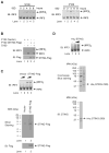
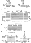
Figure 1. STING Activates IRF3 in an in vitro Reconstitution System
A. ISD activates IRF3 in L929 cells. L929 cells were transfected with ISD for the indicated time. The cytosolic supernatant (S100) was analyzed by native gel electrophoresis to observe the dimerization of endogenous IRF3 (left panel). The high-speed membrane fraction (P100) isolated from ISD stimulated cells was incubated with cytosolic extracts from untreated L929 (S100), and the dimerization of IRF3 was analyzed by native gel electrophoresis (right panel). IB: Immunoblot B. IRF3 activation by the membrane fraction (P100) of STING-expressing cells. Cytosolic extracts from HeLa cells (S100) were incubated with P100 from HEK293T cells overexpressing STING-Flag. IRF3 dimerization was analyzed by native gel electrophoresis. C & D. IRF3 activation by STING. STING-Flag (C) and His6-STING (D) were expressed in and purified from HEK293T and Sf9 cells, respectively, and then incubated with HeLa S100 and ATP followed by native gel electrophoresis. Aliquots of the proteins were stained by silver or Coomassie Blue, or by immunoblotting.
Figure 2
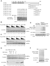
Figure 2. STING C-terminus is Important for IRF3 Activation in vitro
A. Diagrams of full-length and truncated STING used in this study. TM, transmembrane domain. His6-tagged proteins expressed in and purified from E. coli were analyzed by Coomassie Blue staining (lower panel). B & C. His6-STING deletion mutants were incubated with ATP-supplemented HeLa S100, and then IRF3 dimerization was analyzed by native gel electrophoresis. D. His6-STING (341–379) was fractionated by gel filtration on Superdex-200. Each fraction was analyzed by IRF3 dimerization assay (upper panel) and immunoblotting (lower panel). SM: starting material. E. L929 cells stably expressing mouse STING shRNA with or without the simultaneous expression of wild type human STING-Flag were transfected with ISD for the indicated time and then membrane fractions (P100) were resolved by native-PAGE or SDS-PAGE followed by immunoblotting using a STING antibody. Cytosolic extracts (S100) were also immunoblotted with IRF3 antibody after native PAGE. F. Similar to E, except that mouse macrophage RAW 264.7 cells were transfected with ISD to detect the aggregation of endogenous STING.
Figure 3
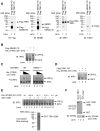
Figure 3. STING Directly Activates TBK1 in vitro
A. MEF cells stably expressing Flag-NEMO-ΔN were used to isolate endogenous TBK1 by virtue of its association with Flag-NEMO-ΔN. The composition of this complex, denoted as Flag-NEMO PD, was analyzed by immunoblotting with the indicated antibodies. HEK293T cells stably overexpressing Flag-TBK1 were used to isolate recombinant TBK1 protein, denoted here as Flag-TBK1 PD. B. Flag-NEMO PD, which contained endogenous TBK1 as shown in A, was incubated with His6-STING (341–379, ~2 μM) and His6-IRF3 (~40 nM) in the presence of ATP. IRF3 dimerization was analyzed by native gel electrophoresis. C. S20 from wild type or NEMO deficient MEFs was incubated with varying concentrations of His6-STING (341–379) (0.375, 0.75 and 1.5 μM) together with 35S-IRF3. IRF3 dimerization was analyzed by native gel electrophoresis followed by autoradiography. D. Similar to B, except that Flag-TBK1 pull-down (PD) complex was used. E. GST-TBK1 was purified from Sf9 cells and analyzed by Coomassie blue staining (lower panel). The purified protein (200 nM) was incubated with His6-IRF3 (200 nM) and varying concentrations of His6-STING (341–379) in the presence of ATP, then IRF3 dimerization was analyzed by native PAGE. F. GST-TBK1 or GST (1 μg) was incubated with 2 μg of His6-STING (341–379) and then pulled down with glutathione Sepharose, followed by immunoblotting. The input represents 10% of the amount of STING used in the pull-down experiments.
Figure 4
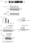
Figure 4. Ser366 and Leu374 of STING are Important for IRF3 Activation
A. Sequence alignment of the carboxy-termini of human, mouse, porcine and bovine STING using Clustal W2. Asterisks indicate residues that were mutated in this study. WT and mutated STING fragments were expressed in and purified from E. coli as His6-tagged proteins and analyzed by Coomassie blue staining (lower panel). Each protein (2 μM) was tested in IRF3 dimerization assay (upper panel). B. HEK293T cells were transiently transfected with full-length STING-Flag (WT) and its mutants (S368A, S366A and L374A) together with an ISRE-luciferase reporter (left panel). The error bars represent the variation ranges of duplicate experiments. The expression of STING proteins was confirmed by immunoblotting (right panel). C. L929 cells stably expressing mouse STING shRNA with or without the simultaneous expression of wild type or mutant (S358A, S366A and L374A) human STING-Flag were transfected with ISD for 4 hours. The dimerization of endogenous IRF3 was analyzed by native gel electrophoresis (left panel). The expression endogenous and rescued STING proteins were confirmed by immunoblotting (right panel). D. L929 cells in which endogenous STING was replaced with wild type or mutant STING-Flag as shown in (C) were transfected with ISD or mock treated. Membrane pellets (P100) from these cells were incubated with His6-IRF3 in the presence of ATP, and IRF3 dimerization was analyzed by native PAGE (upper panel). Each P100 fraction used in this assay was also blotted with a TBK1 antibody (lower panel).
Figure 5
Figure 5. STING Recruits IRF3 to TBK1 to Induce IRF3 Activation
A & B. Wild type or mutant GST-STING (341–379) or GST was incubated with Flag-TBK1 (A) or His6-IRF3 (B) and then pulled down with glutathione Sepharose followed by immunoblotting. The input represents 10% of TBK1 or IRF3 used in the pull-down experiments. C. L929 cells stably expressing STING shRNA and those in which endogenous STING was replaced with WT or mutated STING-Flag were stimulated with ISD for the indicated lengths of time. Cell lysates were immunoprecipitated with anti-Flag agarose and then immunoblotted with the indicated antibodies. Aliquots of the cell lysates were immunoblotted for IRF3 following native PAGE (top). D. Similar to C, except that cell lysates were resolved by SDS-PAGE, followed by immunoblotting with antibodies against TBK1 or TBK1 phosphorylated at Ser172. E. L929 cells, in which STING or TBK1 was depleted by shRNA, were engineered to express STING-Flag using a lentiviral vector. The cells were transfected with ISD for the indicated lengths of time, and then the STING complex was immunoprecipitated using anti-Flag agarose and analyzed by immunoblotting with the indicated antibodies. Aliquots of the cell lysates were immunoblotted with an IRF3 antibody following native PAGE (top).
Figure 6
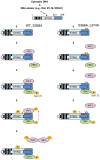
Figure 6. A model of IRF3 Activation by STING
Following detection of cytosolic DNA with a DNA sensor, STING forms oligomers on ER or other intracellular membranes. The C-terminus of STING then recruits IRF3 and TBK1, facilitating IRF3 phosphorylation by TBK1 (left). The mutation of STING at Ser366 or Leu374 does not impair its ability to recruit and activate TBK1, as evidenced by the phosphorylation of TBK1 and STING in ISD-stimulated cells harboring these mutations (right). However, S366A or L374A mutation abolishes the interaction between STING and IRF3, thereby preventing IRF3 phosphorylation by TBK1.

Similar articles

Cited by

References

    1. Takeuchi O, Akira S. Pattern recognition receptors and inflammation. Cell. 2010;140:805–820. - PubMed
    1. Yoneyama M, Fujita T. RNA recognition and signal transduction by RIG-I-like receptors. Immunol Rev. 2009;227:54–65. - PubMed
    1. Kawai T, Takahashi K, Sato S, Coban C, Kumar H, Kato H, Ishii KJ, Takeuchi O, Akira S. IPS-1, an adaptor triggering RIG-I- and Mda5-mediated type I interferon induction. Nat Immunol. 2005;6:981–988. - PubMed
    1. Meylan E, Curran J, Hofmann K, Moradpour D, Binder M, Bartenschlager R, Tschopp J. Cardif is an adaptor protein in the RIG-I antiviral pathway and is targeted by hepatitis C virus. Nature. 2005;437:1167–1172. - PubMed
    1. Seth RB, Sun L, Ea CK, Chen ZJ. Identification and characterization of MAVS, a mitochondrial antiviral signaling protein that activates NF-kappaB and IRF 3. Cell. 2005;122:669–682. - PubMed

Publication types