. 2012 Nov-Dec;88(6):1396-407.
doi: 10.1111/j.1751-1097.2012.01134.x. Epub 2012 Mar 30.

Light-induced retinal degeneration is prevented by zinc, a component in the age-related eye disease study formulation

Affiliations

Light-induced retinal degeneration is prevented by zinc, a component in the age-related eye disease study formulation

Daniel Organisciak et al. Photochem Photobiol. 2012 Nov-Dec.

Abstract

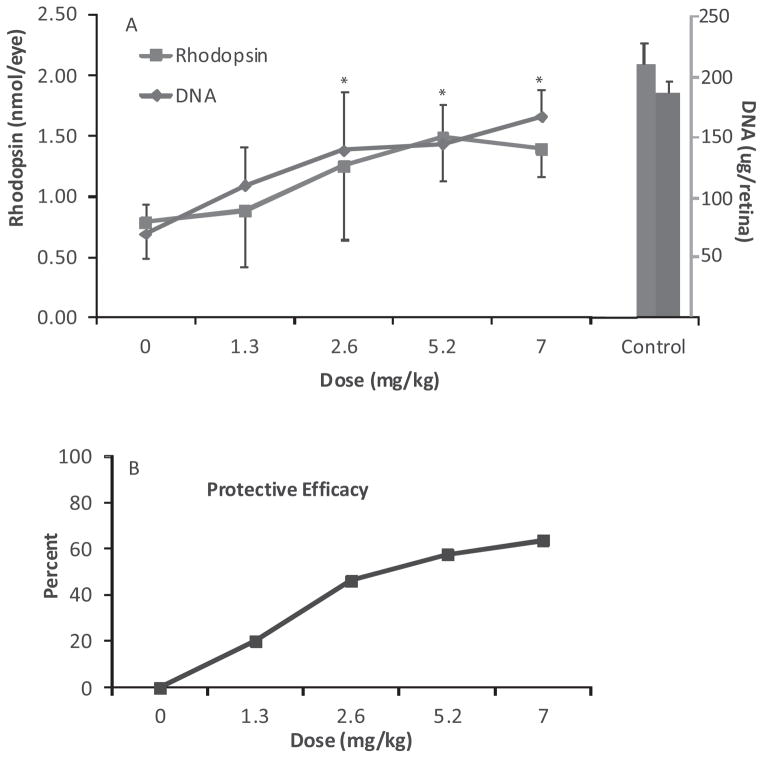
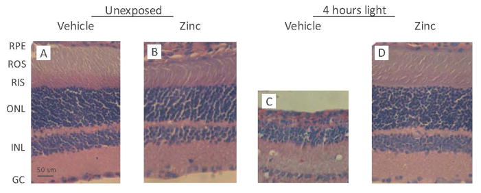
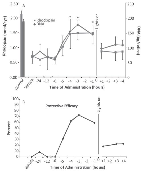
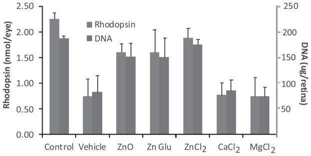
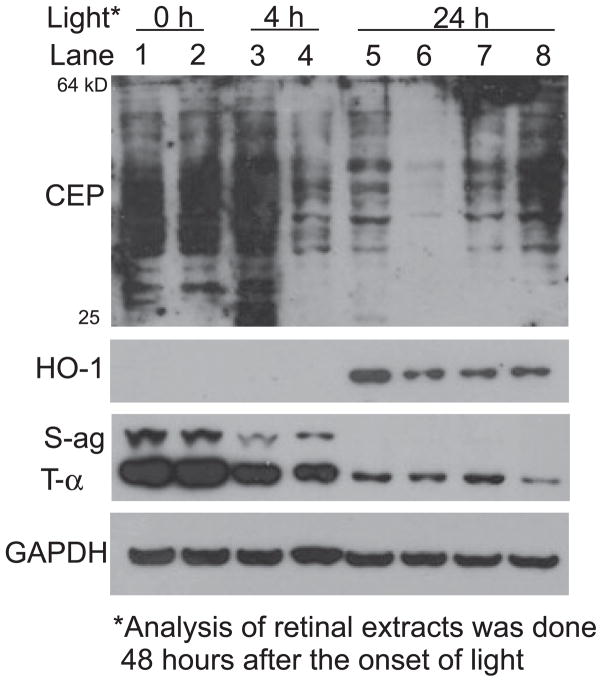
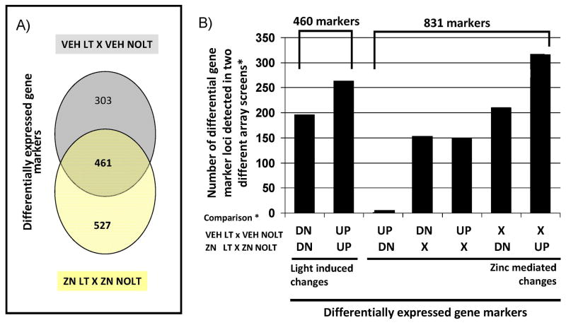
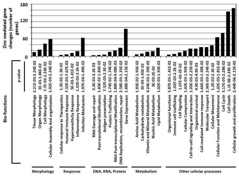
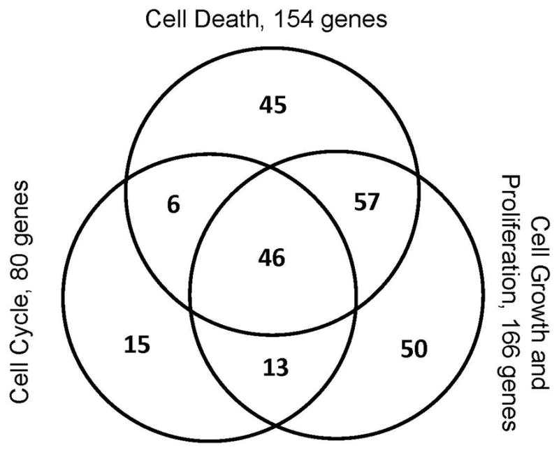
Mineral supplements are often included in multivitamin preparations because of their beneficial effects on metabolism. In this study, we used an animal model of light-induced retinal degeneration to test for photoreceptor cell protection by the essential trace element zinc. Rats were treated with various doses of zinc oxide and then exposed to intense visible light for as long as 8 h. Zinc treatment effectively prevented retinal light damage as determined by rhodopsin and retinal DNA recovery, histology and electrophoretic analysis of DNA damage and oxidized retinal proteins. Zinc oxide was particularly effective when given before light exposure and at doses two- to four-fold higher than recommended by the age-related eye disease study group. Treated rats exhibited higher serum and retinal pigment epithelial zinc levels and an altered retinal gene expression profile. Using an Ingenuity database, 512 genes with known functional annotations were found to be responsive to zinc supplementation, with 45% of these falling into a network related to cellular growth, proliferation, cell cycle and death. Although these data suggest an integrated and extensive regulatory response, zinc induced changes in gene expression also appear to enhance antioxidative capacity in retina and reduce oxidative damage arising from intense light exposure.


Figures

Figure 1
Figure 1
Time line for zinc injections and intense light treatment of rats reared in dim cyclic light or darkness. Following intense light treatment all rats were placed into the dark environment for various periods of time, depending on the measurements to be performed.
Figure 2
Figure 2
Dose response curve for zinc oxide treatment of rats 1 h before intense light exposure. P60 dark reared rats were given zinc oxide, or the aqueous vehicle (IP) and then exposed to light for 4 h. Visual cell recovery was determined by rhodopsin and retinal DNA measurements, performed after a 14 day dark recovery period (* = P < 0.05; A). Data were presented as the mean ± S.D. for n = 8 rats with one eye used for rhodopsin and the fellow eye for DNA measurements. (B) Percent protective efficacy, calculated from the average rhodopsin and retinal DNA values in zinc-treated rats in comparison to vehicle-treated animals.
Figure 3
Figure 3
Representative retinal sections from rats treated with zinc oxide before intense light exposure. Rats were given zinc oxide, at a dose of 5.2 mg kg−1, or vehicle and then kept in darkness (panels A and B), or exposed to light for 4 h (panels C and D). After a 14 day dark recovery period rats were euthanized in a CO2 atmosphere and their eyes enucleated and placed in Karnovsky’s fixative for 24 h. Paraffin-embedded sections were stained with hematoxylin / eosin. The vehicle-treated rat retina (panel C), exhibits distinct losses of photoreceptor nuclei, RPE cell damage and overall thinning compared with unexposed retinas. Zinc treatment (panel D) appears to ameliorate the light-induced changes seen in panel (C). All sections are from the superior hemisphere along the vertical meridian. Abbreviations: RPE, retinal pigment epithelium; ROS, rod outer segments; RIS, rod inner segments; ONL, outer nuclear layer; INL, inner nuclear layer; GC, ganglion cell. Magnification: Bar shown in panel (A) represents 50 μm.
Figure 4
Figure 4
Gel electrophoresis of retinal DNA 48 h after intense light exposure. DNA was extracted and pooled from the retinas of two rats treated with zinc oxide (5.2 mg kg−1), or vehicle, before light exposure. DNA fragmentation was visualized by ethidium bromide staining under UV light. A ladder of lower molecular weight DNA fragments and a variety of higher molecular weight fragments are present in the sample from vehicle-treated rats, whereas both the zinc-treated and dark control rat retinal DNA is retained as a high-molecular weight band near the top of the gel.
Figure 5
Figure 5
Time course of zinc oxide protection in rats exposed to intense visible light. Rats were given a single dose of zinc oxide (5.2 mg kg−1) at various times before or after the onset of light. Two weeks after a 4 h light exposure rhodopsin and retinal DNA were measured to determine the effect of zinc on visual cell recovery. Zinc oxide was effective for the recovery of visual cell markers when given 1–4 h before light and less effective, or ineffective, at earlier times and after light exposure had started. (A) Results represent the mean ± SD for n = 8 rats per time point; * = P < 0.001. (B) Protective efficacy for zinc treatment calculated from the average values for rhodopsin and retinal DNA.
Figure 6
Figure 6
Visual cell recovery in rats previously reared in dim cyclic light. Weanling rats were maintained in a dim cyclic light environment for 40 days and then exposed to intense visible light for 8 h starting at 1:00 A.M. Prior to light treatment rats were given zinc oxide (5.2 mg kg−1) or the aqueous vehicle. Rhodopsin and retinal DNA were measured 2 weeks after light exposure. Zinc treatment was effective in reducing visual cell loss for the 8 h period studied (* = P < 0.01). Results are for a total n = 12–16 animals from two separate experiments and shown as the mean ± SD.
Figure 7
Figure 7
The effects of various zinc salts on visual cell recovery after light exposure. Dark reared rats were given zinc oxide at 5.2 mg kg−1, or an equivalent molar amount of zinc in the form of its gluconate (Glu) 29.1 mg kg−1, or chloride (Cl) salt, 8.7 mg kg−1. One hour later, the rats were exposed to intense visible light for 4 h and rhodopsin and retinal DNA measured 2 weeks later. All forms of zinc were significantly effective in preventing retinal light damage (P < 0.001). Equal molar amounts of the divalent cations MgCl2 (6.1 mg kg−1) and CaCl2 (7.1 mg kg−1) were ineffective in preventing the loss of retinal photoreceptors. Data represents the mean ± SD for n = 6 animals for each condition.
Figure 8
Figure 8
Western analysis of oxidative protein markers and transduction proteins in retinas from light-exposed rats. Dark reared rats were given zinc oxide or vehicle and then exposed to intense light for 4 or 24 h. Retinas were excised 48 h after the onset of light and proteins extracted for gel electrophoresis and western analysis. Each lane contains 20 μg protein extracted from the pooled retinas of two rats. Abbreviations: CEP, carboxyethylpyrrole; HO-1, heme oxygenase-1; S-ag, S-antigen (arrestin); t-α, transducin alpha; GAPDH, glyceraldehyde- 3-phosphate dehydrogenase. Lanes 1 and 2, unexposed rat retinal extracts; lanes 3 and 5, vehicle-treated rat retinal extracts; lanes 4 and 6, zinc oxide treatment (5.2 mg kg−1); lanes 7 and 8, MgCl2 and CaCl2 at equal molar concentration to ZnCl2 (see Fig. 7).
Figure 9
Figure 9
Light- and zinc-mediated effects on the genetic signature of the retina. Dark reared rats were given zinc oxide, at a dose of 5.2 mg kg−1 (ZN), or vehicle (VEH) at 8:00 A.M. and then kept in darkness for 5 h (NOLT), or exposed to light for 4 h (LT) starting at 9:00 A.M. Four treatment groups were generated for gene array analysis using Illumina Genome / Bead Studio software (ZN LT, ZN NOLT, VEH LT and VEH NOLT) and two differential comparisons of pooled data for each treatment group under consideration were made using limma (VEH LT X VEH NOLT and ZN LT X ZN NOLT). A threshold P-value of 0.05 or less (adjusted for multiple testing) was used to define a differentially expressed transcript relative to its control. (A) Venn diagram showing the distribution of differentially expressed genes with respect to VEH LT (relative to VEH NOLT) and ZN LT (relative to ZN NOLT) that are unique to each condition, and shared between the two comparisons. (B) Graph showing the distribution of the differentially expressed genes identified into light-mediated and zinc-mediated changes to the genetic signature of the retina. DN: differentially expressed genes showing a decrease in mRNA expression levels after LT (as compared with NOLT). UP: differentially expressed genes that show an increase in expression levels after LT (as compared with NOLT). X: genes that did not show a change in expression levels after LT (as compared with NOLT).
Figure 10
Figure 10
Most significant biological functions associated with zinc-mediated gene changes. Five hundred and twelve of the 831 gene marker loci defining zinc-mediated gene changes are annotated in the Ingenuity Knowledge Base. Functional analysis of these 512 zinc-mediated differential genes was performed using Ingenuity Pathways Analysis (Ingenuity® Systems, http://www.ingenuity.com). Right-tailed Fisher’s exact test was used to calculate a P-value determining the probability that each biological function assigned to the data set is due to chance alone, the lower the P-value the higher the probability that the gene list is associated with the biological function indicated.
Figure 11
Figure 11
Venn diagram showing the overlap of cell death, cell cycle, cell growth and proliferation associated zinc-mediated differential genes. In total, 232 genes, representing 45% of the 512 genes considered, belong in this gene subset.

Similar articles

Cited by

References

    1. Shahinfar S, Edward DP, Tso MO. A pathological study of photoreceptor cell death in retinal photic injury. Curr Eye Res. 1991;10:47–59. - PubMed
    1. Reme C, Weller M, Szczyensy P, Munz P, Hafezi K, Reinboth J-J, Clausen M. Light-induced apoptosis in the rat retina in vivo. In: Anderson RE, LaVail MM, Hollyfield JG, editors. Degenerative Diseases of the Retina. Plenum Press; New York: 1995. pp. 19–25.
    1. Gordon WC, Casey DM, Lukiw WJ, Bazan NG. DNA damage and repair in light-induced photoreceptor degeneration. Invest Ophthalmol Vis Sci. 2002;43:3511–3521. - PubMed
    1. Organisciak DT, Vaughan DK. Retinal light damage: mechanisms and protection. Prog Retin Eye Res. 2010;29:113–134. - PMC - PubMed
    1. Noell WK, V, Walker S, Kang BS, Berman S. Retinal damage by light in rats. Invest Ophthalmol. 1966;5:450–473. - PubMed

Publication types

LinkOut - more resources