

Targeted focal adhesion kinase activation in cardiomyocytes protects the heart from ischemia/reperfusion injury
- PMID: 22383703
- PMCID: PMC4237311
- DOI: 10.1161/ATVBAHA.112.245134
Targeted focal adhesion kinase activation in cardiomyocytes protects the heart from ischemia/reperfusion injury
Abstract
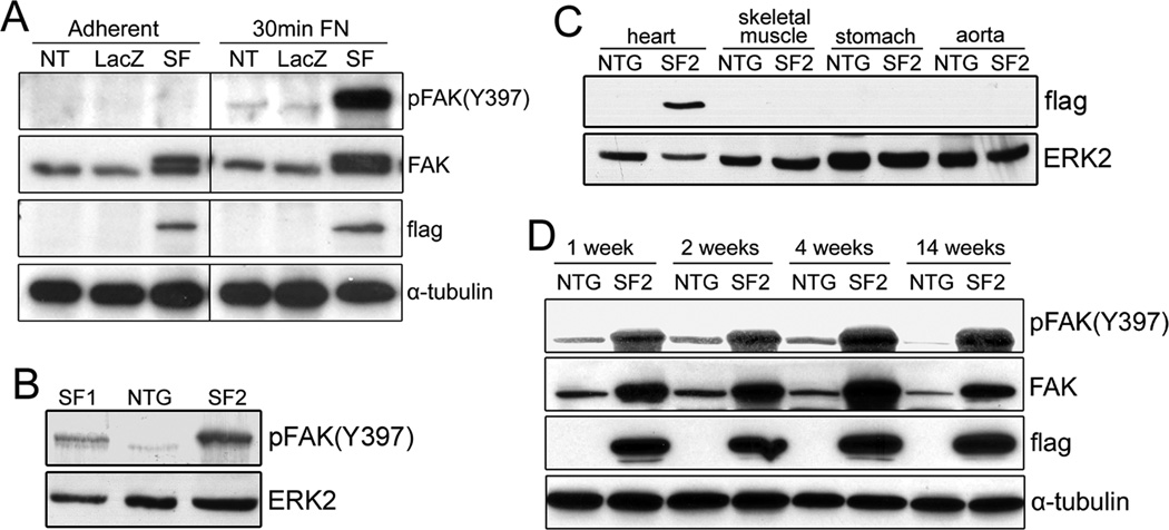
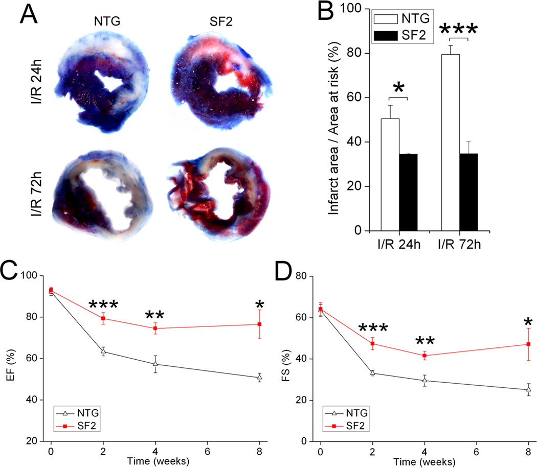
Objective: We previously reported that cardiac-restricted deletion of focal adhesion kinase (FAK) exacerbated myocyte death following ischemia/reperfusion (I/R). Here, we interrogated whether targeted elevation of myocardial FAK activity could protect the heart from I/R injury.
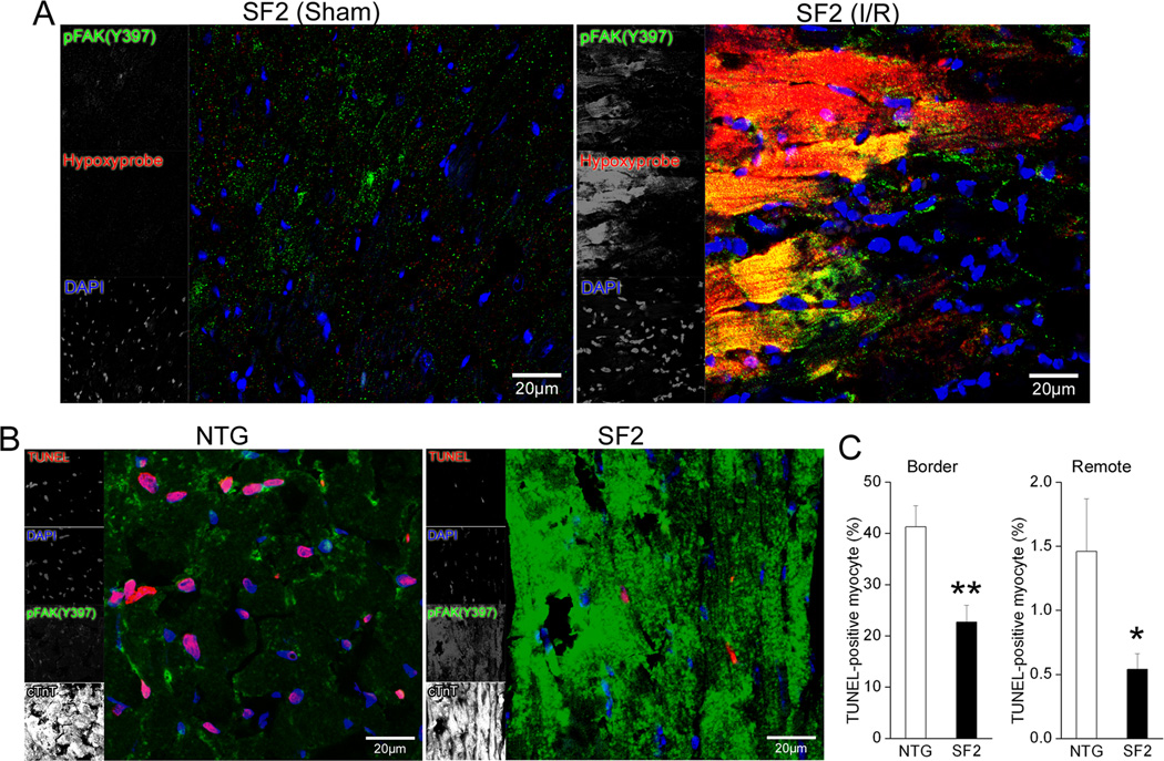
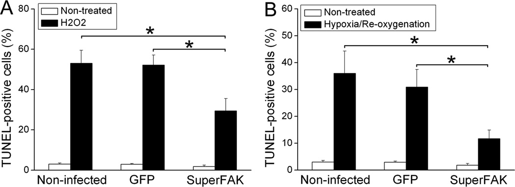
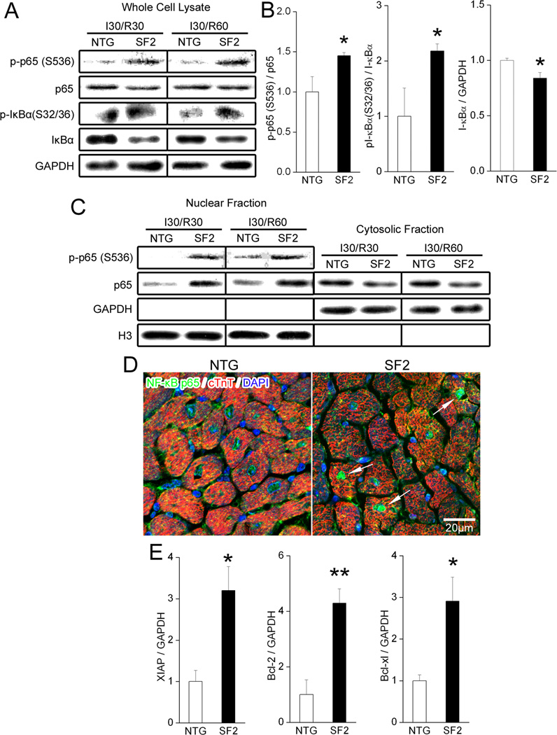
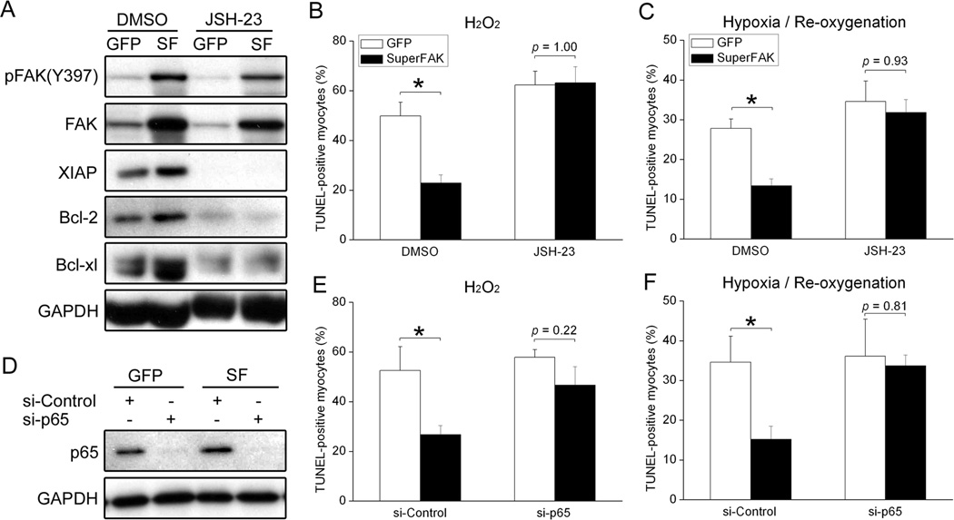
Methods and results: Transgenic mice were generated with myocyte-specific expression of a FAK variant (termed SuperFAK) that conferred elevated allosteric activation. FAK activity in unstressed transgenic hearts was modestly elevated, but this had no discernable effect on anabolic heart growth or cardiac function. Importantly, SuperFAK hearts exhibited a dramatic increase in FAK activity and a reduction in myocyte apoptosis and infarct size 24 to 72 hours following I/R. Moreover, serial echocardiography revealed that the transgenic mice were protected from cardiac decompensation for up to 8 weeks following surgery. Mechanistic studies revealed that elevated FAK activity protected cardiomyocytes from I/R-induced apoptosis by enhancing nuclear factor-κB (NF-κB)-dependent survival signaling during the early period of reperfusion (30 and 60 minutes). Moreover, adenoviral-mediated expression of SuperFAK in cultured cardiomyocytes attenuated H(2)O(2) or hypoxia/reoxygenation-induced apoptosis, whereas blockade of the NF-κB pathway using a pharmacological inhibitor or small interfering RNAs completely abolished the beneficial effect of SuperFAK.
Conclusions: Enhancing cardiac FAK activity attenuates I/R-induced myocyte apoptosis through activation of the prosurvival NF-κB pathway and may represent a novel therapeutic strategy for ischemic heart diseases.
Figures
Similar articles
-
Activation of PKN mediates survival of cardiac myocytes in the heart during ischemia/reperfusion.Circ Res. 2010 Sep 3;107(5):642-9. doi: 10.1161/CIRCRESAHA.110.217554. Epub 2010 Jul 1. Circ Res. 2010. PMID: 20595653 Free PMC article.
-
Overexpression of A kinase interacting protein 1 attenuates myocardial ischaemia/reperfusion injury but does not influence heart failure development.Cardiovasc Res. 2016 Aug 1;111(3):217-26. doi: 10.1093/cvr/cvw161. Epub 2016 Jun 14. Cardiovasc Res. 2016. PMID: 27302402
-
FAK regulates cardiomyocyte survival following ischemia/reperfusion.J Mol Cell Cardiol. 2009 Feb;46(2):241-8. doi: 10.1016/j.yjmcc.2008.10.017. Epub 2008 Nov 5. J Mol Cell Cardiol. 2009. PMID: 19028502 Free PMC article.
-
The Role of NF-κB in Myocardial Ischemia/Reperfusion Injury.Curr Protein Pept Sci. 2022;23(8):535-547. doi: 10.2174/1389203723666220817085941. Curr Protein Pept Sci. 2022. PMID: 35980051 Review.
-
Focal adhesion signaling in heart failure.Pflugers Arch. 2014 Jun;466(6):1101-11. doi: 10.1007/s00424-014-1456-8. Epub 2014 Feb 12. Pflugers Arch. 2014. PMID: 24515292 Free PMC article. Review.
Cited by
-
Artesunate alleviates myocardial ischemia/reperfusion-induced myocardial necrosis in rats and hypoxia/reoxygenation-induced apoptosis in H9C2 cells via regulating the FAK/PI3K/Akt pathway.Ann Transl Med. 2020 Oct;8(20):1291. doi: 10.21037/atm-20-5182. Ann Transl Med. 2020. PMID: 33209871 Free PMC article.
-
ADAM23 in Cardiomyocyte Inhibits Cardiac Hypertrophy by Targeting FAK - AKT Signaling.J Am Heart Assoc. 2018 Sep 18;7(18):e008604. doi: 10.1161/JAHA.118.008604. J Am Heart Assoc. 2018. PMID: 30371220 Free PMC article.
-
Inhibition of ALDH2 by O-GlcNAcylation contributes to the hyperglycemic exacerbation of myocardial ischemia/reperfusion injury.Oncotarget. 2017 Mar 21;8(12):19413-19426. doi: 10.18632/oncotarget.14297. Oncotarget. 2017. PMID: 28038474 Free PMC article.
-
Human fibroblasts display a differential focal adhesion phenotype relative to chimpanzee.Evol Med Public Health. 2016 Mar 23;2016(1):110-6. doi: 10.1093/emph/eow010. Print 2016. Evol Med Public Health. 2016. PMID: 26971204 Free PMC article.
-
MiRNA and TF co-regulatory network analysis for the pathology and recurrence of myocardial infarction.Sci Rep. 2015 Apr 13;5:9653. doi: 10.1038/srep09653. Sci Rep. 2015. PMID: 25867756 Free PMC article.
References
-
- Roger VL, Go AS, Lloyd-Jones DM, Adams RJ, Berry JD, Brown TM, Carnethon MR, Dai S, de Simone G, Ford ES, Fox CS, Fullerton HJ, Gillespie C, Greenlund KJ, Hailpern SM, Heit JA, Ho PM, Howard VJ, Kissela BM, Kittner SJ, Lackland DT, Lichtman JH, Lisabeth LD, Makuc DM, Marcus GM, Marelli A, Matchar DB, McDermott MM, Meigs JB, Moy CS, Mozaffarian D, Mussolino ME, Nichol G, Paynter NP, Rosamond WD, Sorlie PD, Stafford RS, Turan TN, Turner MB, Wong ND, Wylie-Rosett J. Heart disease and stroke statistics--2011 update: a report from the American Heart Association. Circulation. 2011;123:e18–e209. - PMC - PubMed
-
- Yellon DM, Hausenloy DJ. Myocardial reperfusion injury. N Engl J Med. 2007;357:1121–1135. - PubMed
-
- Zhao ZQ, Vinten-Johansen J. Myocardial apoptosis and ischemic preconditioning. Cardiovasc Res. 2002;55:438–455. - PubMed
-
- Matsui T, Tao J, del Monte F, Lee KH, Li L, Picard M, Force TL, Franke TF, Hajjar RJ, Rosenzweig A. Akt activation preserves cardiac function and prevents injury after transient cardiac ischemia in vivo. Circulation. 2001;104:330–335. - PubMed
-
- Muraski JA, Rota M, Misao Y, Fransioli J, Cottage C, Gude N, Esposito G, Delucchi F, Arcarese M, Alvarez R, Siddiqi S, Emmanuel GN, Wu W, Fischer K, Martindale JJ, Glembotski CC, Leri A, Kajstura J, Magnuson N, Berns A, Beretta RM, Houser SR, Schaefer EM, Anversa P, Sussman MA. Pim-1 regulates cardiomyocyte survival downstream of Akt. Nat Med. 2007;13:1467–1475. - PubMed
Publication types
MeSH terms
Substances
Grants and funding
- R01 HL081844-07/HL/NHLBI NIH HHS/United States
- R01 HL081844-06/HL/NHLBI NIH HHS/United States
- R01 HL081844/HL/NHLBI NIH HHS/United States
- HL-081844/HL/NHLBI NIH HHS/United States
- R01 HL081844-03/HL/NHLBI NIH HHS/United States
- R01 HL081844-04/HL/NHLBI NIH HHS/United States
- R01 HL081844-01/HL/NHLBI NIH HHS/United States
- HL-071054/HL/NHLBI NIH HHS/United States
- R01 HL081844-05A1/HL/NHLBI NIH HHS/United States
- R01 HL109607/HL/NHLBI NIH HHS/United States
- R01 HL081844-02/HL/NHLBI NIH HHS/United States
- R01 HL081844-08/HL/NHLBI NIH HHS/United States
- R01 HL071054/HL/NHLBI NIH HHS/United States
LinkOut - more resources
Full Text Sources
Medical
Molecular Biology Databases
Miscellaneous
