

Clinicopathological and biological significance of human voltage-gated proton channel Hv1 protein overexpression in breast cancer
- PMID: 22367212
- PMCID: PMC3340163
- DOI: 10.1074/jbc.M112.345280
Clinicopathological and biological significance of human voltage-gated proton channel Hv1 protein overexpression in breast cancer
Abstract
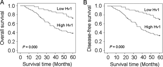
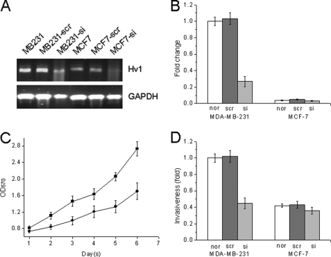
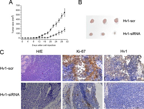
In our previous work, we showed for the first time that the voltage-gated proton channel Hv1 is specifically expressed in highly metastatic human breast tumor tissues and cell lines. However, the contribution of Hv1 to breast carcinogenesis is not well known. In this study, we showed that Hv1 expression was significantly correlated with the tumor size (p = 0.001), tumor classification (p = 0.000), lymph node status (p = 0.000), clinical stage (p = 0.000), and Her-2 status (p = 0.045). High Hv1 expression was associated significantly with shorter overall (p = 0.000) and recurrence-free survival (p = 0.000). In vitro, knockdown of Hv1 expression in the highly metastatic MDA-MB-231 cells decreased the cell proliferation and invasiveness, inhibited the cell proton secretion and intracellular pH recovery, and blocked the cell capacity of acidifying extracellular milieu. Furthermore, the gelatinase activity in MDA-MB-231 cells that suppressed Hv1 was reduced. In vivo, the breast tumor size of the implantation of the MDA-MB-231 xenografts in nude mice that were knocked down by Hv1 was dramatically smaller than that in the control groups. The results demonstrated that the inhibition of Hv1 function via knockdown of Hv1 expression can effectively retard the cancer growth and suppress the cancer metastasis by the decrease of proton extrusion and the down-regulation of gelatinase activity. Based on these results, we came to the conclusion that Hv1 is a potential biomarker for prognosis of breast cancer and a potential target for anticancer drugs in breast cancer therapy.
Figures


Similar articles
-
Specific expression of the human voltage-gated proton channel Hv1 in highly metastatic breast cancer cells, promotes tumor progression and metastasis.Biochem Biophys Res Commun. 2011 Aug 26;412(2):353-9. doi: 10.1016/j.bbrc.2011.07.102. Epub 2011 Jul 29. Biochem Biophys Res Commun. 2011. PMID: 21821008
-
Zn(2+) induces apoptosis in human highly metastatic SHG-44 glioma cells, through inhibiting activity of the voltage-gated proton channel Hv1.Biochem Biophys Res Commun. 2013 Aug 23;438(2):312-7. doi: 10.1016/j.bbrc.2013.07.067. Epub 2013 Jul 24. Biochem Biophys Res Commun. 2013. PMID: 23891691
-
Human voltage-gated proton channel hv1: a new potential biomarker for diagnosis and prognosis of colorectal cancer.PLoS One. 2013 Aug 5;8(8):e70550. doi: 10.1371/journal.pone.0070550. Print 2013. PLoS One. 2013. PMID: 23940591 Free PMC article.
-
Voltage-gated proton channel HV1 in microglia.Neuroscientist. 2014 Dec;20(6):599-609. doi: 10.1177/1073858413519864. Epub 2014 Jan 23. Neuroscientist. 2014. PMID: 24463247 Review.
-
Role of voltage-gated proton channel (Hv1) in cancer biology.Front Pharmacol. 2023 Apr 20;14:1175702. doi: 10.3389/fphar.2023.1175702. eCollection 2023. Front Pharmacol. 2023. PMID: 37153807 Free PMC article. Review.
Cited by
-
The role of Phe150 in human voltage-gated proton channel.iScience. 2022 Oct 20;25(11):105420. doi: 10.1016/j.isci.2022.105420. eCollection 2022 Nov 18. iScience. 2022. PMID: 36388967 Free PMC article.
-
Voltage-Gated Proton Channels in the Tree of Life.Biomolecules. 2023 Jun 24;13(7):1035. doi: 10.3390/biom13071035. Biomolecules. 2023. PMID: 37509071 Free PMC article. Review.
-
Scorpion toxin inhibits the voltage-gated proton channel using a Zn2+ -like long-range conformational coupling mechanism.Br J Pharmacol. 2020 May;177(10):2351-2364. doi: 10.1111/bph.14984. Epub 2020 Mar 3. Br J Pharmacol. 2020. PMID: 31975366 Free PMC article.
-
Biotransformation of zearalenone to non-estrogenic compounds with two novel recombinant lactonases from Gliocladium.BMC Microbiol. 2024 Mar 7;24(1):75. doi: 10.1186/s12866-024-03226-3. BMC Microbiol. 2024. PMID: 38454365 Free PMC article.
-
Interrogation of the intersubunit interface of the open Hv1 proton channel with a probe of allosteric coupling.Sci Rep. 2015 Sep 14;5:14077. doi: 10.1038/srep14077. Sci Rep. 2015. PMID: 26365828 Free PMC article.
References
-
- Gillies R. J., Liu Z., Bhujwalla Z. (1994) 31P MRS measurements of extracellular pH of tumors using 3-aminopropylphosphonate. Am. J. Physiol. 267, C195–C203 - PubMed
-
- Racker E. (1972) Bioenergetics and the problem of tumor growth. Am. Sci. 60, 56–63 - PubMed
-
- Gatenby R. A., Gawlinski E. T. (2003) The glycolytic phenotype in carcinogenesis and tumor invasion. Insights through mathematical models. Cancer Res. 63, 3847–3854 - PubMed
-
- Roos A., Boron W. F. (1981) Intracellular pH. Physiol. Rev. 61, 296–434 - PubMed
Publication types
MeSH terms
Substances
LinkOut - more resources
Full Text Sources
Other Literature Sources
Medical
Research Materials
Miscellaneous
