. 2012;7(2):e30865.
doi: 10.1371/journal.pone.0030865. Epub 2012 Feb 17.

Retinoic acid signaling plays a restrictive role in zebrafish primitive myelopoiesis

Affiliations

Retinoic acid signaling plays a restrictive role in zebrafish primitive myelopoiesis

Dong Liang et al. PLoS One. 2012.

Abstract

Retinoic acid (RA) is known to regulate definitive myelopoiesis but its role in vertebrate primitive myelopoiesis remains unclear. Here we report that zebrafish primitive myelopoiesis is restricted by RA in a dose dependent manner mainly before 11 hpf (hours post fertilization) when anterior hemangioblasts are initiated to form. RA treatment significantly reduces expressions of anterior hemangioblast markers scl, lmo2, gata2 and etsrp in the rostral end of ALPM (anterior lateral plate mesoderm) of the embryos. The result indicates that RA restricts primitive myelopoiesis by suppressing formation of anterior hemangioblasts. Analyses of ALPM formation suggest that the defective primitive myelopoiesis resulting from RA treatment before late gastrulation may be secondary to global loss of cells for ALPM fate whereas the developmental defect resulting from RA treatment during 10-11 hpf should be due to ALPM patterning shift. Overexpressions of scl and lmo2 partially rescue the block of primitive myelopoiesis in the embryos treated with 250 nM RA during 10-11 hpf, suggesting RA acts upstream of scl to control primitive myelopoiesis. However, the RA treatment blocks the increased primitive myelopoiesis caused by overexpressing gata4/6 whereas the abolished primitive myelopoiesis in gata4/5/6 depleted embryos is well rescued by 4-diethylamino-benzaldehyde, a retinal dehydrogenase inhibitor, or partially rescued by knocking down aldh1a2, the major retinal dehydrogenase gene that is responsible for RA synthesis during early development. Consistently, overexpressing gata4/6 inhibits aldh1a2 expression whereas depleting gata4/5/6 increases aldh1a2 expression. The results reveal that RA signaling acts downstream of gata4/5/6 to control primitive myelopoiesis. But, 4-diethylamino-benzaldehyde fails to rescue the defective primitive myelopoiesis in either cloche embryos or lycat morphants. Taken together, our results demonstrate that RA signaling restricts zebrafish primitive myelopoiesis through acting downstream of gata4/5/6, upstream of, or parallel to, cloche, and upstream of scl.


Conflict of interest statement

Competing Interests: The authors have declared that no competing interests exist.

Figures

Figure 1
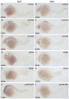
Figure 1. Excessive RA inhibits primitive myelopoiesis in zebrafish embryos in a dose dependent manner.
All embryos are positioned anterior left and lateral front. Embryos were treated with vehicle DMSO (A, G), 6.25 nM (B, H), 12.5 nM (C, I), 25 nM (D, J) and 50 nM RA (E, K) respectively from 1–2-cell stage until 26 hpf or microinjected with cyp26a1-MO, cyp26b1-MO and cyp26c1-MO together at 1–2-cell stage (F, L). They were then examined for expressions of myeloid markers lcp1 (A–F) and mpx (G–L) at 26 hpf by whole mount in situ hybridization. The number shown in the lower left-hand corner of each panel is the number of embryos exhibiting the typical phenotype shown in the panel to the number of embryos totally observed.
Figure 2
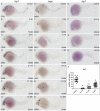
Figure 2. RA restricts the primitive myelopoiesis mainly before 11 hpf.
All embryos are positioned anterior left and lateral front. Embryos were treated with vehicle DMSO (A, H, O) or with 50 nM RA (B–G, I–N) from 3 to 5 (B, I), 5 to 7 (C, J), 7 to 9 (D, K), 9 to 11 (E, L), 11 to 13 (F, M) and 13 to 26 hpf (G, N), or with 250 nM RA form 10 to 11 (P), 11 to 13 (Q), 13 to 22 hpf (R), respectively. They were then examined for expressions of myeloid markers lcp1 (A–G, O–R) and mpx (H–N) at 26 hpf (A–N) or 22 hpf (O–R) by whole mount in situ hybridization. The number shown in the lower left-hand corner of each panel is the number of embryos exhibiting the typical phenotype shown in the panel to the number of embryos totally observed. The typical embryos expressing lcp1+ cells at 22 hpf were shown in O–R. The scatter plot (S) shows the number of lcp1+ cells counted from each of the embryos at 22 hpf with different treatment (control; 10–11 hpf RA treatment; 11–13 hpf RA treatment and 13–22 hpf RA treatment).
Figure 3
Figure 3. RA restricts the formation of anterior hemangioblasts in zebrafish embryos.
All flat-mounted embryos are positioned anterior left and dorsal front. Embryos were treated with vehicle DMSO (A, D, G, J, M), 50 nM RA from 1–2-cell stage to 14 hpf (B, E, H, K, N) or 250 nM RA from 10 to 11 hpf (C, F, I, L, O), respectively. They were then examined for expressions of pu.1 (A–C), scl (D–F), lmo2 (G–I), etsrp (J–L), and gata2 (M–O) in the rostral end of ALPM at 14 hpf by whole mount in situ hybridization. Expression of myoD in somites is used for staging. Bracket indicates the location of RBI. The number shown in the lower left-hand corner of each panel is the number of embryos exhibiting the typical phenotype shown in the panel to the number of embryos totally observed.
Figure 4
Figure 4. ALPM is lost in the embryos treated with 50 nM RA from 1–2-cell stage to 11 hpf but not eliminated in the ones treated with 250 nM RA during 10–11 hpf.
All embryos including flat-mounted embryos (A–C, H–O) and whole-mounted embryos (D–G) are positioned anterior left and dorsal front (A–C, H–O) or lateral front (D–G). Embryos treated with vehicle DMSO (A), 50 nM RA from 1–2-cell stage to 11 hpf (B), and 250 nM RA during 10–11 hpf (C) were examined for hoxb5b expression at 11 hpf (A–C). Embryos treated with 50 nM RA treatment from 1–2-cell stage to 11 hpf displayed ectopically expression of hoxb5b in ALPM (B) but the ones treated with 250 nM RA during 10–11 hpf did not exhibit this ectopical expression (C). Compared with control embryos (D, F), overexpressions of hoxb5b by microinjecting embryos at 1–2-cell stage with hoxb5b mRNA significantly suppressed expressions of lcp1 (E) and mpx (G) at 24 hpf. Embryos treated with vehicle DMSO (H, J), 50 nM RA from 1–2-cell stage to 14 hpf (I) or 250 nM RA during 10–11 hpf (K) were examined for expressions of ALPM marker gata4 at 14 hpf. The location of ALPM at 11 hpf (A–C) or at 14 hpf (H, J, K) is indicated by bracket. Embryos treated with vehicle DMSO (L), 250 nM RA during 10–11 hpf (M, O), or microinjected with scl/lmo2 mRNA (N, O) were examined for expressions of cardiac marker hand2 at 14 hpf. Expression of myoD in somites (H–O) was used for staging and ntl expression was used for labeling embryonic axial mesoderm (J–O). The length between the anterior end of gata4 expression domain (J, K) or hand 2 expression domain (L–N) and the anterior end of ntl expression domain and the length between the posterior end of gata4 expression domain (J, K) or hand 2 expression domain (L–N) and the anterior end of ntl expression domain are marked by two different brackets. Red line denotes the anterior level of ntl expression domain (J–O). Arrow indicates the most anterior end of notochord marked by expression of ntl (J–O). sc: spinal cord; PLPM: posterior lateral plate mesoderm. The number shown in the lower left-hand corner of each panel is the number of embryos exhibiting the typical phenotype shown in the panel to the number of embryos totally observed.
Figure 5
Figure 5. Overexpressions of scl and lmo2 into RA-treated zebrafish embryos partially rescue the defective primitive myelopoiesis.
Both flat-mounted embryos (A–H) and whole-mounted embryos (I–P) are positioned anterior left and dorsal front (A–H) or lateral front (I–P). Embryos were treated with vehicle DMSO (A, E, I, M), 250 nM RA during 10 to 11 hpf (B, F, J, N), or microinjected with scl and lmo2 mRNA at 1–2-cell stage (C, G, K, O), or microinjected with scl and lmo2 mRNA at 1–2-cell stage and then treated with 250 nM RA during 10 to 11 hpf (D, H, L, P), respectively. They were then examined for expressions of hemangioblast markers etsrp (A–D) and gata2 (E–H) at 14 hpf, and myeloid markers lcp1 (I–L) and mpx (M–P) at 24 hpf by whole mount in situ hybridization. Expression of myoD in somites was used for staging (A–H). Bracket indicates the location of RBI (A, B, E, F), and ALPM (C, D, G, H). The number shown in the lower left-hand corner of each panel is the number of embryos exhibiting the typical phenotype shown in the panel to the number of embryos totally observed.
Figure 6
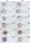
Figure 6. DEAB cannot rescue the defective primitive myelopoiesis in cloche or lycat knockdown embryos.
All embryos are positioned anterior left and lateral front. Wild type siblings (A, G), cloche (B, H) and the embryos microinjected with lycat-MO at 1–2-cell stage (E, K) were treated with vehicle DMSO whereas cloche (C, I), cloche siblings (D, J) and lycat-MO knockdown (F, L) embryos were treated with 10 µM DEAB from 1–2-cell stage until 26 hpf. They were then examined for expressions of myeloid markers lcp1 (A–F) and mpx (G–L) at 26 hpf by whole mount in situ hybridization. The number shown in the lower left-hand corner of each panel is the number of embryos exhibiting the typical phenotype shown in the panel to the number of embryos totally observed.
Figure 7
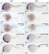
Figure 7. RA inhibits primitive myelopoiesis by acting downstream of gata4/5/6.
All embryos are positioned anterior left and lateral front. Embryos were microinjected with gata5-MO and gata6-MO together (B, C, G, H) at 1–2-cell stage and then treated with 10 µM DEAB (C, H) or vehicle DMSO (B, G) immediately, or microinjected with gata4 mRNA and gata6 mRNA together (D, E, I, J) and then treated with 250 nM RA (E, J) or vehicle DMSO (D, I) from 10 to 11 hpf, respectively. They were then examined for expressions of myeloid markers lcp1 (A–E) and mpx (F–J) at 24 hpf by whole mount in situ hybridization. The number shown in the lower left-hand corner of each panel is the number of embryos exhibiting the typical phenotype shown in the panel to the number of embryos totally observed.
Figure 8
Figure 8. aldh1a2 is one of downstream target genes of gata4/5/6 to affect zebrafish primitive myelopoiesis.
Embryos are positioned animal pole top and ventral front (A–I), anterior top and dorsal front (J–O), and anterior left and lateral front (R–W). Embryos were microinjected with control MO (A, D, G, J, M, R, U), gata5-MO plus gata6-MO (B, E, H, K, N), gata4 mRNA plus gata6 mRNA (C, F, I, L, O), gata5-MO and gata6-MO plus control MO (S, V), gata5-MO and gata6-MO plus aldh1a2 MO (T, W) at 1–2-cell stage, respectively. They were then examined for expressions of aldh1a2 at 5 hpf (A–C), 7 hpf (D–F), 9 hpf (G–I), 11 hpf (J–L) and 13 hpf (M–O), lcp1 (R–T) and mpx at 24 hpf (U–W) by whole mount in situ hybridization, respectively. qRT-PCR was performed to confirm the relative expression level changes of aldh1a2 in gata4/5/6 depleted embryos (P) or in gata4/6 overexpressed embryos (Q) at 5, 7, 9, 11, 13 hpf, and those of lcp1 and mpx at 24 hpf (X). The number shown in the lower left-hand corner of each panel (R–W) is the number of embryos exhibiting the typical phenotype shown in the panel to the number of embryos totally observed. *: P<0.05; **: P<0.01.

Similar articles

Cited by

References

    1. Galloway JL, Zon LI. Ontogeny of hematopoiesis: examining the emergence of hematopoietic cells in the vertebrate embryo. Curr Top Dev Biol. 2003;53:139–158. - PubMed
    1. Davidson AJ, Zon LI. The ‘definitive’ (and ‘primitive’) guide to zebrafish hematopoiesis. Oncogene. 2004;23:7233–7246. - PubMed
    1. Hsia N, Zon LI. Transcriptional regulation of hematopoietic stem cell development in zebrafish. Exp Hematol. 2005;33:1007–1014. - PubMed
    1. Bertrand JY, Chi NC, Santoso B, Teng S, Stainier DY, et al. Haematopoietic stem cells derive directly from aortic endothelium during development. Nature. 2010;464:108–111. - PMC - PubMed
    1. Sumanas S, Gomez G, Zhao Y, Park C, Choi K, et al. Interplay among Etsrp/ER71, Scl, and Alk8 signaling controls endothelial and myeloid cell formation. Blood. 2008;111:4500–4510. - PMC - PubMed

Publication types

MeSH terms

LinkOut - more resources