

Human alpha defensin 5 expression in the human kidney and urinary tract
- PMID: 22359618
- PMCID: PMC3281003
- DOI: 10.1371/journal.pone.0031712
Human alpha defensin 5 expression in the human kidney and urinary tract
Abstract
Background: The mechanisms that maintain sterility in the urinary tract are incompletely understood. Recent studies have implicated the importance of antimicrobial peptides (AMP) in protecting the urinary tract from infection. Here, we characterize the expression and relevance of the AMP human alpha-defensin 5 (HD5) in the human kidney and urinary tract in normal and infected subjects.
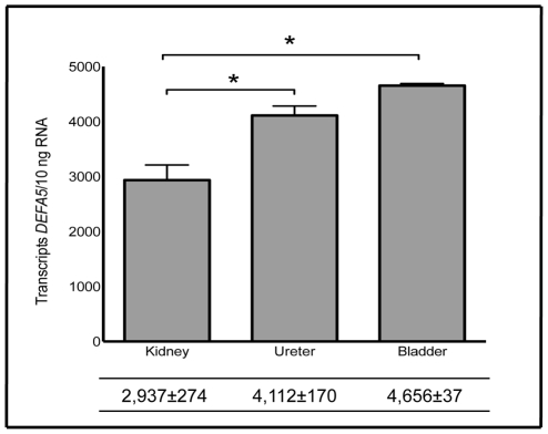
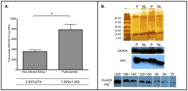
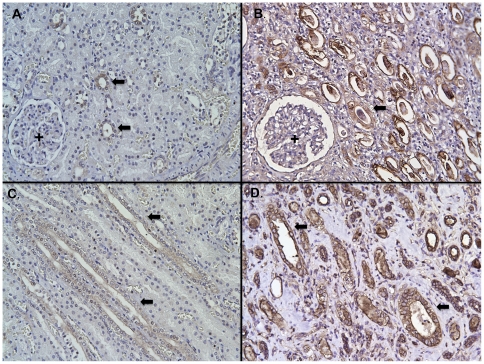
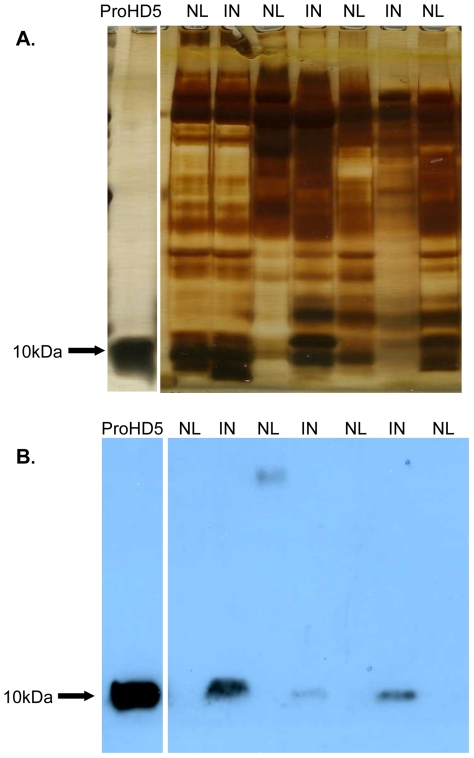
Methodology/principal findings: Using RNA isolated from human kidney, ureter, and bladder tissue, we performed quantitative real-time PCR to show that DEFA5, the gene encoding HD5, is constitutively expressed throughout the urinary tract. With pyelonephritis, DEFA5 expression significantly increased in the kidney. Using immunoblot analysis, HD5 production also increased with pyelonephritis. Immunostaining localized HD5 to the urothelium of the bladder and ureter. In the kidney, HD5 was primarily produced in the distal nephron and collecting tubules. Using immunoblot and ELISA assays, HD5 was not routinely detected in non-infected human urine samples while mean urinary HD5 production increased with E.coli urinary tract infection.
Conclusions/significance: DEFA5 is expressed throughout the urinary tract in non-infected subjects. Specifically, HD5 is expressed throughout the urothelium of the lower urinary tract and in the collecting tubules of the kidney. With infection, HD5 expression increases in the kidney and levels become detectable in the urine. To our knowledge, our findings represent the first to quantitate HD5 expression and production in the human kidney. Moreover, this is the first report to detect the presence of HD5 in infected urine samples. Our results suggest that HD5 may have an important role in maintaining urinary tract sterility.
Conflict of interest statement
Figures


Similar articles
-
Ribonuclease 7 is a potent antimicrobial peptide within the human urinary tract.Kidney Int. 2011 Jul;80(2):174-80. doi: 10.1038/ki.2011.109. Epub 2011 Apr 27. Kidney Int. 2011. PMID: 21525852
-
Ribonuclease 7, an antimicrobial peptide upregulated during infection, contributes to microbial defense of the human urinary tract.Kidney Int. 2013 Apr;83(4):615-25. doi: 10.1038/ki.2012.410. Epub 2013 Jan 9. Kidney Int. 2013. PMID: 23302724 Free PMC article.
-
An endogenous ribonuclease inhibitor regulates the antimicrobial activity of ribonuclease 7 in the human urinary tract.Kidney Int. 2014 May;85(5):1179-91. doi: 10.1038/ki.2013.395. Epub 2013 Oct 9. Kidney Int. 2014. PMID: 24107847 Free PMC article.
-
Amplifying renal immunity: the role of antimicrobial peptides in pyelonephritis.Nat Rev Nephrol. 2015 Nov;11(11):642-55. doi: 10.1038/nrneph.2015.105. Epub 2015 Jul 7. Nat Rev Nephrol. 2015. PMID: 26149835 Review.
-
Intercalated cell function, kidney innate immunity, and urinary tract infections.Pflugers Arch. 2024 Apr;476(4):565-578. doi: 10.1007/s00424-024-02905-4. Epub 2024 Jan 16. Pflugers Arch. 2024. PMID: 38227050 Review.
Cited by
-
Application of enzyme-linked immunosorbent assay to detect antimicrobial peptides in human intestinal lumen.J Immunol Methods. 2024 Feb;525:113599. doi: 10.1016/j.jim.2023.113599. Epub 2023 Dec 9. J Immunol Methods. 2024. PMID: 38081407 Free PMC article.
-
Multifaceted immune functions of human defensins and underlying mechanisms.Semin Cell Dev Biol. 2019 Apr;88:163-172. doi: 10.1016/j.semcdb.2018.02.023. Epub 2018 Mar 13. Semin Cell Dev Biol. 2019. PMID: 29501617 Free PMC article. Review.
-
The human alpha defensin HD5 neutralizes JC polyomavirus infection by reducing endoplasmic reticulum traffic and stabilizing the viral capsid.J Virol. 2014 Jan;88(2):948-60. doi: 10.1128/JVI.02766-13. Epub 2013 Nov 6. J Virol. 2014. PMID: 24198413 Free PMC article.
-
The role of the antimicrobial peptide cathelicidin in renal diseases.Pediatr Nephrol. 2015 Aug;30(8):1225-32. doi: 10.1007/s00467-014-2895-3. Epub 2014 Aug 27. Pediatr Nephrol. 2015. PMID: 25159719 Review.
-
Expression and function of human ribonuclease 4 in the kidney and urinary tract.Am J Physiol Renal Physiol. 2021 May 1;320(5):F972-F983. doi: 10.1152/ajprenal.00592.2020. Epub 2021 Apr 5. Am J Physiol Renal Physiol. 2021. PMID: 33818125 Free PMC article.
References
-
- Weichhart T, Haidinger M, Horl WH, Saemann MD. Current concepts of molecular defence mechanisms operative during urinary tract infection. European journal of clinical investigation. 2008;38(Suppl 2):29–38. - PubMed
-
- Zasloff M. Antimicrobial peptides, innate immunity, and the normally sterile urinary tract. J Am Soc Nephrol. 2007;18:2810–2816. - PubMed
-
- Lehrer RI, Lichtenstein AK, Ganz T. Defensins: antimicrobial and cytotoxic peptides of mammalian cells. Annu Rev Immunol. 1993;11:105–128. - PubMed
-
- Valore EV, Ganz T. Posttranslational processing of defensins in immature human myeloid cells. Blood. 1992;79:1538–1544. - PubMed
-
- Liu L, Zhao C, Heng HH, Ganz T. The human beta-defensin-1 and alpha-defensins are encoded by adjacent genes: two peptide families with differing disulfide topology share a common ancestry. Genomics. 1997;43:316–320. - PubMed
Publication types
MeSH terms
Substances
Grants and funding
LinkOut - more resources
Full Text Sources
Other Literature Sources
Medical
Molecular Biology Databases
