. 2012 Apr 1;188(7):3278-93.
doi: 10.4049/jimmunol.1100271. Epub 2012 Feb 22.

Tcra enhancer activation by inducible transcription factors downstream of pre-TCR signaling

Affiliations

Tcra enhancer activation by inducible transcription factors downstream of pre-TCR signaling

Beatriz del Blanco et al. J Immunol. .

Abstract

The Tcra enhancer (Eα) is essential for pre-TCR-mediated activation of germline transcription and V(D)J recombination. Eα is considered an archetypical enhanceosome that acts through the functional synergy and cooperative binding of multiple transcription factors. Based on dimethylsulfate genomic footprinting experiments, there has been a long-standing paradox regarding Eα activation in the absence of differences in enhancer occupancy. Our data provide the molecular mechanism of Eα activation and an explanation of this paradox. We found that germline transcriptional activation of Tcra is dependent on constant phospholipase Cγ, as well as calcineurin- and MAPK/ERK-mediated signaling, indicating that inducible transcription factors are crucially involved. NFAT, AP-1, and early growth response factor 1, together with CREB-binding protein/p300 coactivators, bind to Eα as part of an active enhanceosome assembled during pre-TCR signaling. We favor a scenario in which the binding of lymphoid-restricted and constitutive transcription factors to Eα prior to its activation forms a regulatory scaffold to recruit factors induced by pre-TCR signaling. Thus, the combinatorial assembly of tissue- and signal-specific transcription factors dictates the Eα function. This mechanism for enhancer activation may represent a general paradigm in tissue-restricted and stimulus-responsive gene regulation.


Conflict of interest statement

CONFLICT OF INTEREST

The authors declare that they have no conflict of interest.

Figures

Figure 1
Figure 1. Eα structure
Diagram depicts the location of the Tα1-Tα4 regions as well as the described constitutively bound factors.
Figure 2
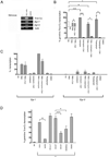
Figure 2. Both the calcineurin/Ca2+- and MAPK/ERK-mediated signaling pathways are required for the induction of germline Tcra transcription
(A) Analysis by RT-PCR of germline Tcra-Cα, Egr-1, and Egr-3 transcripts in unstimulated (−) Scid.adh cells or in cells stimulated with TAC mAb or control Ab (control). These results are representative of three experiments. (B, C) Analysis by quantitative RT-PCR of germline Tcra-Cα, Egr-1, and Egr-3 transcripts in unstimulated cells (NS) or cells stimulated with the indicated stimuli in the presence or absence of CsA. The effect of the DMSO vehicle control was also analyzed. (B) Transcript levels were normalized to Actb in each sample and expressed as the percentage of the level of germline Tcra-Cα transcripts in ionomcycin-treated cells. Data represent the mean ± s.e.m. of three determinations from two independent experiments. Paired sample t-tests were used to determine the statistical significance between the levels of transcription present in the unstimulated cells versus cells stimulated with ionomycin, thapsigargin or PMA (***P<0.0005, *P=0.045, and *P=0.0244, respectively) and to determine the statistical significance of the effect of CsA treatment on the expression of germline Tcra-Cα transcripts in ionomycin-, PMA+ionomycin-, and thapsigargin-stimulated cells (***P<0.0005, *P=0.0161, and *P=0.0445, respectively). (C) Expression levels of the transcripts were normalized to Actb in each sample and are expressed as the percentage of the level of Egr-1 transcripts in the PMA+ionomcycin treated cells. Data are represented as the mean ± s.e.m. of three determinations from two independent experiments. (D) Analysis by real time RT-PCR of germline Tcra-Cα transcripts in stimulated cells in the presence or absence of the indicated inhibitors. Paired sample t-tests were used to determine the statistical significance of the effect of EGTA versus untreated cells (**P=0.0069). Paired t-tests were also used to analyze the effect of the PUO126 and PD98059 treatments compared to the effect of UO124 treatment on the expression of germline Tcra-Cα transcripts (***P<0.0005 and *P=0.0152, respectively).
Figure 3
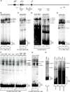
Figure 3. NFAT, AP-1, and Egr-1 factors bind specifically to several sites within the Tα1-Tα2 and flanking regions
(A) The diagram depicts the NFAT, AP-1, and Egr binding sites present within Eα and in the flanking regions. (B–D) Analysis of the binding of NFAT to 5′ Tα1 (B), Tα1 (C), and Tα2 (D) by EMSAs. (E) Analysis of the Ets-1 binding to 5′ Tα1 by EMSA. (F) Analysis of AP-1 and CREB binding to Tα1 by EMSA. (G) Analysis of AP-1 and P-CREB binding to Tα1 by EMSA. (H) Analysis of Egr-1 binding to 5′ Tα1, Tα1-Tα2, and 3′ Tα4 by EMSAs. Radiolabeled probes were incubated with nuclear extracts from unstimulated (NS) or PMA+ionomycin stimulated (S) cells (B–G) or rEgr-1 (H) in the presence of control or specific Abs. The complexes containing NFAT1-, NFAT-2, Ets-1, JunB, JunD, CREB, and Egr-1 are indicated. NFAT1*, NFAT-2*, CREB*, P-CREB*, and Egr-1* denote complexes formed in the presence of specific Abs.
Figure 4
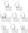
Figure 4. NFAT2, AP-1, Egr-1, and p300 factors, together with GATA-3, E47, Ets-1, and CREB, are part of the active Eα enhanceosome in Scid.adh cells
Chromatin preparations from unstimulated cells (NS) or cells stimulated with PMA+ionomycin (S) for 3.5 h were immunoprecipitated with the indicated Abs: GATA-3 (A), NFAT2 (B), JunB and JunD (C), Egr-1 (D), p300 (E), E47 (F), Ets-1 (G), and CREB (H). DNA purified from the Ab-bound fractions was used as a template for quantitative PCR to evaluate the presence of the Eα and Oct2 sequences. The values obtained from the Eα and Oct2 signals were normalized to that of the input, and expressed as the relative enrichment of the indicated factor binding versus the signals obtained from the control Abs. Data represent the mean ± s.e.m. of three determinations from two to three independent experiments. Paired sample t-tests were used to determine the statistical significance between the values of the ChIPs from unstimulated versus stimulated cells (GATA-3 *P=0.0101, NFAT2 **P=0.0054, JunB **P=0.0094, JunD **P=0.0073, Egr-1 ***P<0.0005, p300 **P=0.0036, and Ets-1 ***P<0.0005).
Figure 5
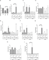
Figure 5. Inducible transcription factors are transiently recruited to Eα during thymocyte development
(A) Analysis of Cα transcripts by quantitative RT-PCR in Rxβ thymocytes in the absence (none) or presence of U73122 or DMSO control. Expression levels of Cα transcripts were normalized to that of Actb. Data represent the mean ± s.e.m. of three determinations from three independent experiments. Paired sample t-tests were used to determine the statistical significance between the transcription levels present in the DMSO- and U73122-treated cells, *P=0.0243. (B–G) Chromatin preparations from total Rag2−/− (R) or Rxβ thymocytes were immunoprecipitated with the indicated Abs: E47 (B), NFAT1 (C), NFAT2 (D), JunD and JunB (E), Egr-1 (F), and Ets-1 (G). DNA purified from the Ab-bound fractions was used as a template for quantitative PCR to evaluate the presence of Eα and Oct2 sequences. The Eα and Oct2 signal levels were normalized to the input and expressed as the relative enrichment of the indicated factor binding to signals obtained with control Abs. Data represent the mean ± s.e.m. of three determinations from two to four independent experiments. Paired sample t-tests were used to determine the statistical significance between the ChIP values obtained using Rag2−/− and Rxβ thymocytes (E47 ***P<0.0005 and Ets-1 ***P<0.0005). (H–J) Chromatin preparations from total Rag2−/− (R) or CD3ε mAb-injected Rag2−/− (R/CD3ε) thymocytes were immunoprecipitated with the indicated Abs: E47 (H) and JunD (I). Data represent the mean ± s.e.m. of three to four determinations from two independent experiments. Paired sample t-tests were used to determine statistical significance between the values of ChIPs for JunD obtained in Rag2−/− and CD3ε Ab-injected Rag2−/− thymocytes (P=*0.0284).
Figure 6
Figure 6. Eα-transcriptional induction is dependent on calcineurin-mediated signaling
(A) The diagram depicts the structure of Eα440 and Eα386, indicating the location of the NFAT and AP-1 binding sites present and the location of the Tα1, Tα2, Tα3, and Tα4 regions. (B, C) The indicated enhancers, Eα440 or Eα386, were cloned into enhancer-dependent test constructs where LUC is transcribed from the indicated promoters. The 3xNFAT-LUC and 9xNFAT-LUC reporter constructs were used as controls. The constructs were electroporated into Jurkat cells together with a reference plasmid containing the renilla luciferase reporter. Cells were unstimulated (NS) or PMA+ionomycin stimulated (S) for 24 h. The LUC activity levels were normalized to the renilla luciferase levels from the control plasmid. Data represent the mean ± s.e.m. of 6–11 determinations. Paired sample t-tests were used to determine for statistical significance between the levels of relative firefly/renilla luciferase activity in the unstimulated versus stimulated Jurkat cells (Jα49-Eα386 *P=0.0119, Jα49p-Eα440 **P=0.0061, TEA-Eα386 *P=0.0189, TEA-Eα440 *P=0.0347, 3xNFAT **P=0.0013, 9xNFAT *P=0.0336, and Vδ1-Eα440 *P=0.0407) and between relative the firefly/renilla luciferase activity present in untreated and CsA-treated stimulated Jurkat cells (Jα49-Eα386 *P=0.0493, Jα49p-Eα440 *P=0.0374, TEA-Eα386 *P=0.0479, TEA-Eα440 *P=0.0148, 3xNFAT *P=0.0463, and 9xNFAT *P=0.0295).
Figure 7
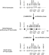
Figure 7. Model for the assembly of different Eα enhanceosomes during thymocyte development
The described constitutively bound factors are indicated by white ovals, the potentially bound NFAT factors are indicated by black ovals, the putatively bound AP-1 factors are indicated by grey ovals, and the Egr binding sites are indicated by black lines. The locations of Tα1, Tα2, Tα3, and Tα4 are indicated. In DN3a thymocytes, only the constitutive transcription factors would occupy their binding sites at the Eα. After β-selection during the first proliferative phase, the induced NFAT, AP-1, and Egr-1 factors will occupy their sites within the enhancer in DN3b, DN4, and early proliferating DP thymocytes. This would result in the recruitment of the HATs, CBP, and p300, and productive Eα-TEA/Jα49p interactions that activate germline transcription. During the second non-proliferative phase in small resting DP thymocytes, when V(D)J recombination at Tcra occurs, Eα might be occupied by the constitutive factors, and it remains active through the assembly of a new enhanceosome characterized by strong E-47 binding to E-box-III and Ets-1 binding to Tα2, and the recruitment of CBP/p300.

Similar articles

Cited by

References

    1. Taghon T, Yui MA, Pant R, Diamond RA, E V. Developmental and molecular characterization of emerging β- and γδ-selected pre-T cells in the adult mouse thymus. Immunity. 2006;24:53–64. - PubMed
    1. Ciofani M, Schmitt TM, Ciofani A, Michie AM, Cuburu N, Aublin A, Maryanski JL, Zúñiga-Pflucker JC. Obligatory role for cooperative signaling by pre-TCR and Notch during thymocyte differentiation. J. Immunol. 2004;172:5230–5239. - PubMed
    1. Carter JH, Lefebvre JM, Wiest DL, Tourtellote WG. Redundant role for early growth response transcriptional regulators in thymocyte differentiation and survival. J. Immunol. 2007;178:6796–6805. - PubMed
    1. Aifantis I, Gounari F, Scorrano L, Borowski C, von Boehmer H. Constitutive pre-TCR signaling promotes differentiation through Ca2+ mobilization and activation of NF-kB and NFAT. Nat. Immunol. 2001;2:403–409. - PubMed
    1. Canté-Barrett K, Winslow MM, Crabtree GB. Selective role of NFATc3 in positive selection of thymocytes. J. Immunol. 2007;179:103–110. - PubMed

Publication types

MeSH terms