Order versus Disorder: in vivo bone formation within osteoconductive scaffolds
- PMID: 22355786
- PMCID: PMC3281274
- DOI: 10.1038/srep00274
Order versus Disorder: in vivo bone formation within osteoconductive scaffolds
Abstract
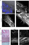
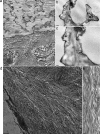
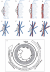
In modern biomaterial design the generation of an environment mimicking some of the extracellular matrix features is envisaged to support molecular cross-talk between cells and scaffolds during tissue formation/remodeling. In bone substitutes chemical biomimesis has been particularly exploited; conversely, the relevance of pre-determined scaffold architecture for regenerated bone outputs is still unclear. Thus we aimed to demonstrate that a different organization of collagen fibers within newly formed bone under unloading conditions can be generated by differently architectured scaffolds. An ordered and confined geometry of hydroxyapatite foams concentrated collagen fibers within the pores, and triggered their self-assembly in a cholesteric-banded pattern, resulting in compact lamellar bone. Conversely, when progenitor cells were loaded onto nanofibrous collagen-based sponges, new collagen fibers were distributed in a nematic phase, resulting mostly in woven isotropic bone. Thus specific biomaterial design relevantly contributes to properly drive collagen fibers assembly to target bone regeneration.
Figures


Similar articles
-
Hydroxyapatite reinforced collagen scaffolds with improved architecture and mechanical properties.Acta Biomater. 2015 Apr;17:16-25. doi: 10.1016/j.actbio.2015.01.031. Epub 2015 Jan 30. Acta Biomater. 2015. PMID: 25644451
-
Anisotropic Cryostructured Collagen Scaffolds for Efficient Delivery of RhBMP-2 and Enhanced Bone Regeneration.Materials (Basel). 2019 Sep 24;12(19):3105. doi: 10.3390/ma12193105. Materials (Basel). 2019. PMID: 31554158 Free PMC article.
-
Fabrication of pre-determined shape of bone segment with collagen-hydroxyapatite scaffold and autogenous platelet-rich plasma.J Mater Sci Mater Med. 2009 Jan;20(1):23-31. doi: 10.1007/s10856-008-3507-1. Epub 2008 Jul 24. J Mater Sci Mater Med. 2009. PMID: 18651114
-
Woven bone overview: structural classification based on its integral role in developmental, repair and pathological bone formation throughout vertebrate groups.Eur Cell Mater. 2019 Oct 1;38:137-167. doi: 10.22203/eCM.v038a11. Eur Cell Mater. 2019. PMID: 31571191 Review.
-
Scaffolds Fabricated from Natural Polymers/Composites by Electrospinning for Bone Tissue Regeneration.Adv Exp Med Biol. 2018;1078:49-78. doi: 10.1007/978-981-13-0950-2_4. Adv Exp Med Biol. 2018. PMID: 30357618 Review.
Cited by
-
Calcite as a Precursor of Hydroxyapatite in the Early Biomineralization of Differentiating Human Bone-Marrow Mesenchymal Stem Cells.Int J Mol Sci. 2021 May 6;22(9):4939. doi: 10.3390/ijms22094939. Int J Mol Sci. 2021. PMID: 34066542 Free PMC article.
-
Design Strategies and Biomimetic Approaches for Calcium Phosphate Scaffolds in Bone Tissue Regeneration.Biomimetics (Basel). 2022 Aug 13;7(3):112. doi: 10.3390/biomimetics7030112. Biomimetics (Basel). 2022. PMID: 35997432 Free PMC article. Review.
-
Advances in the Fabrication of Scaffold and 3D Printing of Biomimetic Bone Graft.Ann Biomed Eng. 2021 Apr;49(4):1128-1150. doi: 10.1007/s10439-021-02752-9. Epub 2021 Mar 5. Ann Biomed Eng. 2021. PMID: 33674908 Review.
-
Microfluidic Technology for the Production of Well-Ordered Porous Polymer Scaffolds.Polymers (Basel). 2020 Aug 19;12(9):1863. doi: 10.3390/polym12091863. Polymers (Basel). 2020. PMID: 32825098 Free PMC article. Review.
-
Transportation conditions for prompt use of ex vivo expanded and freshly harvested clinical-grade bone marrow mesenchymal stromal/stem cells for bone regeneration.Tissue Eng Part C Methods. 2014 Mar;20(3):239-51. doi: 10.1089/ten.TEC.2013.0250. Epub 2013 Aug 20. Tissue Eng Part C Methods. 2014. PMID: 23845029 Free PMC article.
References
-
- Barrilleaux B., Phinney D. G., Prockop D. J. & O'Connor K. C. Review: ex vivo engineering of living tissues with adult stem cells. Tissue Eng 12, 3007–3019 (2006). - PubMed
-
- Bianco P. & Robey P. G. Stem cells in tissue engineering. Nature 414, 118–121 (2001). - PubMed
-
- Cancedda R., Bianchi G., Derubeis A. & Quarto R. Cell therapy for bone disease: a review of current status. Stem Cells 21, 610–619 (2003). - PubMed
-
- Livingston T. L. et al. Mesenchymal stem cells combined with biphasic calcium phosphate ceramics promote bone regeneration. J Mater Sci Mater Med 14, 211–218 (2003). - PubMed
LinkOut - more resources
Full Text Sources

