Inherent structural disorder and dimerisation of murine norovirus NS1-2 protein
- PMID: 22347381
- PMCID: PMC3274520
- DOI: 10.1371/journal.pone.0030534
Inherent structural disorder and dimerisation of murine norovirus NS1-2 protein
Abstract
Human noroviruses are highly infectious viruses that cause the majority of acute, non-bacterial epidemic gastroenteritis cases worldwide. The first open reading frame of the norovirus RNA genome encodes for a polyprotein that is cleaved by the viral protease into six non-structural proteins. The first non-structural protein, NS1-2, lacks any significant sequence similarity to other viral or cellular proteins and limited information is available about the function and biophysical characteristics of this protein. Bioinformatic analyses identified an inherently disordered region (residues 1-142) in the highly divergent N-terminal region of the norovirus NS1-2 protein. Expression and purification of the NS1-2 protein of Murine norovirus confirmed these predictions by identifying several features typical of an inherently disordered protein. These were a biased amino acid composition with enrichment in the disorder promoting residues serine and proline, a lack of predicted secondary structure, a hydrophilic nature, an aberrant electrophoretic migration, an increased Stokes radius similar to that predicted for a protein from the pre-molten globule family, a high sensitivity to thermolysin proteolysis and a circular dichroism spectrum typical of an inherently disordered protein. The purification of the NS1-2 protein also identified the presence of an NS1-2 dimer in Escherichia coli and transfected HEK293T cells. Inherent disorder provides significant advantages including structural flexibility and the ability to bind to numerous targets allowing a single protein to have multiple functions. These advantages combined with the potential functional advantages of multimerisation suggest a multi-functional role for the NS1-2 protein.
Conflict of interest statement
Figures


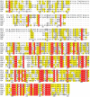
 . Proteins (or regions of proteins) shown to the left of the boundary line are predicted to be intrinsically disordered. Proteins to the right of the boundary line are predicted to be structured. NS1-2 regions (•); N-terminal caspase cleavage product (casp), truncated NS1-2 protein (trunc), full-length NS1-2 (full), middle ordered region (ord). The other MNV-1 non-structural proteins (∇) are numbered 2–7. Structural proteins are indicated by □.


Similar articles
-
Noroviruses Co-opt the Function of Host Proteins VAPA and VAPB for Replication via a Phenylalanine-Phenylalanine-Acidic-Tract-Motif Mimic in Nonstructural Viral Protein NS1/2.mBio. 2017 Jul 11;8(4):e00668-17. doi: 10.1128/mBio.00668-17. mBio. 2017. PMID: 28698274 Free PMC article.
-
Transcriptomic analysis of human norovirus NS1-2 protein highlights a multifunctional role in murine monocytes.BMC Genomics. 2017 Jan 5;18(1):39. doi: 10.1186/s12864-016-3417-4. BMC Genomics. 2017. PMID: 28056773 Free PMC article.
-
Reverse genetics mediated recovery of infectious murine norovirus.J Vis Exp. 2012 Jun 24;(64):4145. doi: 10.3791/4145. J Vis Exp. 2012. PMID: 22760450 Free PMC article.
-
Murine norovirus protein NS1/2 aspartate to glutamate mutation, sufficient for persistence, reorients side chain of surface exposed tryptophan within a novel structured domain.Proteins. 2014 Jul;82(7):1200-9. doi: 10.1002/prot.24484. Epub 2013 Dec 26. Proteins. 2014. PMID: 24273131 Free PMC article.
-
Nonstructural protein 4 of human norovirus self-assembles into various membrane-bridging multimers.J Biol Chem. 2024 Sep;300(9):107724. doi: 10.1016/j.jbc.2024.107724. Epub 2024 Aug 28. J Biol Chem. 2024. PMID: 39214299 Free PMC article.
Cited by
-
Lagovirus Non-structural Protein p23: A Putative Viroporin That Interacts With Heat Shock Proteins and Uses a Disulfide Bond for Dimerization.Front Microbiol. 2022 Jul 7;13:923256. doi: 10.3389/fmicb.2022.923256. eCollection 2022. Front Microbiol. 2022. PMID: 35923397 Free PMC article.
-
Membrane alterations induced by nonstructural proteins of human norovirus.PLoS Pathog. 2017 Oct 27;13(10):e1006705. doi: 10.1371/journal.ppat.1006705. eCollection 2017 Oct. PLoS Pathog. 2017. PMID: 29077760 Free PMC article.
-
Murine norovirus replication induces G0/G1 cell cycle arrest in asynchronously growing cells.J Virol. 2015 Jun;89(11):6057-66. doi: 10.1128/JVI.03673-14. Epub 2015 Mar 25. J Virol. 2015. PMID: 25810556 Free PMC article.
-
Norovirus NS1/2 protein increases glutaminolysis for efficient viral replication.PLoS Pathog. 2024 Jul 8;20(7):e1011909. doi: 10.1371/journal.ppat.1011909. eCollection 2024 Jul. PLoS Pathog. 2024. PMID: 38976719 Free PMC article.
-
Foodborne viruses: Detection, risk assessment, and control options in food processing.Int J Food Microbiol. 2018 Nov 20;285:110-128. doi: 10.1016/j.ijfoodmicro.2018.06.001. Epub 2018 Jun 8. Int J Food Microbiol. 2018. PMID: 30075465 Free PMC article. Review.
References
-
- Fankhauser RL, Monroe SS, Noel JS, Humphrey CD, Bresee JS, et al. Epidemiologic and molecular trends of Norwalk-like viruses associated with outbreaks of gastroenteritis in the United States. J Infect Dis. 2002;186:1–7. - PubMed
-
- Kirkwood CD, Streitberg R. Calicivirus shedding in children after recovery from diarrhoeal disease. J Clin Virol. 2008;43:346–348. - PubMed
-
- Zintz C, Bok K, Parada E, Barnes-Eley M, Berke T, et al. Prevalence and genetic characterization of caliciviruses among children hospitalized for acute gastroenteritis in the United States. Infect Genet Evol. 2005;5:281–290. - PubMed
Publication types
MeSH terms
Substances
Grants and funding
LinkOut - more resources
Full Text Sources
Medical
Miscellaneous

