

doi: 10.1038/nsmb.2233.
Crystal structure of an asymmetric trimer of a bacterial glutamate transporter homolog
Affiliations
- PMID: 22343718
- PMCID: PMC3633560
- DOI: 10.1038/nsmb.2233
Item in Clipboard
Crystal structure of an asymmetric trimer of a bacterial glutamate transporter homolog
Nat Struct Mol Biol.
.
Abstract
We report a structure of a trimeric glutamate transporter homolog from Pyrococcus horikoshii with two protomers in an inward facing state and the third in an intermediate conformation between the outward and inward facing states. The intermediate shows a cavity in the thinnest region of the transporter, which is potentially accessible to extracellular and cytoplasmic solutions. Our findings suggest a structural principle by which transport intermediates may mediate uncoupled permeation of polar solutes.
Figures

Crystal structure of the asymmetric trimer. a, b, Averaged 2fo-fc electron density maps at 2.5 σ (dark grey mesh) and anomalous difference Fourier maps at 5 σ (green mesh) for the IFS and the iOFS, respectively. c, Surface representations of the trimer viewed within the membrane plane. Trimerization and transport domains are colored wheat and blue, respectively. d, Cartoon representation of the protomers in the iOFS (left) and the IFS (right). The third protomer is omitted for clarity; HP1 and HP2 are yellow and red, respectively; and bound L-aspartate and sodium ions are shown as spheres.
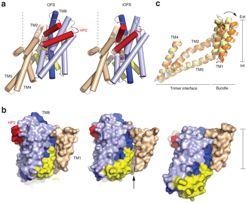
Conformational transition in the iOFS. a, Cylinder representation of GltPh viewed from the membrane plane in the OFS (left) and iOFS (right). The color scheme is as in Figure 1. TM8 is dark blue, and bound substrates are emphasized as spheres. Dashed lines, normal to the membrane plane, highlight the transport domain orientation differences. b, Superimposition of the trimerization domains in the OFS (yellow), iOFS (light orange) and IFS (orange). c, Surface representation of the protomers in the OFS (left), iOFS (center), and IFS (right) colored as in a. An arrow marks the enlarged crevice between the transport and trimerization domains.
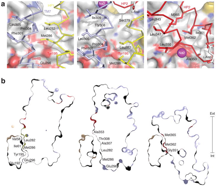
Cavity at the domain interface in the iOFS. a, The interface is viewed from within the transport domain in the OFS (left), iOFS (center) and IFS (right). The trimerization domain is in surface representation and colored by atom type. Interacting transport domain elements are in ribbon representation and colored light blue (TM7), yellow (HP1) and red (HP2). The rest of the transport domain is omitted for clarity. The side chains making contact with the trimerization domain and bound L-aspartate are emphasized as sticks, and sodium ion in Na2 site is shown as a sphere. b, Thin cross-sections of the protomers approximately through Ser65 and Tyr195 and normal to the membrane plane. Residues lining the domain interface are labeled.
Similar articles
-
Transport mechanism of a bacterial homologue of glutamate transporters.Nature. 2009 Dec 17;462(7275):880-5. doi: 10.1038/nature08616. Epub 2009 Nov 18. Nature. 2009. PMID: 19924125 Free PMC article.
-
Structure of a glutamate transporter homologue from Pyrococcus horikoshii.Nature. 2004 Oct 14;431(7010):811-8. doi: 10.1038/nature03018. Nature. 2004. PMID: 15483603
-
Accessing a transporter structure.Nature. 2004 Oct 14;431(7010):752-3. doi: 10.1038/431752a. Nature. 2004. PMID: 15483590 No abstract available.
-
Transport mechanism of a glutamate transporter homologue GltPh.Biochem Soc Trans. 2016 Jun 15;44(3):898-904. doi: 10.1042/BST20160055. Biochem Soc Trans. 2016. PMID: 27284058 Free PMC article. Review.
-
The dual-function glutamate transporters: structure and molecular characterisation of the substrate-binding sites.Biochim Biophys Acta. 2002 Sep 10;1555(1-3):92-5. doi: 10.1016/s0005-2728(02)00260-8. Biochim Biophys Acta. 2002. PMID: 12206897 Review.
Cited by
-
High-speed atomic force microscopy reveals a three-state elevator mechanism in the citrate transporter CitS.Proc Natl Acad Sci U S A. 2022 Feb 8;119(6):e2113927119. doi: 10.1073/pnas.2113927119. Proc Natl Acad Sci U S A. 2022. PMID: 35101979 Free PMC article.
-
Structural ensemble of a glutamate transporter homologue in lipid nanodisc environment.Nat Commun. 2020 Feb 21;11(1):998. doi: 10.1038/s41467-020-14834-8. Nat Commun. 2020. PMID: 32081874 Free PMC article.
-
Symport and antiport mechanisms of human glutamate transporters.Nat Commun. 2023 May 4;14(1):2579. doi: 10.1038/s41467-023-38120-5. Nat Commun. 2023. PMID: 37142617 Free PMC article.
-
Large domain movements through the lipid bilayer mediate substrate release and inhibition of glutamate transporters.Elife. 2020 Nov 6;9:e58417. doi: 10.7554/eLife.58417. Elife. 2020. PMID: 33155546 Free PMC article.
-
Electrogenic Steps Associated with Substrate Binding to the Neuronal Glutamate Transporter EAAC1.J Biol Chem. 2016 May 27;291(22):11852-64. doi: 10.1074/jbc.M116.722470. Epub 2016 Apr 4. J Biol Chem. 2016. PMID: 27044739 Free PMC article.
References
-
- Danbolt NC. Prog Neurobiol. 2001;65:1–105. - PubMed
-
- Fairman WA, Vandenberg RJ, Arriza JL, Kavanaugh MP, Amara SG. Nature. 1995;375:599–603. - PubMed
-
- Wadiche JI, Arriza JL, Amara SG, Kavanaugh MP. Neuron. 1995;14:1019–27. - PubMed
-
- Veruki ML, Morkve SH, Hartveit E. Nat Neurosci. 2006;9:1388–96. - PubMed
Publication types
MeSH terms
Substances
Associated data
- Actions
- Actions
Grants and funding
LinkOut - more resources
Full Text Sources
Other Literature Sources
Molecular Biology Databases
