. 2012 May;32(5):860-73.
doi: 10.1038/jcbfm.2011.202. Epub 2012 Feb 15.

Guanine nucleotide-binding protein Gαi2: a new partner of claudin-5 that regulates tight junction integrity in human brain endothelial cells

Affiliations

Guanine nucleotide-binding protein Gαi2: a new partner of claudin-5 that regulates tight junction integrity in human brain endothelial cells

Anny-Claude Luissint et al. J Cereb Blood Flow Metab. 2012 May.

Abstract

The blood-brain barrier (BBB) selectively controls the exchanges between the blood and the brain: it is formed by tight junctions (TJs) between adjacent microvascular endothelial cells. The transmembrane protein claudin-5 is known as a key TJ protein at the BBB, although, the molecular mechanisms by which it regulates TJ tightness are poorly understood. To identify putative claudin-5 partners that contribute to TJ integrity, claudin-5-enriched membrane microdomains were prepared by cell fractionation, using the human brain endothelial cell line hCMEC/D3 and claudin-5 immunoprecipitates were submitted to tandem mass spectrometry. Because a high concentration of mannitol is known to transiently destabilize TJs, this analysis was performed in basal conditions, after mannitol treatment, and after recovery of TJ integrity. We here demonstrate that the G-protein subunit αi2 (Gαi2) interacts with claudin-5 and that association is correlated with TJ integrity in hCMEC/D3 cells; also, a selective expression of Gαi2 is observed in human brain vasculature in situ. Moreover, small interfering RNA-mediated depletion of Gαi2 or claudin-5 in hCMEC/D3 cells similarly increases their paracellular permeability and delays TJ recovery after mannitol treatment. Altogether, our results identify Gαi2 as a novel claudin-5 partner required for TJ integrity in brain endothelial cells.


Figures
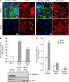
Figure 1
Figure 1
Claudin-5 knockdown increases paracellular permeability in hCMEC/D3 cells. (A) Immunofluorescence staining of hCMEC/D3 cells, treated by either nontargeting small interfering RNAs (siRNAs) (siCtrl) or siRNAs against claudin-5 (siCld5) at confluence. Labeling with claudin-5 monoclonal antibody and zonula occudens (ZO)-1 polyclonal antibodies (a, b, e, and f) or claudin-5 polyclonal antibodies and VE-cadherin monoclonal antibody (c, d, g, and h). Nuclei in blue were labeled with DAPI. The arrow (b) and arrowhead (c) point to continuous and punctuated ZO-1 staining of cell–cell junctions, respectively. Scale bars represent 10 μm. (B, upper panel) hCMEC/D3 cells treated with siCtrl (white bars) or two individual siCld5 (gray bars) were grown to confluence on Transwell inserts and permeability assays to Lucifer Yellow (LY) were performed: permeability coefficients (Pe) are presented. Results are mean values±s.d. of Pe values of triplicates in one representative experiment (out of three independent experiments). (B, lower panel) Western blot analysis of claudin-5 expression in hCMEC/D3 cells, either treated with siCtrl or with two individual siCld5. Cell extracts were prepared from two duplicate filters (F1/F2), then proteins were separated by sodium dodecyl sulfate polyacrylamide gel electrophoresis (SDS-PAGE) and immunoblotting (IB) was performed with anti-claudin-5 or anti-actin monoclonal antibodies. (C) hCMEC/D3 cells, treated with siCtrl (white bars) or siCld5 #1 (gray bars) were grown to confluence on Transwell inserts. Permeability assays were performed as indicated: to LY (457 Da), 4 kDa FITC-dextran or 70 kDa FITC-dextran; permeability coefficients (Pe) are presented. Results are mean Pe values±s.d. of triplicates in one representative experiment (out of three independent experiments). ***P≤0.005 compared with siCtrl-treated cells, not significant (N.S.).
Figure 2
Figure 2
Hyperosmolar concentration of mannitol induces a transient increase of endothelial permeability to Lucifer Yellow (LY). hCMEC/D3 cells were grown at confluence on Transwell inserts. Cells were treated with 1 M mannitol for 30 minutes, then medium was changed to let the cells recover for up to 48 hours. Permeability to LY was measured directly after mannitol treatment (white bars) and after 24 or 48 hours recovery (gray bars), as indicated. Results are mean Pe values±s.d. from three independent experiments performed in triplicates. ***P<0.005 compared with untreated cells, not significant (N.S.).
Figure 3
Figure 3
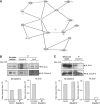
G-protein subunit αi2 (Gαi2) interacts with claudin-5 and belongs to a multiprotein complex. (A) A connectivity map of claudin-5, extracted from the whole claudin-5/CLDN5 interactome network (see Supplementary File) established with the Ingenuity software, from molecular interactions validated and published in the literature. Gray nodes represent proteins identified by mass spectrometry in the present study as claudin-5 partners. White nodes represent additional proteins, not identified in the present study: F-actin, zonula occudens (ZO)-1 (TJP1), and occludin (OCLN). Solid lines indicate direct protein–protein interactions; dotted lines indicate indirect interactions. A putative direct interaction between claudin-5 and Gαi2 is suggested by a question mark. GNAI2, Gαi2; CTNND1, p120 catenin; CDH5, VE-cadherin, JUP, γcatenin; ACTB, β-actin; MPDZ, multiple PDZ domain protein (MUPP-1); STOM, stomatin; Pkc(s), PKC protein kinases. (B, C) hCMEC/D3 cells were untreated or treated with 1 M mannitol for 30 minutes. Caveolae fractions (B) were isolated as described in Materials and methods in each condition. Whole cell lysates were solubilized in immunoprecipitation buffer (see Materials and methods). Immunoprecipitations (IP) were performed with mouse monoclonal antibodies directed against claudin-5 or Gαi2 as indicated. Immune complexes were immunoblotted (IB) with anti-claudin-5 or anti-Gαi2 antibodies, as indicated. Results are representative of three independent experiments. Arrows and arrowheads indicate the electrophoretic mobility of claudin-5 and Gαi2, respectively. Immunoblots were scanned and histograms in the lower panels represent the relative intensity of the indicated protein band after mannitol treatment versus without treatment.
Figure 4
Figure 4
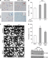
G-protein subunit αi2 (Gαi2) is expressed in human brain endothelium in situ and contributes to the integrity of endothelial junctions. (A) Immunohistochemical detection of claudin-5 and Gαi2 in human central nervous system. Claudin-5 and Gαi2 are detected in the endothelium of blood capillaries in the cortical gray matter and in the white matter. The insets show a small artery (a) and a small venule (b). Asterisks (*) indicate the vessel lumens. The insets show a venula (c) and a small artery (d). The scale bar is 10 μm for all images and 100 μm for the inset in image d. (B) hCMEC/D3 cells were treated for 6 or 24 hours with pertussis toxin (PTX at 10 ng/mL), an inhibitor of G-protein αi GTPase activity. Permeability assays were then performed with Lucifer Yellow (LY). Results are expressed as percentage of control permeability (untreated cells: Pe (LY)=2.86±0.04 × 10⁻³ cm/min) and are mean values±s.d. of triplicate Pe values in one representative experiment of three independent experiments. (C) Immunostaining of Gαi2 (a, c, and e) and zonula occudens (ZO)-1 (b, d, and f) at the cell membrane of hCMEC/D3 cells, treated with either nontargeting small interfering RNAs (siRNAs) (siCtrl) or siRNAs against Gαi2 (siGαi2). Cells were grown to confluence, fixed, permeabilized, and simultaneously incubated with anti-Gαi2 polyclonal antibodies and anti-ZO-1 monoclonal antibodies. Nuclei staining is not specific. The same fields are shown for ZO-1 and Gαi2 staining. Scale bars represent 10 μm. (D, upper panel) LY permeability assays performed with hCMEC/D3 cells treated with siCtrl (white bars) or two individual siGαi2 (gray bars). Results are expressed as percentage of control permeability (control siRNA-treated cells: Pe (LY)=0.92±0.02 × 10⁻³ cm/min) and are mean values±s.d. of triplicate Pe values in one representative experiment of three independent experiments. (D, lower panel) Expression level of Gαi2 in hCMEC/D3 cells in the same conditions as above. Whole cell lysates were prepared from 2 duplicate filters (F1, F2) and proteins were separated by sodium dodecyl sulfate polyacrylamide gel electrophoresis (SDS-PAGE) and detected by immunoblotting with anti-Gαi2 or anti-actin antibodies as protein loading controls. ***P⩽0.001, **P⩽0.05 compared with control cells.
Figure 5
Figure 5
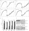
G-protein subunit αi2 (Gαi2) or claudin-5 depletion delays the recovery of cell–cell junction integrity after 1 M mannitol treatment. hCMEC/D3 cells treated with nontargeting small interfering RNAs (siRNAs) (siCtrl) or siRNAs against claudin-5 (siCld5), Gαi2 (siGαi2), or both siCld5+siGαi2 were grown in a 96-well plate of an xCELLigence system (Roche). The impedance measurement (cell index: CI) was monitored in real time and plots were produced using the RTCA Software. At t=70 hours, cells were treated with 1 M mannitol (asterisk). After 30 minutes, the medium was changed for a recovery period (up to 4 days). (AD) Plots show CI recordings during the whole experiment period (7 days) with asterisk representing time where mannitol was added. Arrowheads (1 to 4) point to times of analysis (as shown in (E)): cell confluence (t=56 hours) and various recovery times (t=84, 112, 164 hours). (E) CI values at the times indicated above (arrowheads 1 to 4 in (AD)). Results are mean CI values±s.d. (n=3 wells) from one representative experiment out of three independent experiments. (F) Expression level of claudin-5 and Gαi2 proteins in hCMEC/D3 cells either treated with siCtrl, siCld5, siGαi2, siCld5+siGαi2, or siCtrl at a double concentration (2 × ) as control. Whole cell lysates were prepared in each condition and proteins were separated by sodium dodecyl sulfate polyacrylamide gel electrophoresis (SDS-PAGE) and detected by immunoblotting (IB) with anti-claudin-5, anti-Gαi2, or anti-VE-cadherin antibodies. #P<0.005 compared with siCtrl-treated cells; ***P<0.001; not significant (N.S.).

Similar articles

Cited by

References

    1. Abbott NJ, Patabendige AA, Dolman DE, Yusof SR, Begley DJ. Structure and function of the blood-brain barrier. Neurobiol Dis. 2010;37:13–25. - PubMed
    1. Adamson P, Wilbourn B, Etienne-Manneville S, Calder V, Beraud E, Milligan G, Couraud PO, Greenwood J. Lymphocyte trafficking through the blood-brain barrier is dependent on endothelial cell heterotrimeric G-protein signaling. FASEB J. 2002;16:1185–1194. - PubMed
    1. Bruckener KE, el Baya A, Galla HJ, Schmidt MA. Permeabilization in a cerebral endothelial barrier model by pertussis toxin involves the PKC effector pathway and is abolished by elevated levels of cAMP. J Cell Sci. 2003;116:1837–1846. - PubMed
    1. Coureuil M, Mikaty G, Miller F, Lecuyer H, Bernard C, Bourdoulous S, Dumenil G, Mege RM, Weksler BB, Romero IA, Couraud PO, Nassif X. Meningococcal type IV pili recruit the polarity complex to cross the brain endothelium. Science. 2009;325:83–87. - PMC - PubMed
    1. Coyne CB, Gambling TM, Boucher RC, Carson JL, Johnson LG. Role of claudin interactions in airway tight junctional permeability. Am J Physiol Lung Cell Mol Physiol. 2003;285:L1166–L1178. - PubMed

Publication types

MeSH terms

LinkOut - more resources