

Humanin, a cytoprotective peptide, is expressed in carotid atherosclerotic [corrected] plaques in humans
- PMID: 22328926
- PMCID: PMC3273477
- DOI: 10.1371/journal.pone.0031065
Humanin, a cytoprotective peptide, is expressed in carotid atherosclerotic [corrected] plaques in humans
Erratum in
- PLoS One. 2012;7(3). doi:10.1371/annotation/1a60b239-181f-4d5f-9a37-3d3b04949954
Abstract
Objective: The mechanism of atherosclerotic plaque progression leading to instability, rupture, and ischemic manifestation involves oxidative stress and apoptosis. Humanin (HN) is a newly emerging endogenously expressed cytoprotective peptide. Our goal was to determine the presence and localization of HN in carotid atherosclerotic plaques.
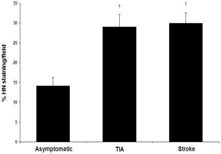
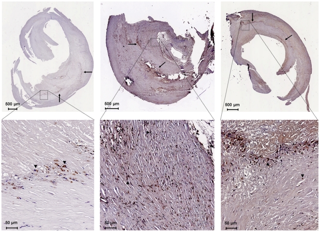
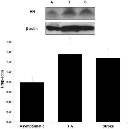
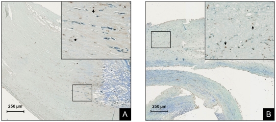
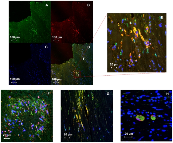
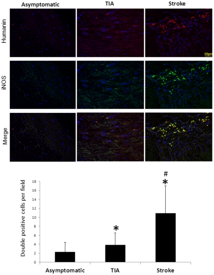
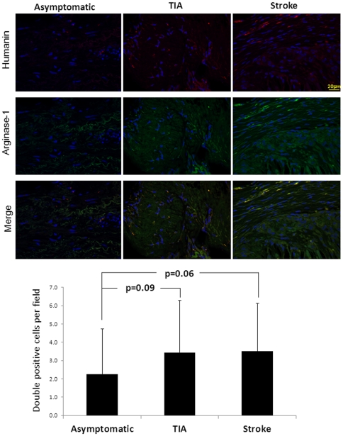
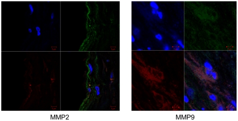
Methods and results: Plaque specimens from 34 patients undergoing carotid endarterectomy were classified according to symptomatic history. Immunostaining combined with digital microscopy revealed greater expression of HN in the unstable plaques of symptomatic compared to asymptomatic patients (29.42±2.05 vs. 14.14±2.13% of plaque area, p<0.0001). These data were further confirmed by immunoblot (density of HN/β-actin standard symptomatic vs. asymptomatic 1.32±0.14 vs. 0.79±0.11, p<0.01). TUNEL staining revealed a higher proportion of apoptotic nuclei in the plaques of symptomatic patients compared to asymptomatic (68.25±3.61 vs. 33.46±4.46% of nuclei, p<0.01). Double immunofluorescence labeling revealed co-localization of HN with macrophages (both M1 and M2 polarization), smooth muscle cells, fibroblasts, and dendritic cells as well as with inflammatory markers MMP2 and MMP9.
Conclusions: The study demonstrates a higher expression of HN in unstable carotid plaques that is localized to multiple cell types within the plaque. These data support the involvement of HN in atherosclerosis, possibly as an endogenous response to the inflammatory and apoptotic processes within the atheromatous plaque.
Conflict of interest statement
Figures
Similar articles
-
Association between 5-lipoxygenase expression and plaque instability in humans.Arterioscler Thromb Vasc Biol. 2005 Aug;25(8):1665-70. doi: 10.1161/01.ATV.0000172632.96987.2d. Epub 2005 Jun 2. Arterioscler Thromb Vasc Biol. 2005. PMID: 15933245
-
Factors associated with apoptosis in symptomatic and asymptomatic carotid atherosclerotic plaques.Int J Immunopathol Pharmacol. 2005 Oct-Dec;18(4):645-53. doi: 10.1177/039463200501800405. Int J Immunopathol Pharmacol. 2005. PMID: 16388711 Clinical Trial.
-
Carotid atherosclerotic plaques in patients with transient ischemic attacks and stroke have unstable characteristics compared with plaques in asymptomatic and amaurosis fugax patients.J Vasc Surg. 2005 Dec;42(6):1075-81. doi: 10.1016/j.jvs.2005.08.009. J Vasc Surg. 2005. PMID: 16376194
-
Insulin-like growth factor-I receptors in atherosclerotic plaques of symptomatic and asymptomatic patients with carotid stenosis: effect of IL-12 and IFN-gamma.Am J Physiol Heart Circ Physiol. 2007 Feb;292(2):H1051-7. doi: 10.1152/ajpheart.00801.2006. Epub 2006 Oct 13. Am J Physiol Heart Circ Physiol. 2007. PMID: 17040964
-
The role of humanin in natural stress tolerance: An underexplored therapeutic avenue.Biochim Biophys Acta Gen Subj. 2022 Jan;1866(1):130022. doi: 10.1016/j.bbagen.2021.130022. Epub 2021 Oct 7. Biochim Biophys Acta Gen Subj. 2022. PMID: 34626747 Review.
Cited by
-
Circulating humanin levels are associated with preserved coronary endothelial function.Am J Physiol Heart Circ Physiol. 2013 Feb 1;304(3):H393-7. doi: 10.1152/ajpheart.00765.2012. Epub 2012 Dec 7. Am J Physiol Heart Circ Physiol. 2013. PMID: 23220334 Free PMC article.
-
From Mitochondria to Atherosclerosis: The Inflammation Path.Biomedicines. 2021 Mar 5;9(3):258. doi: 10.3390/biomedicines9030258. Biomedicines. 2021. PMID: 33807807 Free PMC article. Review.
-
Humanin prevents intra-renal microvascular remodeling and inflammation in hypercholesterolemic ApoE deficient mice.Life Sci. 2012 Sep 4;91(5-6):199-206. doi: 10.1016/j.lfs.2012.07.010. Epub 2012 Jul 20. Life Sci. 2012. PMID: 22820173 Free PMC article.
-
Effects of Mitochondrial-Derived Peptides (MDPs) on Mitochondrial and Cellular Health in AMD.Cells. 2020 Apr 29;9(5):1102. doi: 10.3390/cells9051102. Cells. 2020. PMID: 32365540 Free PMC article. Review.
-
MOTS-c: A Mitochondrial-Encoded Regulator of the Nucleus.Bioessays. 2019 Sep;41(9):e1900046. doi: 10.1002/bies.201900046. Epub 2019 Aug 5. Bioessays. 2019. PMID: 31378979 Free PMC article. Review.
References
-
- Ross R. Atherosclerosis–an inflammatory disease. N Engl J Med. 1999;340:115–126. - PubMed
-
- Galkina E, Ley K. Leukocyte influx in atherosclerosis. Curr Drug Targets. 2007;8:1239–1248. - PubMed
-
- Hansson GK, Libby P. The immune response in atherosclerosis: a double-edged sword. Nat Rev Immunol. 2006;6:508–519. - PubMed
Publication types
MeSH terms
Substances
Grants and funding
- R01 ES020812/ES/NIEHS NIH HHS/United States
- AG034430/AG/NIA NIH HHS/United States
- R01 GM090311/GM/NIGMS NIH HHS/United States
- R01 AG031750/AG/NIA NIH HHS/United States
- R00 HL88048/HL/NHLBI NIH HHS/United States
- 1 TL1 RR024152/RR/NCRR NIH HHS/United States
- TL1 RR024152/RR/NCRR NIH HHS/United States
- GM090311/GM/NIGMS NIH HHS/United States
- R01 AG034430/AG/NIA NIH HHS/United States
- R01 HL092954/HL/NHLBI NIH HHS/United States
- HL92954/HL/NHLBI NIH HHS/United States
- R00 HL088048/HL/NHLBI NIH HHS/United States
- AG31750/AG/NIA NIH HHS/United States
LinkOut - more resources
Full Text Sources
Medical
Miscellaneous
