. 2012 Feb;8(2):e1002497.
doi: 10.1371/journal.ppat.1002497. Epub 2012 Feb 2.

Enhancement of chemokine function as an immunomodulatory strategy employed by human herpesviruses

Affiliations

Enhancement of chemokine function as an immunomodulatory strategy employed by human herpesviruses

Abel Viejo-Borbolla et al. PLoS Pathog. 2012 Feb.

Abstract

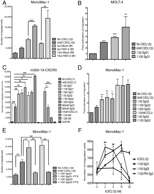
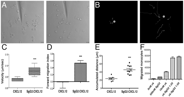
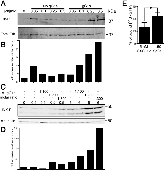
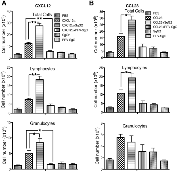
Herpes simplex virus (HSV) types 1 and 2 are highly prevalent human neurotropic pathogens that cause a variety of diseases, including lethal encephalitis. The relationship between HSV and the host immune system is one of the main determinants of the infection outcome. Chemokines play relevant roles in antiviral response and immunopathology, but the modulation of chemokine function by HSV is not well understood. We have addressed the modulation of chemokine function mediated by HSV. By using surface plasmon resonance and crosslinking assays we show that secreted glycoprotein G (SgG) from both HSV-1 and HSV-2 binds chemokines with high affinity. Chemokine binding activity was also observed in the supernatant of HSV-2 infected cells and in the plasma membrane of cells infected with HSV-1 wild type but not with a gG deficient HSV-1 mutant. Cell-binding and competition experiments indicate that the interaction takes place through the glycosaminoglycan-binding domain of the chemokine. The functional relevance of the interaction was determined both in vitro, by performing transwell assays, time-lapse microscopy, and signal transduction experiments; and in vivo, using the air pouch model of inflammation. Interestingly, and in contrast to what has been observed for previously described viral chemokine binding proteins, HSV SgGs do not inhibit chemokine function. On the contrary, HSV SgGs enhance chemotaxis both in vitro and in vivo through increasing directionality, potency and receptor signaling. This is the first report, to our knowledge, of a viral chemokine binding protein from a human pathogen that increases chemokine function and points towards a previously undescribed strategy of immune modulation mediated by viruses.


Conflict of interest statement

The authors have declared that no competing interests exist.

Figures

Figure 1
Figure 1. Cloning, expression and purification of HSV gG.
(A) Schematic representation of SgG1 and SgG2 constructs used in this study. A fragment of the extracellular domain of both gG1 and gG2 was amplified and cloned into a baculovirus-expression vector. The putative signal peptide from gG was substituted by the honeybee melittin signal peptide. The position of the amino acid residues is indicated. The dashed lines indicate the fragment of the extracellular domain included in the construct. Abbreviations: SP, signal peptide; Tmb, Transmembrane domain; His, histidine tag; HM, honeybee melittin signal peptide; ED, extracellular domain. (B) SDS-PAGE followed by coomassie staining showing purified SgG1 (left panel) and SgG2 (right panel). (C) Western blots showing the detection of SgG1 and SgG2 with an anti-histidine (left panel), an anti-gG1 (middle panel) or an anti-gG2 (right panel) antibody. Molecular masses are shown in kilodaltons (kDa).
Figure 2
Figure 2. HSV-1 and HSV-2 gGs bind chemokines.
(A) Sensorgrams depicting the interaction between chemokines and SgG1 (left) or SgG2 (right). The indicated chemokines were injected at a 100 nM concentration. The arrow indicates the end of injection. All curves were analyzed with the BiaEvaluation software and represent the interaction of the chemokine after subtraction of the blank curve. Only 4 out of 11–12 positive interactions are shown. Abbreviations: Diff. Resp., Differential response; R.U., response units; s, seconds (B, C) Crosslinking assays showing the interaction of HSV-SgGs with [125I]-hCXCL10 (B) and [125I]-hCCL25 (C). Recombinant purified HSV-SgGs were incubated with iodinated chemokine and crosslinked with EGS (for [125I]-hCCL25) or BS3 (for [125I]-hCXCL10). The samples were resolved by SDS-PAGE, fixed and visualized by autoradiography. (D) Crosslinking assay between [125I]-hCXCL12α and SgG2 in the presence of increasing concentrations of cold hCXCL12α. Molecular masses are indicated in kDa. SgG-chemokine complexes are indicated with arrows and crosslinked chemokine dimers are marked with asterisks. Abbreviations: CRD, CrmB-cysteine rich domain.
Figure 3
Figure 3. HSV-1 and HSV-2 gG expressed during infection bind chemokines.
(A) Graph showing binding of radio-iodinated hCXCL10 to the surface of HSV-1 infected cells. Binding is observed at 14–16 h.p.i., only when cells are infected with wt HSV-1 but not when infected with a HSV-1ΔgG mutant. (B) Crosslinking assay showing the interaction between [125I]-hCXCL12α and HSV-2 gG in the supernatant of HSV-2 infected cells. The arrows point to the crosslinked complex. Abbreviations: h.p.i, hours post-infection. **P<0.01.
Figure 4
Figure 4. Determination of the chemokine domain involved in the interaction with HSV gGs.
Heparin competition of chemokine binding to SgG1 and SgG2. hCXCL12α was injected at a concentration of 100 nM alone or in combination with the indicated increasing concentrations of heparin. The value of chemokine binding without heparin was considered 100%. All curves were analyzed with the BiaEvaluation software and represent the interaction of the chemokine after subtraction of the HBS-EP curve. The error bars represent the standard error of three independent experiments. *P<0.05; P<0.001.
Figure 5
Figure 5. HSV SgGs enhance chemokine-mediated cell migration.
MonoMac-1 cells (A, D, E, F), MOLT-4 (B), m300-19-hCXCR5 (C) cells were incubated with the specified chemokine in Transwell plates. The effect of mock- or HSV-2-infected supernatant (Mock SN or HSV-2 SN, respectively) (A), purified SgG1 (B, C, E, F), SgG2 (C–F), M3 (C) and PRV-SgG (F) was analyzed. The number of migrated cells or the fold activation of migration is depicted. (C) SgG1 or SgG2 require the presence of the chemokine to enhance migration since addition of either of them without chemokine did not have any effect on chemotaxis. (D) Binding of HSV SgGs to the chemokine is necessary for the enhancement in chemotaxis. Representation of the fold activation of migration observed when cells were incubated with either hCXCL12β or hCCL2 in the absence or presence of increasing concentrations of HSV-2 gGs. (E) Addition of pertussis toxin (PTX) inhibits SgG-mediated enhancement of chemotaxis. Graph showing the effect of PTX addition on HSV SgGs enhancement of hCXCL12β-mediated chemotaxis. The number of migrated MonoMac-1 cells is represented. (F) HSV SgGs displace the hCXCL12β chemotactic curve towards lower concentrations of chemokine. MonoMac-1 cells were incubated with increasing concentrations of hCXCL12β in the absence or presence of a 1∶100 molar ratio of HSV SgGs or PRV-SgG. (A–F) Error bars indicate standard deviation values obtained from triplicate samples (A, C, E, F). One representative experiment of at least three is shown. In B and D, error bars represent the standard deviation in the fold activation obtained using three independent experiments performed in duplicate. *P<0.05; **P<0.01; ***P<0.001.
Figure 6
Figure 6. Analysis of SgG2 induced enhancement of chemotaxis by time-lapse video microscopy.
(A) Selected frames from Videos S1 and S2 showing migration towards CXCL12 (left) or CXCL12-SgG2 (right). (B) Migrating cells were tracked and their progressive trajectories were plotted according to the recorded xy coordinates. Orange dots indicate the real position of the micropipette dispensing CXCL12 (left) or CXCL12-SgG2 (right). Each line represents the path followed by one cell during 10 min at the initial phase of chemotaxis. (C) The velocity of cell movement, (D) forward migration index and (E) total traveled distance by cells migrating towards the micropipette dispensing CXCL12 or CXCL12-SgG2 were plotted. Representative data from 6 cells (CXCL12) and 10 cells (CXCL12-SgG2) migrating at the initial time period are shown. Time-lapse videos were analyzed using Image J 1.43 software. The trajectories of the tracks, velocities, FMI and distances traveled were calculated using Manual Tracking and Chemotaxis Tool version 1.01 plugging. The analysis of 1 representative video out of three is shown. (F) Representation of migrated monocytes in the presence of CXCL12 alone or in combination with SgG2 using the transwell technology. 1 representative experiment out of three is shown. Error bars indicate standard error values. *:P<0.05; **:P<0.01; ***:P<0.001.
Figure 7
Figure 7. HSV SgG enhances chemokine-mediated signaling.
(A) Western blots showing activation of ERK (p-ERK, top blot) and loading control (Total ERK, bottom blot) in MonoMac-1 cells incubated with CXCL12 alone or with a constant 1∶200 molar ratio of chemokine∶SgG1. (C) Western blot showing the effect of increasing concentrations of HSV SgG1 on chemokine-mediated JNK phosphorylation (top blot). As loading control, the blots were stripped and incubated with anti-alpha-tubulin (bottom blot). (B and D) Graphs depicting the results obtained after performing a densitometer analysis of the blots. The densities obtained from each of the lanes in the MAPKs blots were normalized to the loading controls and later to the mock sample. (E) Graph showing the percentage of [35S]-GTPγ binding to CXCR4 mediated by CXCL12 alone or with SgG2 (considering no CXCL12 as 100%). The results of combining three independent experiments performed in duplicate are shown. Error bars indicate standard deviation values. * P<0.05.
Figure 8
Figure 8. HSV-2 SgG enhances chemokine-mediated cell migration in vivo.
CXCL12α (A) or CCL28 (B) were injected into dorsal air pouches in mice alone or in combination with HSV-2 SgG or PRV SgG. Cell migration into the air cavity was monitored. Cells were extracted and identified by flow cytometry with specific markers. The number of total leukocytes (top), lymphocytes (middle) and granulocyte cells (bottom graph) is represented. Data are mean and SEM from 5–6 mice per group and are representative of 2–3 separate experiments. *:P<0.05; **:P<0.001.

Similar articles

Cited by

References

    1. Wutzler P, Doerr HW, Farber I, Eichhorn U, Helbig B, et al. Seroprevalence of herpes simplex virus type 1 and type 2 in selected German populations-relevance for the incidence of genital herpes. J Med Virol. 2000;61:201–207. - PubMed
    1. Paz-Bailey G, Ramaswamy M, Hawkes SJ, Geretti AM. Herpes simplex virus type 2: epidemiology and management options in developing countries. Sex Transm Infect. 2007;83:16–22. - PMC - PubMed
    1. Kimberlin DW. Management of HSV encephalitis in adults and neonates: diagnosis, prognosis and treatment. Herpes. 2007;14:11–16. - PubMed
    1. Kimberlin D. Herpes simplex virus, meningitis and encephalitis in neonates. Herpes. 2004;11(Suppl 2):65A–76A. - PubMed
    1. Baggiolini M. Chemokines and leukocyte traffic. Nature. 1998;392:565–568. - PubMed

Publication types

MeSH terms