Interleukin-1β modulates smooth muscle cell phenotype to a distinct inflammatory state relative to PDGF-DD via NF-κB-dependent mechanisms
- PMID: 22318995
- PMCID: PMC3339851
- DOI: 10.1152/physiolgenomics.00160.2011
Interleukin-1β modulates smooth muscle cell phenotype to a distinct inflammatory state relative to PDGF-DD via NF-κB-dependent mechanisms
Abstract
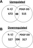
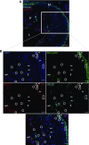
Smooth muscle cell (SMC) phenotypic modulation in atherosclerosis and in response to PDGF in vitro involves repression of differentiation marker genes and increases in SMC proliferation, migration, and matrix synthesis. However, SMCs within atherosclerotic plaques can also express a number of proinflammatory genes, and in cultured SMCs the inflammatory cytokine IL-1β represses SMC marker gene expression and induces inflammatory gene expression. Studies herein tested the hypothesis that IL-1β modulates SMC phenotype to a distinct inflammatory state relative to PDGF-DD. Genome-wide gene expression analysis of IL-1β- or PDGF-DD-treated SMCs revealed that although both stimuli repressed SMC differentiation marker gene expression, IL-1β distinctly induced expression of proinflammatory genes, while PDGF-DD primarily induced genes involved in cell proliferation. Promoters of inflammatory genes distinctly induced by IL-1β exhibited over-representation of NF-κB binding sites, and NF-κB inhibition in SMCs reduced IL-1β-induced upregulation of proinflammatory genes as well as repression of SMC differentiation marker genes. Interestingly, PDGF-DD-induced SMC marker gene repression was not NF-κB dependent. Finally, immunofluorescent staining of mouse atherosclerotic lesions revealed the presence of cells positive for the marker of an IL-1β-stimulated inflammatory SMC, chemokine (C-C motif) ligand 20 (CCL20), but not the PDGF-DD-induced gene, regulator of G protein signaling 17 (RGS17). Results demonstrate that IL-1β- but not PDGF-DD-induced phenotypic modulation of SMC is characterized by NF-κB-dependent activation of proinflammatory genes, suggesting the existence of a distinct inflammatory SMC phenotype. In addition, studies provide evidence for the possible utility of CCL20 and RGS17 as markers of inflammatory and proliferative state SMCs within atherosclerotic plaques in vivo.
Figures


Similar articles
-
PDGF-DD, a novel mediator of smooth muscle cell phenotypic modulation, is upregulated in endothelial cells exposed to atherosclerosis-prone flow patterns.Am J Physiol Heart Circ Physiol. 2009 Feb;296(2):H442-52. doi: 10.1152/ajpheart.00165.2008. Epub 2008 Nov 21. Am J Physiol Heart Circ Physiol. 2009. PMID: 19028801 Free PMC article.
-
Neutralization of S100A4 induces stabilization of atherosclerotic plaques: role of smooth muscle cells.Cardiovasc Res. 2022 Jan 7;118(1):141-155. doi: 10.1093/cvr/cvaa311. Cardiovasc Res. 2022. PMID: 33135065 Free PMC article.
-
Platelet-derived growth factor-BB and Ets-1 transcription factor negatively regulate transcription of multiple smooth muscle cell differentiation marker genes.Am J Physiol Heart Circ Physiol. 2004 Jun;286(6):H2042-51. doi: 10.1152/ajpheart.00625.2003. Epub 2004 Jan 29. Am J Physiol Heart Circ Physiol. 2004. PMID: 14751865
-
Transcriptional regulation of vascular smooth muscle cell proliferation, differentiation and senescence: Novel targets for therapy.Vascul Pharmacol. 2022 Oct;146:107091. doi: 10.1016/j.vph.2022.107091. Epub 2022 Jul 25. Vascul Pharmacol. 2022. PMID: 35896140 Review.
-
Selective transcription in response to an inflammatory stimulus.Cell. 2010 Mar 19;140(6):833-44. doi: 10.1016/j.cell.2010.01.037. Cell. 2010. PMID: 20303874 Free PMC article. Review.
Cited by
-
The Notch pathway attenuates interleukin 1β (IL1β)-mediated induction of adenylyl cyclase 8 (AC8) expression during vascular smooth muscle cell (VSMC) trans-differentiation.J Biol Chem. 2012 Jul 20;287(30):24978-89. doi: 10.1074/jbc.M111.292516. Epub 2012 May 21. J Biol Chem. 2012. PMID: 22613711 Free PMC article.
-
"How to Release or Not Release, That Is the Question." A Review of Interleukin-1 Cellular Release Mechanisms in Vascular Inflammation.J Am Heart Assoc. 2024 Mar 5;13(5):e032987. doi: 10.1161/JAHA.123.032987. Epub 2024 Feb 23. J Am Heart Assoc. 2024. PMID: 38390810 Free PMC article. Review.
-
Myocardin regulates vascular smooth muscle cell inflammatory activation and disease.Arterioscler Thromb Vasc Biol. 2015 Apr;35(4):817-28. doi: 10.1161/ATVBAHA.114.305218. Epub 2015 Jan 22. Arterioscler Thromb Vasc Biol. 2015. PMID: 25614278 Free PMC article.
-
IL-1 in Abdominal Aortic Aneurysms.J Cell Immunol. 2023;5(2):22-31. doi: 10.33696/immunology.5.163. J Cell Immunol. 2023. PMID: 37476160 Free PMC article.
-
Lipoprotein(a) the Insurgent: A New Insight into the Structure, Function, Metabolism, Pathogenicity, and Medications Affecting Lipoprotein(a) Molecule.J Lipids. 2020 Feb 1;2020:3491764. doi: 10.1155/2020/3491764. eCollection 2020. J Lipids. 2020. PMID: 32099678 Free PMC article. Review.
References
-
- Abi-Younes S, Sauty A, Mach F, Sukhova GK, Libby P, Luster AD. The stromal cell-derived factor-1 chemokine is a potent platelet agonist highly expressed in atherosclerotic plaques. Circ Res 86: 131–138, 2000. - PubMed
-
- Amento EP, Ehsani N, Palmer H, Libby P. Cytokines and growth factors positively and negatively regulate interstitial collagen gene expression in human vascular smooth muscle cells. Arterioscler Thromb 11: 1223–1230, 1991. - PubMed
-
- Banai S, Wolf Y, Golomb G, Pearle A, Waltenberger J, Fishbein I, Schneider A, Gazit A, Perez L, Huber R, Lazarovichi G, Rabinovich L, Levitzki A, Gertz SD. PDGF-receptor tyrosine kinase blocker AG1295 selectively attenuates smooth muscle cell growth in vitro and reduces neointimal formation after balloon angioplasty in swine. Circulation 97: 1960–1969, 1998. - PubMed
-
- Bergsten E, Uutela M, Li X, Pietras K, Ostman A, Heldin CH, Alitalo K, Eriksson U. PDGF-D is a specific, protease-activated ligand for the PDGF beta-receptor. Nat Cell Biol 3: 512–516, 2001. - PubMed
Publication types
MeSH terms
Substances
Grants and funding
LinkOut - more resources
Full Text Sources
Molecular Biology Databases

