

PAX2 loss by immunohistochemistry occurs early and often in endometrial hyperplasia
- PMID: 22317873
- PMCID: PMC4646427
- DOI: 10.1097/PGP.0b013e318226b376
PAX2 loss by immunohistochemistry occurs early and often in endometrial hyperplasia
Abstract
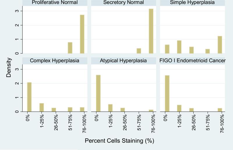
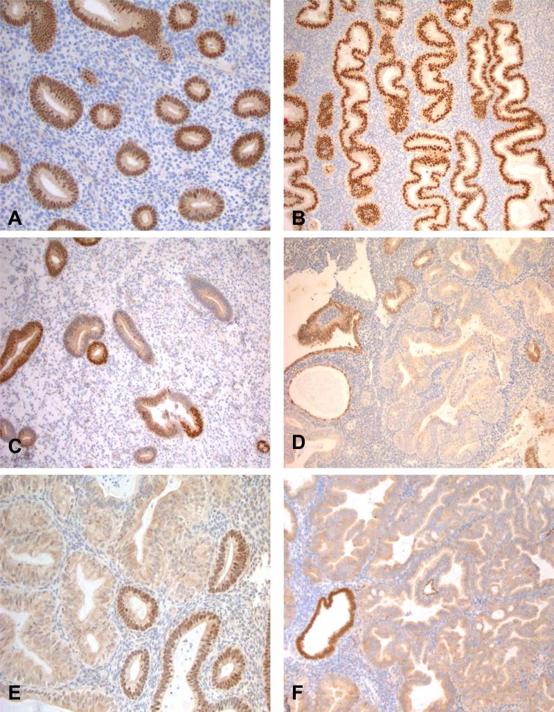
Immunohistochemical markers to assist in the diagnosis and classification of hyperplastic endometrial epithelial proliferations would be of diagnostic use. To examine the possible use of PAX2 as a marker of hyperplastic endometrium, cases of normal endometrium, simple and complex hyperplasia without atypia, atypical hyperplasia, and International Federation of Gynecology and Obstetrics (FIGO) grade 1 endometrioid carcinomas were stained for PAX2. Two hundred and six endometrial samples were available for interpretation of PAX2 staining. The percentage of cases with complete PAX2 loss (0% of cells staining) increased with increasing severity of hyperplasia: 0% of normal proliferative and secretory endometrium (n=28), 17.4% of simple hyperplasia (n=23), 59.0% of complex hyperplasia (n=83), 74.1% of atypical hyperplasia (n=54), and 73.3% of FIGO grade 1 endometrioid cancers (n=15). Partial loss of PAX2 expression did occur in normal endometrium (17.9%) but in smaller proportions of tissue and was less frequent than in simple hyperplasia (47.8% with partial loss), complex hyperplasia (32.5%), atypical hyperplasia (22.2%), and FIGO grade 1 carcinomas (20.0%). Uniform PAX2 expression was rare in complex (8.4%) and atypical hyperplasia (3.7%) and carcinoma (6.7%). When evaluating loss of PAX2 in histologically normal endometrium adjacent to lesional endometrium in a given case, statistically significant differences in staining were observed for simple hyperplasia (P=0.011), complex hyperplasia (P<0.001), atypical hyperplasia (P<0.001), and FIGO grade 1 endometrioid cancer (P=0.003). In summary, PAX2 loss seems to occur early in the development of endometrial precancers and may prove useful in some settings as a diagnostic marker in determining normal endometrium from complex and atypical hyperplasia and low-grade carcinomas. However, it is not useful in distinguishing between these diagnostic categories.
Figures
Similar articles
-
New concepts for an old problem: the diagnosis of endometrial hyperplasia.Hum Reprod Update. 2017 Mar 1;23(2):232-254. doi: 10.1093/humupd/dmw042. Hum Reprod Update. 2017. PMID: 27920066 Free PMC article. Review.
-
BCL-2 and PAX2 Expressions in EIN which Had Been Previously Diagnosed as Non-Atypical Hyperplasia.Pathol Oncol Res. 2019 Apr;25(2):471-476. doi: 10.1007/s12253-017-0378-0. Epub 2017 Dec 21. Pathol Oncol Res. 2019. PMID: 29270778
-
Pax2 expression in simultaneously diagnosed WHO and EIN classification systems.Int J Gynecol Pathol. 2015 Jan;34(1):40-6. doi: 10.1097/PGP.0000000000000185. Int J Gynecol Pathol. 2015. PMID: 25473752
-
PAX2 in endometrial carcinogenesis and in differential diagnosis of endometrial hyperplasia: A systematic review and meta-analysis of diagnostic accuracy.Acta Obstet Gynecol Scand. 2019 Mar;98(3):287-299. doi: 10.1111/aogs.13512. Epub 2019 Jan 8. Acta Obstet Gynecol Scand. 2019. PMID: 30511741
-
[Endometrial hyperplasias: histology, classification, prognostic significance and therapy].Zentralbl Gynakol. 1997;119(6):251-9. Zentralbl Gynakol. 1997. PMID: 9312959 Review. German.
Cited by
-
DNA methylation promotes paired box 2 expression via myeloid zinc finger 1 in endometrial cancer.Oncotarget. 2016 Dec 20;7(51):84785-84797. doi: 10.18632/oncotarget.12626. Oncotarget. 2016. PMID: 27764784 Free PMC article.
-
Could Obesity be a Triggering Factor for Endometrial Tubal Metaplasia to be a Precancerous Lesion?J Obes. 2020 Mar 20;2020:2825905. doi: 10.1155/2020/2825905. eCollection 2020. J Obes. 2020. PMID: 32300484 Free PMC article.
-
Biomarkers of progestin therapy resistance and endometrial hyperplasia progression.Am J Obstet Gynecol. 2012 Jul;207(1):36.e1-8. doi: 10.1016/j.ajog.2012.05.012. Epub 2012 May 16. Am J Obstet Gynecol. 2012. PMID: 22727345 Free PMC article.
-
New concepts for an old problem: the diagnosis of endometrial hyperplasia.Hum Reprod Update. 2017 Mar 1;23(2):232-254. doi: 10.1093/humupd/dmw042. Hum Reprod Update. 2017. PMID: 27920066 Free PMC article. Review.
-
Clinicopathological and immunohistochemical characterization of papillary proliferation of the endometrium: A single institutional experience.Oncotarget. 2016 Jun 28;7(26):39197-39206. doi: 10.18632/oncotarget.10049. Oncotarget. 2016. PMID: 27322430 Free PMC article.
References
-
- Beutler HK, Dockerty MB, Randall LM. Precancerous lesions of the endometrium. Am J Obstet Gynecol. 1963;86:433–43. - PubMed
-
- Campbell PE, Barter RA. The significance of a typical endometrial hyperplasia. J Obstet Gynaecol Br Commonw. 1961;68:668–72. - PubMed
-
- Gore H, Hertig AT. Carcinoma in situ of the endometrium. Am J Obstet Gynecol. 1966;94(1):134–55. - PubMed
-
- Gusberg SB, Kaplan AL. Precursors of Corpus Cancer. Iv. Adenomatous Hyperplasia as Stage O Carcinoma of the Endometrium. Am J Obstet Gynecol. 1963;87:662–78. - PubMed
-
- Hendrickson MKR. Surgical Pathology of the Uterine Corpus Vol. WB Saunders Co.; Philadelphia: 1980.
Publication types
MeSH terms
Substances
Grants and funding
LinkOut - more resources
Full Text Sources
