

Fibroblast growth factor 19 expression correlates with tumor progression and poorer prognosis of hepatocellular carcinoma
- PMID: 22309595
- PMCID: PMC3293719
- DOI: 10.1186/1471-2407-12-56
Fibroblast growth factor 19 expression correlates with tumor progression and poorer prognosis of hepatocellular carcinoma
Abstract
Background: Although fibroblast growth factor 19 (FGF19) can promote liver carcinogenesis in mice, its involvement in human hepatocellular carcinoma (HCC) has not been well investigated. FGF19, a member of the FGF family, has unique specificity for its receptor FGFR4. This study aimed to clarify the involvement of FGF19 in the development of HCC.
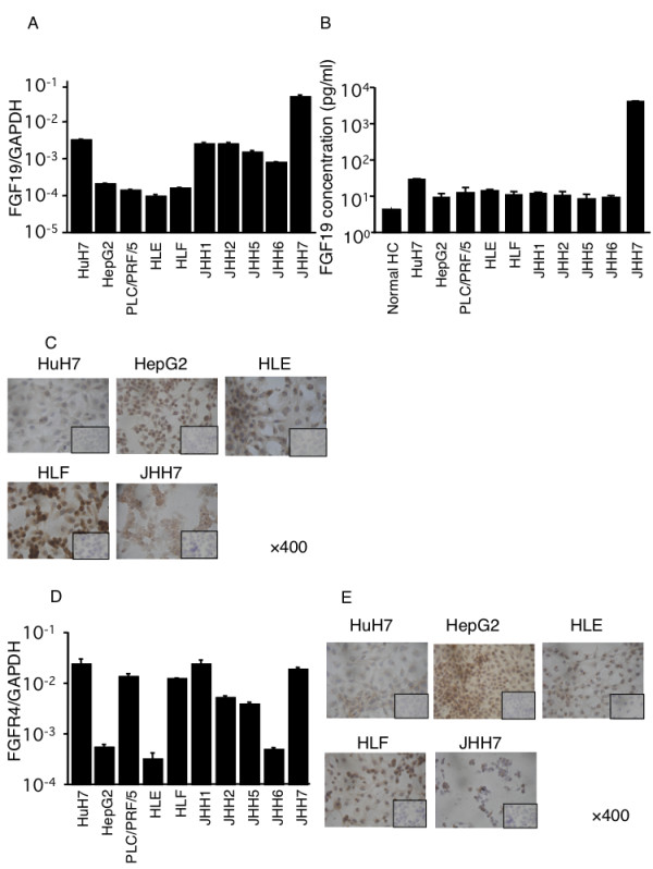
Methods: We investigated human FGF19 and FGFR4 expression in 40 hepatocellular carcinoma specimens using quantitative real-time reverse transcription polymerase chain reaction (RT-PCR) analysis and immunohistochemistry. Moreover, we examined the expression and the distribution of FGF19 and FGFR4 in 5 hepatocellular carcinoma cell lines (HepG2, HuH7, HLE, HLF, and JHH7) using RT-PCR and immunohistochemistry. To test the role of the FGF19/FGFR4 system in tumor progression, we used recombinant FGF19 protein and small interfering RNA (siRNA) of FGF19 and FGFR4 to regulate their concentrations.
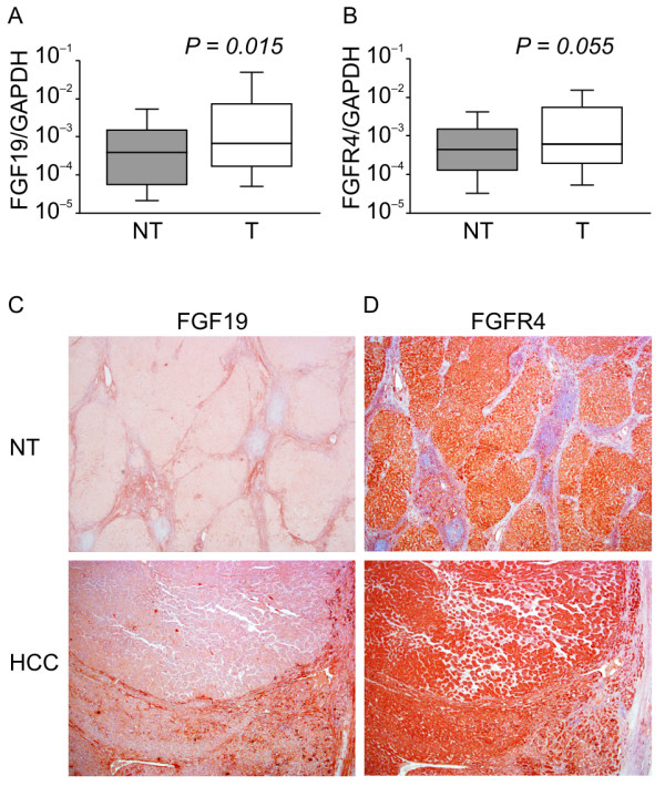
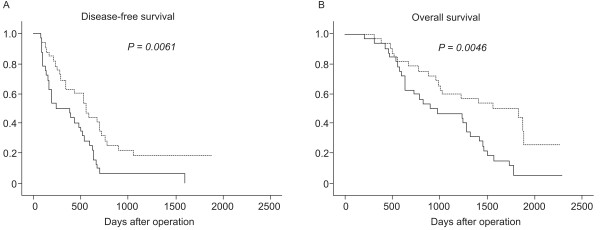
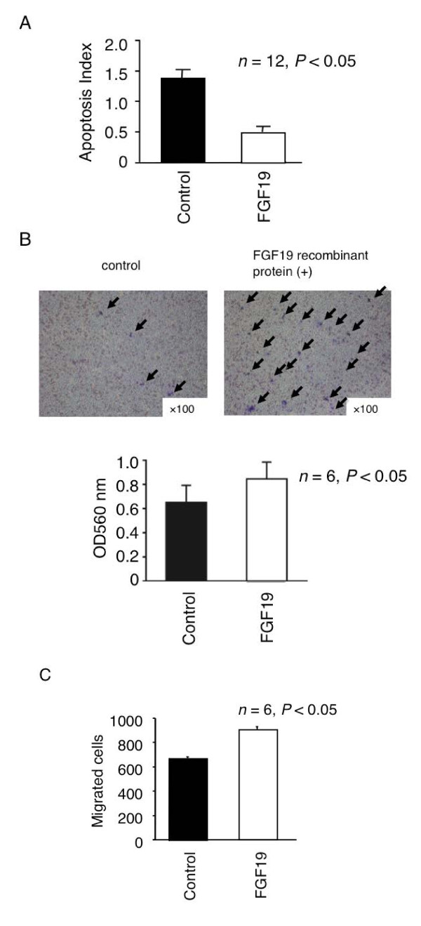
Results: We found that FGF19 was significantly overexpressed in HCCs as compared with corresponding noncancerous liver tissue (P < 0.05). Univariate and multivariate analyses revealed that the tumor FGF19 mRNA expression was an independent prognostic factor for overall and disease-free survival. Moreover, we found that the FGF19 recombinant protein could increase the proliferation (P < 0.01, n = 12) and invasion (P < 0.01, n = 6) capabilities of human hepatocellular carcinoma cell lines and inhibited their apoptosis (P < 0.01, n = 12). Inversely, decreasing FGF19 and FGFR4 expression by siRNA significantly inhibited proliferation and increased apoptosis in JHH7 cells (P < 0.01, n = 12). The postoperative serum FGF19 levels in HCC patients was significantly lower than the preoperative levels (P < 0.01, n = 29).
Conclusions: FGF19 is critically involved in the development of HCCs. Targeting FGF19 inhibition is an attractive potential therapeutic strategy for HCC.
Figures


Similar articles
-
Klotho-beta and fibroblast growth factor 19 expression correlates with early recurrence of resectable hepatocellular carcinoma.Liver Int. 2019 Sep;39(9):1682-1691. doi: 10.1111/liv.14055. Epub 2019 Jul 11. Liver Int. 2019. PMID: 30698907
-
Up-regulation of fibroblast growth factor 19 and its receptor associates with progression from fatty liver to hepatocellular carcinoma.Oncotarget. 2016 Aug 9;7(32):52329-52339. doi: 10.18632/oncotarget.10750. Oncotarget. 2016. PMID: 27447573 Free PMC article.
-
Fibroblast Growth Factor 19-Mediated Up-regulation of SYR-Related High-Mobility Group Box 18 Promotes Hepatocellular Carcinoma Metastasis by Transactivating Fibroblast Growth Factor Receptor 4 and Fms-Related Tyrosine Kinase 4.Hepatology. 2020 May;71(5):1712-1731. doi: 10.1002/hep.30951. Epub 2020 Feb 10. Hepatology. 2020. PMID: 31529503
-
FGF19-FGFR4 Signaling in Hepatocellular Carcinoma.Cells. 2019 Jun 4;8(6):536. doi: 10.3390/cells8060536. Cells. 2019. PMID: 31167419 Free PMC article. Review.
-
Targeted inhibition of the FGF19-FGFR4 pathway in hepatocellular carcinoma; translational safety considerations.Liver Int. 2014 Jul;34(6):e1-9. doi: 10.1111/liv.12462. Epub 2014 Jan 24. Liver Int. 2014. PMID: 24393342 Review.
Cited by
-
Emerging roles of FGF signaling in hepatocellular carcinoma.Transl Cancer Res. 2016 Feb;5(1):1-6. Transl Cancer Res. 2016. PMID: 27226954 Free PMC article. No abstract available.
-
Insights Into Mechanisms of Tumor and Immune System Interaction: Association With Wound Healing.Front Oncol. 2019 Oct 25;9:1115. doi: 10.3389/fonc.2019.01115. eCollection 2019. Front Oncol. 2019. PMID: 31709183 Free PMC article.
-
Altered Microbiota Diversity and Bile Acid Signaling in Cirrhotic and Noncirrhotic NASH-HCC.Clin Transl Gastroenterol. 2020 Mar;11(3):e00131. doi: 10.14309/ctg.0000000000000131. Clin Transl Gastroenterol. 2020. PMID: 32352707 Free PMC article.
-
Quantitative liver proteomics identifies FGF19 targets that couple metabolism and proliferation.PLoS One. 2017 Feb 8;12(2):e0171185. doi: 10.1371/journal.pone.0171185. eCollection 2017. PLoS One. 2017. PMID: 28178326 Free PMC article.
-
Targeting FGF19/FGFR4 Pathway: A Novel Therapeutic Strategy for Hepatocellular Carcinoma.Diseases. 2015 Oct 28;3(4):294-305. doi: 10.3390/diseases3040294. Diseases. 2015. PMID: 28943626 Free PMC article. Review.
References
MeSH terms
Substances
LinkOut - more resources
Full Text Sources
Other Literature Sources
Medical
Miscellaneous
