. 2012 Jun;14(6):882-901.
doi: 10.1111/j.1462-5822.2012.01763.x. Epub 2012 Feb 28.

PRMT1 methylates the single Argonaute of Toxoplasma gondii and is important for the recruitment of Tudor nuclease for target RNA cleavage by antisense guide RNA

Affiliations

PRMT1 methylates the single Argonaute of Toxoplasma gondii and is important for the recruitment of Tudor nuclease for target RNA cleavage by antisense guide RNA

Alla Musiyenko et al. Cell Microbiol. 2012 Jun.

Abstract

Argonaute (Ago) plays a central role in RNA interference in metazoans, but its status in lower organisms remains ill-defined. We report on the Ago complex of the unicellular protozoan, Toxoplasma gondii (Tg), an obligatory pathogen of mammalian hosts. The PIWI-like domain of TgAgo lacked the canonical DDE/H catalytic triad, explaining its weak target RNA cleavage activity. However, TgAgo associated with a stronger RNA slicer, a Tudor staphylococcal nuclease (TSN), and with a protein Arg methyl transferase, PRMT1. Mutational analysis suggested that the N-terminal RGG-repeat domain of TgAgo was methylated by PRMT1, correlating with the recruitment of TSN. The slicer activity of TgAgo was Mg(2+)-dependent and required perfect complementarity between the guide RNA and the target. In contrast, the TSN activity was Ca(2+) -dependent and required an imperfectly paired guide RNA. Ago knockout parasites showed essentially normal growth, but in contrast, the PRMT1 knockouts grew abnormally. Chemical inhibition of Arg-methylation also had an anti-parasitic effect. These results suggest that the parasitic PRMT1 plays multiple roles, and its loss affects the recruitment of a more potent second slicer to the parasitic RNA silencing complex, the exact mechanism of which remains to be determined.


Figures

Fig. 1
Fig. 1
Putative domains of TgAgo based on sequence homology. In comparing the TgAgo primary structure with its closest homologue, mouse Ago1, and with human Ago2, only the identical residues were shaded. Note the relatively unique sequence of the TgAgo PIWI (including the inserts), which is even more pronounced in the less conserved PAZ domain. The helices (red arrow) and β-strands (blue cylinders) of the PAZ domain are indicated, and the corresponding residues are coloured similarly. The TgAgo structures were predicted (Rost et al., 2004), whereas the two others were from experimentally determined structures (Lingel et al., 2003; Song et al., 2003). Note the lack of the conserved catalytic triad (DDH, red colour with *) in TgAgo, but the closest charged residues in TgAgo, shown in red, were tested by mutation (Fig. 2B). The Tg R787 residue, also important for activity and conserved in all Ago proteins, is marked with #. The underlined Q633 and H634 in HsAgo2 were mutated previously (Liu et al., 2004), as described under Results.
Fig. 2
Fig. 2
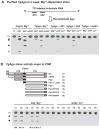
Recombinant TgAgo has weak Mg2+-dependent slicer activity that maps to PIWI. A. Biochemical properties of the TgAgo slicer activity. Standard slicer assay using recombinant His-tagged TgAgo (or human Ago2), T7-transcribed target RNA, continuously labelled with α-32P-UTP, and synthetic antisense (AS) guide RNA was performed in vitro as described in Experimental procedures. The hAgo2 reactions were incubated for 20 min; the no-enzyme control (N) and TgAgo reactions were for 30 min (30 min). Two different AS sequences (AS1, AS2) and the 2 nt centrally mismatched version of AS2 (mAS2) have been described. FL denotes full-length (uncleaved) substrate RNA, and the open and closed circles denote AS1- and AS2-guided cleavage products respectively. Reactions that lacked Mg2+ or contained Ca2+ instead, or had ATP added, are so indicated. B. Preliminary mutational analysis to define TgAgo domains. Various recombinant TgAgo mutants, numbered (1)–(8), refer to the sequence description in Fig. 1, and were employed in cleavage reactions as in panel (A), using AS2 or mAS2 as the guide RNA and the corresponding divalent cation. The intensity of the two product bands were added and calculated as percentage of all bands. This number for wild-type Ago was taken as 100, and other numbers expressed as its percentage; average of three reactions with standard errors are shown. Absence of a number means that no product could be detected. Note that loss of PIWI or single mutations of any of the three residues (see Fig. 1, red with * and #; mutants 6, 7, 8) completely abolished activity.
Fig. 3
Fig. 3
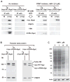
Native T. gondii Ago-containing complex. A. Left panels: The native complex containing TgAgo was immunoprecipitated from transgenic parasites expressing either full length (WT) or RGG domain (the first 65 amino acids)-deleted (ΔRGG) myc-tagged Ago, and the indicated proteins in the complex (including myc-Ago) were detected by Western using specific antibodies as described in Experimental procedures. Note the absence of the three accessory proteins in the ΔRGG Ago precipitates. NI, non-immune mouse serum. Right panels: Essentially identical analysis was performed with the same recombinant parasite strains grown in the presence of 15 μM of the PRMT inhibitor, AMI-1 (sc-222202; Santa Cruz Biotechnology) (Cheng et al., 2004). In these cells, TSN was absent from even the wild-type Ago complex, suggesting that its association was Ago methylation-dependent. B. Western analysis. Total parasite extracts (i.e. solubilized by boiling in SDS-PAGE sample buffer without immunoprecipitation) were analysed by Western blotting to detect the indicated proteins, which shows similar expression of these proteins in wild type and ΔRGG Ago-expressing parasites. C. Confirmation of AMI-1 activity. Tg-infected HFF cells were incubated in the presence of 0, 2, 10, 20 μM AMI-1 and total cell proteins were analysed by SDS-PAGE and Western blotting using a pan dimethyl-Arg antibody mixture. Parallel western for actin shows equal sample loading.
Fig. 4
Fig. 4
Methylation of TgAgo by PRMT1 in vitro. Methylation assay was carried out as described under Experimental procedures. Left: 1 μg of purified His-tagged proteins were examined by SDS-PAGE and Coomassie Blue (R250) staining to check for purity (Ago, TgAgo, full length; PRMT1, TgPRMT1; ΔRGG, TgAgo with RGG domain deleted, as in Fig. 2B). Right: Half of each methylation reaction (10 μl, containing 1 μg TgAgo as substrate) was analysed in SDS-PAGE and fluorography to detect the methylated Ago. One control reaction, in which TgAgo was incubated with radioactive AdoMet but no PRMT1, showed no methylation. Reactions 1, 2 and 3 contained AMI-1 (10 μM), sinefungin (4 μM) and chaetocin (20 μM), respectively (Fan et al., 2009), of which the first two caused significant inhibition.
Fig. 5
Fig. 5
Biochemically distinct dual slicer activities in T. gondii RISC. Tg RISC, containing myc-Ago, was immunoprecipitated from parasite cells using myc antibody as described in Fig. 3, and employed in slicer activity assay programmed with either perfectly matched AS2 or mismatched mAS2 guide RNA and with the indicated divalent cations. Reactions were incubated for 30 or 60 min as shown. In the rightmost panel, the effect of 0.5 mM TSN inhibitor (pdTp) or the control non-inhibitor (dTp) was tested in selected reaction mixtures (a, b, c, d). Note that reactions ‘c’ and ‘d’ were specifically inhibited by pdTp, but not by dTp. N, No-enzyme control shows the substrate RNA only.
Fig. 6
Fig. 6
The two slicer activities of Tg RISC correspond to Ago and TSN. A. Ago activity in TSN-free RISC. Tg RISC, containing myc-tagged TgAgo but free of TSN (Fig. 3A), was isolated by immunoprecipitations from parasites treated with PRMT inhibitor (AMI-1) and used in slicer assay as described for Fig. 5. The guide RNA was either perfectly matched (AS2) or mismatched (mAS2). All reactions were incubated for 60 min. N, No-enzyme control. The divalent cations in various reactions were: Mg2+ (a, a′), Ca2+ (b, b′), Mg2+ plus Ca2+ (c, c′). Two reactions contained TSN inhibitor, pdTp, but were not inhibited (when compared with the corresponding inhibitor-free reactions). Note that all functional reactions that showed activity here showed only weak, Mg2+-dependent, and perfectly matched guide-dependent activity, similar to recombinant TgAgo (e.g. Fig. 2). B. Protein profile of Tg RISC, containing myc-tagged, catalytically defective E884A Ago. Note that the mutant Ago in the RISC is nonetheless methylated and associated with TSN. C. TSN activity in Ago-defective RISC. The Ago-defective RISC from panel B was tested for slicer activity as in panel (A). Labels are the same as in panel (A). Where indicated, reactions were incubated for 30 min or 60 min; all others were for 60 min. Reactions were inhibited by TSN inhibitor (pdTp), but not by the control non-inhibitor (dTp), both at 0.5 mM.
Fig. 7
Fig. 7
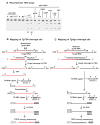
Slicer activity of recombinant TgTSN and mapping of cleavage sites. A. In vitro TSN assay. Bacterially made, purified, His-tagged TgTSN was employed in slicer assay as described for Fig. 6A and B. The activity was weaker than the TSN activity of the RISC purified from in vivo (last lane), and could not be improved by adding 50 ng of recombinant TgAgo or Tg extract (representative data from a range of 10–100 ng tested, with no effect). The R365A mutant TSN lost the slicer activity, but the C366P mutant did not. All reactions were for 60 min except where noted. TSN inhibitor (pdTp) and control non-inhibitor (dTp) were used as in Fig. 6C. B. Mapping of TgTSN cleavage site (details in Experimental procedures). One of the mismatched guide RNAs (described in Results) with 2 nt mismatch (GA) and the relevant portion of target sequence are shown for illustration. The two fragments of the target RNA are in red and black. Synthetic primers of known sequence were used; thus, the last nucleotide at 3′ end of the red sequence (G), before primer 2 starts, denotes the cleavage site. C. Mapping of TgAgo cleavage site. The overall plan is similar to that of TSN, but the ligations are different due to the 3′-OH and 5′-P ends created by Ago. Note that the red fragment can also ligate to Primer 2 but will not ligate to Primer 1, and therefore this fragment will not undergo exponential amplification in the PCR step.
Fig. 8
Fig. 8
Analysis of the role of Ago and PRMT1 in parasitic cell division in both RH and ΔKu80 backgrounds. Tet-regulated Ago and PRMT1 knockout parasites were constructed and used to infect HFF cells as described in Experimental procedures (Mital et al., 2005; Sheiner et al., 2011). (A–C): TATi-RH strain. (A, B): Western blot; lane C, parent parasite; lane H, heterodiploid (or merodiploid), expressing both native and myc-tagged recombinant proteins, to which anhydro-tetracycline (Atc) was added and parasites harvested at 0, 4, 8 and 12 h later. Total protein from approximately 2 × 10⁷ parasites was analysed in Western using polyclonal Ago and PRMT1-reactive antibodies, which shows relatively rapid loss of (A) Ago and (B) PRMT1. Note that recombinant proteins are myc-tagged and their expression is turned off by Atc. (C): Pictures representative of majority of PV for wild type, Ago KO and PRMT1 KO parasites, immunostained (Green) with SAG1 (surface) antibody at 30 h post infection. The parasite counts in representative parasitophorous vacuoles are noted with the variation shown in PRMT strains. In the last panel (D), 20 μM AMI-1, a PRMT inhibitor (Fig. 3), was used on wild-type Tg-infected cells, which also inhibited parasite growth. (E–G): TATi-ΔKu80 strain. (E): schematic of the promoter knockout, recombinant construct. (F, G): Western for Ago and PRMT1, respectively; lane C, Parent TATi-ΔKu80, no recombinant gene; other lanes represent promoter-KO, recombinant-expressing parasite. H. Similar to panel (C) above, immunostained at 24 h post infection.
Fig. 9
Fig. 9
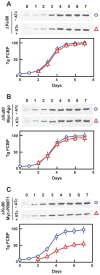
Intracellular growth of Ago KO and PRMT1 KO parasites. Total parasite growth was measured by immunoblot of the infected HFF cell protein using antibody against Tg FCBP protein. (A) Parent TATi-ΔKu80 strain; (B) Atc-regulated Ago1 KO; and (C) Atc-regulated PRMT1 KO. The numbers on top indicate days post infection. The faint FCBP band on ‘day 0’ was contributed by the input parasites. A representative ECL blot for each is shown, and the densitometric scans represent mean from three experiments with standard error bars. In the plots, O, no KO (without ATc); Δ, KO (with ATc).
Fig. 10
Fig. 10
Quantification of plaque forming ability, invasion and egress of Ago KO and PRMT1 KO parasites the TATi-ΔKu80 background. The recombinant genes were shut down by Atc, where mentioned. Plaque assay was done as described in Experimental procedures and (A) representative pictures and (B) number plots are shown. C. Number of PV formed at 36 h p.i. D. Number of unlysed PV remaining after 50 h p.i., representing lack of egress. Note the slow growth of PRMT1 KO by all criteria. B–D. Mean ± standard error (bars), n = 3 experiments, Student’s t-test, P < 0.05 vs. control (*).

Similar articles

Cited by

References

    1. Adams B, Musiyenko A, Kumar R, Barik S. A novel class of dual-family immunophilins. J Biol Chem. 2005;280:24308–24314. - PMC - PubMed
    1. Al Riyahi A, Al-Anouti F, Al-Rayes M, Ananvoranich S. Single argonaute protein from Toxoplasma gondii is involved in the double-stranded RNA induced gene silencing. Int J Parasitol. 2006;36:1003–1014. - PubMed
    1. Barik S. An intronic microRNA silences genes that are functionally antagonistic to its host gene. Nucleic Acids Res. 2008;36:5232–5241. - PMC - PubMed
    1. Baum J, Papenfuss AT, Mair GR, Janse CJ, Vlachou D, Waters AP, et al. Molecular genetics and comparative genomics reveal RNAi is not functional in malaria parasites. Nucleic Acids Res. 2009;37:3788–3798. - PMC - PubMed
    1. Blackwell E, Zhang X, Ceman S. Arginines of the RGG box regulate FMRP association with polyribosomes and mRNA. Hum Mol Genet. 2010;19:1314–1323. - PMC - PubMed

Publication types

MeSH terms