. 2012 Apr;86(8):4139-50.
doi: 10.1128/JVI.06327-11. Epub 2012 Feb 1.

Annexin A2 is involved in the formation of hepatitis C virus replication complex on the lipid raft

Affiliations

Annexin A2 is involved in the formation of hepatitis C virus replication complex on the lipid raft

Vikas Saxena et al. J Virol. 2012 Apr.

Abstract

The hepatitis C virus (HCV) RNA replicates in hepatic cells by forming a replication complex on the lipid raft (detergent-resistant membrane [DRM]). Replication complex formation requires various viral nonstructural (NS) proteins as well as host cellular proteins. In our previous study (C. K. Lai, K. S. Jeng, K. Machida, and M. M. Lai, J. Virol. 82:8838-8848, 2008), we found that a cellular protein, annexin A2 (Anxa2), interacts with NS3/NS4A. Since NS3/NS4A is a membranous protein and Anxa2 is known as a lipid raft-associated scaffold protein, we postulate that Anxa2 helps in the formation of the HCV replication complex on the lipid raft. Further studies showed that Anxa2 was localized at the HCV-induced membranous web and interacted with NS4B, NS5A, and NS5B and colocalized with them in the perinuclear region. The silencing of Anxa2 decreased the formation of membranous web-like structures and viral RNA replication. Subcellular fractionation and bimolecular fluorescence complementation analysis revealed that Anxa2 was partially associated with HCV at the lipid raft enriched with phosphatidylinositol-4-phosphate (PI4P) and caveolin-2. Further, the overexpression of Anxa2 in HCV-nonsusceptible HEK293 cells caused the enrichment of HCV NS proteins in the DRM fraction and increased the colony-forming ability of the HCV replicon. Since Anxa2 is known to induce the formation of the lipid raft microdomain, we propose that Anxa2 recruits HCV NS proteins and enriches them on the lipid raft to form the HCV replication complex.


Figures
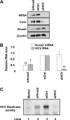
Fig 1
Fig 1
Effect of siRNA-mediated knockdown of Anxa2 on HCV replication. Naïve Huh7.5 (A and B) or HCVrep-HA (C) cells were transfected with the indicated siRNA. siRNA-transfected Huh7.5 cells then were infected with HCV JC1 virus. At 3 days after infection, cells were harvested for immunoblotting and the qRT-PCR determination of RNA. (A) Cell lysates were subjected to 10% SDS-PAGE, followed by Western blotting using anti-NS5A, anti-core, anti-Anxa2, or anti-β-actin antibody. (B) Quantitative RT-PCR of Anxa2 and HCV RNA. The RNA quantity was normalized against GAPDH RNA. Anxa2 and HCV RNA level in cells treated with different siRNA relative to those in siCtrl-treated cells are shown. Representative data (means ± standard deviations) from three independent experiments are shown. *, P < 0.05; **, P < 0.01. (C) In vitro RNA replication of siRNA-transfected HCVrep-HA cells. At 6 days after siRNA transfection, crude replication complex was harvested and analyzed for in vitro replicase activity using [α-32P]UTP as the label as described in Materials and Methods. In vitro-transcribed labeled RNA from the parental replicon pUC-Rep/S1179I served as the RNA size marker (lane 1).
Fig 2
Fig 2
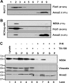
Association of Anxa2 protein with detergent-resistant membrane fractions. (A) Huh7.5 naïve cells and (B) HCVrep-HA replicon cells were harvested at 24 h after seeding. Cell lysates were subjected to 1% Triton X-100 treatment and fractionated by discontinuous OptiPrep gradient centrifugation as described in Materials and Methods. Fractions were analyzed on a 10% SDS-PAGE gel, followed by immunoblotting with monoclonal antibodies against Anxa2, Flot1, and NS5A. Fractions are numbered from 1 to 9 in order from top to bottom (light to heavy). Results of Western blotting were quantified by PhosphorImager counting. The percentage of each protein in the DRM fraction (lanes 1 to 4) relative to the total signal intensity of the protein in all 9 fractions is indicated in parentheses. (C) Protection of HCV NSPs and Anxa2 by membranes against protease digestion. The postnuclear supernatant (PNS) from HCV-EV71I-Luc replicon cells were loaded as such (lane 1) or treated with 0.5% Triton X-100 (lanes 4 and 5 and lanes 8 and 9) or left untreated (lanes 2 and 3 and lanes 6 and 7), and this was followed by no digestion (lane 2 to 5) or treatment with proteinase K (lane 6 to 9). Thus, treated PNSs were fractionated into a 10,000 × g pellet (P) and supernatant (S) and analyzed for the presence of HCV NS5A, vimentin, and Anxa2. The positions of protein size markers are indicated on the left, and the arrow indicates the degraded form of Anxa2.
Fig 3
Fig 3
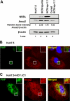
Anxa2 colocalized with the PI4P-rich lipid environment in HCV-infected cells. (A) Huh7.5 naïve, HCVrep-HA, HCVrep-1.1, and cured HCVrep-1.1 cells were harvested at 24 h after seeding, and equal amounts of total protein content were subjected to 10% SDS-PAGE followed by Western blotting with anti-NS5A, anti-Anxa2, and anti-β-actin antibodies. Results of Western blotting were quantified by PhosphorImager counting. Relative band intensities of Anxa2 compared to that of Huh7.5 naïve cells as a ratio of Anxa2 and β-actin are shown. Huh7.5 naïve cells (B) and HCV-JC1-infected cells (C) were costained with an anti-Anxa2 rabbit polyclonal antibody (red) and a PI4P mouse monoclonal antibody (green). Nuclear DNA was stained with DAPI (blue). Images shown were collected sequentially with a confocal laser-scanning microscope and merged to demonstrate colocalization (yellow merge fluorescence). Enlargements of the sections, indicated by the white squares, are shown in the inset panels.
Fig 4
Fig 4
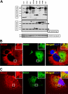
Anxa2 interacts with HCV nonstructural proteins. (A) Huh7.5 cells were transfected with plasmids encoding HA-tagged HCV NS proteins or GST. At 48 h posttransfection, cell lysates were immunoprecipitated (IP) using anti-Anxa2 monoclonal antibodies. The immunoprecipitates and 5% of the total cell lysates were subjected to 10% SDS-PAGE followed by Western blotting with anti-HA and anti-Anxa2 antibodies. (Upper panel) Anti-HA immunoblot showing the input proteins (arrows) (5% total). (Middle panel) Anti-HA immunoblot showing HCV NSP coimmunoprecipitated by anti-Anxa2 antibody. (Lower panel) Anxa2 protein immunoprecipitated by anti-Anxa2 antibody. Protein molecular mass markers are indicated on the left side. Filled arrowheads indicate the IgG heavy (55-kDa) and light (25-kDa) chains. (B and C) Colocalization of NS4B (B) and NS5A (C) with Anxa2 in HCV-JC1-infected Huh7.5 cells. Images shown were collected sequentially with a confocal laser-scanning microscope and merged to demonstrate colocalization. Enlargements of the sections, from different areas of the cells, indicated by the white squares are shown in the inset panels.
Fig 5
Fig 5
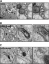
Immunoelectron microscopy of NS5A and Anxa2 on the membranous web. HCV JC1-infected Huh7.5 cells were labeled with antibody against NS5A alone (A) or with both NS5A and Anxa2 (B and C). NS5A was tethered to 12-nm and Anxa2 to 6-nm gold particles. Shown are consecutively enlarged views of the membranous web. Arrows show NS5A, and arrowheads show Anxa2 in electron-dense granules on the membrane of DMV. N, nucleus. DMV, double-membrane vesicles. MMV, multiple-membrane vesicles.
Fig 6
Fig 6
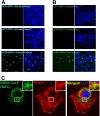
BiFC showed that Anxa2 interacts with NS5A at the Cav2-containing lipid raft. HEK293 cells were cotransfected with cCFP-fused GST or NS5A with nCerulean-Anxa2 (A) or Cav2-nVenus (B). BiFC constructs were fixed and stained with DAPI and visualized by confocal microscopy at 48 h posttransfection. (C) Huh7.5 cells cotransfected with cCFP-NS5A and Cav2-nVenus BiFC constructs were fixed and immunostained with anti-Anxa2 (red) monoclonal antibody. Cellular DNA was labeled with DAPI (blue). Enlarged views of sections of images are shown (inset).
Fig 7
Fig 7
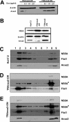
Anxa2 promotes the enrichment of HCV NS5A in DRM fraction. (A) TRAnxa2-OFF and TRAnxa2-ON cells were cultured in the presence of the indicated amounts of doxycycline (Dox). After 48 h, cell lysates were resolved by SDS-PAGE and then immunoblotted with a monoclonal antibody against Anxa2. Immunoblotting against β-actin serves as a loading control. (B) Huh7.5 cells, TRAnxa2-OFF, and TRAnxa2-ON cells were transfected with a plasmid encoding HA-tagged NS5A protein (NS5A-HA). At 48 h posttransfection, cells were analyzed for the expression of NS5A and Anxa2. Huh7.5 (C), TRAnxa2-OFF (D), and TRAnxa2-ON (E) cells were transfected with NS5A-HA and subjected to 1% Triton X-100 treatment and fractionated by discontinuous OptiPrep gradient centrifugation as described in Materials and Methods. Fractions were separated on a 10% SDS-PAGE gel, followed by immunoblotting with monoclonal antibodies against HA tag (depicting NS5A), Flot1, and Anxa2. Fractions are numbered from 1 to 9 in order from top to bottom (light to heavy). NS5A* represents a longer exposure of the same film shown for NS5A.
Fig 8
Fig 8
Silencing of Anxa2 inhibits membranous web formation. Huh7.5 cells were treated with control siRNAs for 2 days and then infected with HCV JC1. Cells were fixed and processed for electron microscopy at the third day postinfection. The consecutive enlargement of the boxed areas are shown from left to right. Note the very heterogeneous membranous web (MW) in control cells. Scale bars are given in the lower left of each panel. N, nucleus; DMV, double-membrane vesicles; MMV, multiple membrane vesicles; ER, endoplasmic reticulum; m, mitochondria.
Fig 9
Fig 9
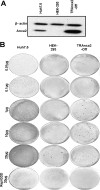
Colony formation of HCV 1b subgenomic RNA replicon in Huh7.5, HEK293, and TRAnxa2-OFF cell lines. (A) The cell lines were analyzed by immunoblotting for the expression of Anxa2. (B) In vitro-transcribed wild-type or GDD mutant (Rep/GDD) subgenomic replicon RNAs in the indicated amounts were electroporated into each cell line, and cells were cultured with G418 for 12 to 15 days before staining with crystal violet.
Fig 10
Fig 10
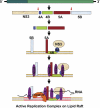
Hypothetical model for the mechanism of formation of a HCV replication complex on a lipid raft. HCV polyprotein is stepwise processed into intermediate and individual viral proteins. After NS3 and NS5B are proteolytically cleaved from the polyprotein, the uncleaved NS4AB-5A is recruited to the membrane by the intrinsic membrane-anchoring character of NS4B (15). NS3 becomes associated with this membrane by binding to its cofactor, NS4A. NS5B, being a membranous protein, also localizes to the membrane. Anxa2 can interact with NS3/NS4A, NS4B, NS5A, and NS5B. Membrane binding of HCV protein induces calcium ion flux (19), and Anxa2, in response to this flux, interacts with signal phospholipid, which initiates raft clustering. Thus, Anxa2 creates a lipid raft around these membrane-anchored proteins. NS5B, through its interaction with hVAP-33 (15), is brought into this complex, and, in this manner, the HCV RC is established. How HCV RNA enters into this complex is not yet known.

Similar articles

Cited by

References

    1. Aizaki H, Choi KS, Liu M, Li YJ, Lai MM. 2006. Polypyrimidine-tract-binding protein is a component of the HCV RNA replication complex and necessary for RNA synthesis. J. Biomed. Sci. 13:469–480 - PubMed
    1. Aizaki H, Lee KJ, Sung VM, Ishiko H, Lai MM. 2004. Characterization of the hepatitis C virus RNA replication complex associated with lipid rafts. Virology 324:450–461 - PubMed
    1. Alter MJ. 2007. Epidemiology of hepatitis C virus infection. World J. Gastroenterol. 13:2436–2441 - PMC - PubMed
    1. Ayala-Sanmartin J, Henry JP, Pradel LA. 2001. Cholesterol regulates membrane binding and aggregation by annexin 2 at submicromolar Ca(2+) concentration. Biochim. Biophys. Acta 1510:18–28 - PubMed
    1. Babiychuk EB, Draeger A. 2000. Annexins in cell membrane dynamics. Ca(2+)-regulated association of lipid microdomains. J. Cell Biol. 150:1113–1124 - PMC - PubMed

LinkOut - more resources