Review
. 2012:726:441-65.
doi: 10.1007/978-1-4614-0980-9_20.

Assembly and architecture of HIV

Affiliations
Review

Assembly and architecture of HIV

Barbie K Ganser-Pornillos et al. Adv Exp Med Biol. 2012.

Abstract

HIV forms spherical, membrane-enveloped, pleomorphic virions, 1,000-1,500 Å in diameter, which contain two copies of its single-stranded, positive-sense RNA genome. Virus particles initially bud from host cells in a noninfectious or immature form, in which the genome is further encapsulated inside a spherical protein shell composed of around 2,500 copies of the virally encoded Gag polyprotein. The Gag molecules are radially arranged, adherent to the inner leaflet of the viral membrane, and closely associated as a hexagonal, paracrystalline lattice. Gag comprises three major structural domains called MA, CA, and NC. For immature virions to become infectious, they must undergo a maturation process that is initiated by proteolytic processing of Gag by the viral protease. The new Gag-derived proteins undergo dramatic rearrangements to form the mature virus. The mature MA protein forms a "matrix" layer and remains attached to the viral envelope, NC condenses with the genome, and approximately 1,500 copies of CA assemble into a new cone-shaped protein shell, called the mature capsid, which surrounds the genomic ribonucleoprotein complex. The HIV capsid conforms to the mathematical principles of a fullerene shell, in which the CA subunits form about 250 CA hexamers arrayed on a variably curved hexagonal lattice, which is closed by incorporation of exactly 12 pentamers, seven pentamers at the wide end and five at the narrow end of the cone. This chapter describes our current understanding of HIV's virion architecture and its dynamic transformations: the process of virion assembly as orchestrated by Gag, the architecture of the immature virion, the virus maturation process, and the structure of the mature capsid.


Figures

Fig. 1
Fig. 1
Schematic of the HIV-1 replication cycle, emphasizing the stages discussed in this chapter. See text for details. (Adapted from Ganser-Pornillos et al. (2008), with permission from Elsevier.)
Fig. 2
Fig. 2
Organization of the immature and mature HIV-1 virions. (a) Schematic tertiary structural model of full-length HIV-1 Gag. Individual domains are in different colors and are labeled on the left. This color scheme is maintained throughout the chapter. (b) Schematic model of the immature virion. (c) Schematic model of the mature virion. Images of (d) immature and (e) mature virions preserved in vitreous ice. (Reprinted from Ganser-Pornillos et al. (2008), with permission from Elsevier.)
Fig. 3
Fig. 3
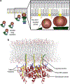
The MA domain functions in binding and targeting of Gag to the plasma membrane. (a) Schematic showing soluble Gag proteins in a “folded-over” conformation, and membrane-bound assembling Gag molecules in a “beads-on-a-string” configuration. The boxed region illustrates the “myristyl switch” mechanism of membrane binding. (b) Structural model of the MA trimer bound to the lipid bilayer. [(b) Reprinted from Ganser-Pornillos et al. (2008), with permission from Elsevier.]
Fig. 4
Fig. 4
The immature Gag lattice. (a) Side view and (b) top view of a low-resolution model of two interacting Gag hexamers (Wright et al. 2007). The CANTD, CACTD, and SP1 layers are colored green, cyan, and gray, respectively. (Model used to generate images courtesy of Elizabeth Wright.) (c) Global map of the immature HIV-1 lattice (Briggs et al. 2009). The Gag hexamers are represented by hexagons and colored according to symmetry cross-correlation on a scale from low (red) to high (green). (Lattice map images courtesy of John Briggs.)
Fig. 5
Fig. 5
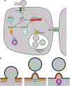
Retrovirus budding. (a) Schematic representation of the cellular ESCRT pathway. Known interactions between ESCRT complexes and ESCRT-associated proteins are indicated by the black double-headed arrows. Red arrows connect viral late domains or native cellular recruitment factors with their corresponding complex. Recruitment factors (viral Gag protein, ESCRT-0, and CEP55) are colored in green shades. Although it is known that recruitment of NEDD4L to viral budding sites leads to the eventual recruitment of ESCRT-III, the precise mechanism by which this occurs is not known. (b) Schematic representation of the membrane fission mechanism catalyzed by ESCRT-III proteins and VPS4. (See text for details.)
Fig. 6
Fig. 6
Proteolytic processing of Gag during maturation. (a) Schematic showing the HIV-1 Gag proteolysis cascade. Cleavage events that generate the mature CA termini are indicated by the red arrows. (b) Secondary and tertiary structural changes at the N-terminal and C-terminal ends of the CA domain. The depicted conformations of the MA/CA and CA/SP1 junctions in Gag are based on a variety of structural, biochemical, and mutagenesis studies. In mature CA, the Nterminal 13 residues are folded into a β-hairpin (colored yellow), and the C-terminal 11 residues are disordered. (Structure images reprinted from Ganser-Pornillos et al. (2008), with permission from Elsevier.)
Fig. 7
Fig. 7
Morphologies of representative mature capsids of different orthoretroviruses. Images and fullerene models of Moloney murine leukemia virus (Mo-MLV, a gammaretrovirus), Mason-Pfizer monkey virus (MPMV, a betaretrovirus), and HIV-1 (a lentivirus) are shown. In all cases, the capsids are organized as fullerene structures that incorporate 12 pentamers (red) to close the hexagonal lattice. (Virus images reproduced from Ganser-Pornillos et al. (2004), with permission from American Society for Microbiology.)
Fig. 8
Fig. 8
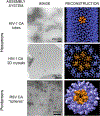
Gallery of in vitro assembly systems for retroviral capsids: helical tubes, two-dimensional hexagonal crystals, and icosahedral particles. (Images of HIV-1 assemblies reprinted from (Ganser-Pornillos et al. 2008), with permission from Elsevier. Image of RSV particles courtesy of Rebecca Craven. Reconstructed images of HIV-1 tubes reprinted from (Li et al. 2000), with permission from Macmillan Publishers Ltd. (Map used to generate T=1 image courtesy of Alasdair Steven.)
Fig. 9
Fig. 9
Organization of the mature HIV-1 capsid. (a) Structural model of the complete capsid, with the CANTD hexamers in green, CANTD pentamers in light green, and CACTD dimers in cyan. (b) Two views of the X-ray structure of the HIV-1 CA hexamer (Pornillos et al. 2009). (c) Two views of the X-ray structure of the HIV-1 CA pentamer (Pornillos et al. 2011).
Fig. 10
Fig. 10
Quasi-equivalence in the pentameric and hexameric CANTD rings. Top views of the (a) hexameric and (b) pentameric CANTD rings, with each subunit in a different color. Subunits in the pentamer and hexamer are shown in darker and lighter shades, respectively. The angles subtended by adjacent domains are shown explicitly for the blue and orange subunits. One of the repeating three-helix units is outlined in black. (c) Close-up view of the pentameric and hexameric repeat units, superimposed on helices 1 and 3 of the blue subunit. The aliphatic residues that form a small hydrophobic core are shown explicitly and labeled. (d) The “rotation” between adjacent subunits, in going from the hexamer to the pentamer. The approximate position of the rotation axis is indicated by the red dot. Note that this axis is parallel to neither the pentameric nor hexameric symmetry axes. (Reprinted from Pornillos et al. (2011), with permission from Macmillan Publishers Ltd.)
Fig. 11
Fig. 11
Proposed mechanisms for generating curvature in the mature retroviral capsid. (a) Structural model of the capsid, showing a line of connected rings. The CANTD hexamers are colored in green, and CACTD dimers are in cyan. One pentamer is shown on the left, with the CANTD colored light green. The two regions that are proposed to contribute to curvature generation are indicated by the black box (NTD–CTD interface) and the blue box (CTD–CTD dimer interface). (b) Flexion at the intermolecular NTD–CTD interface occurs about molecular pivots composed of helix-capping hydrogen bonds. Shown are 17 independent X-ray structures of the NTD–CTD interface, superimposed on the CANTD domain. Rigid-body movement of CACTD relative to CANTD is indicated by the red double-headed arrow. Hydrogen-bonded side chains are shown explicitly. Hydrogen bonds are indicated as yellow lines. (c) Superposition of the X-ray structure (colored light blue) (Worthylake et al. 1999) and NMR structure (colored cyan) (Byeon et al. 2009) of the isolated full-affinity CACTD dimer. The structures show distinct configurations of the dimer. The red double-headed arrows indicate putative slippage or rotation about the dimer symmetry axis (red oval).

Similar articles

Cited by

References

    1. Accola MA, Höglund S, Göttlinger HG (1998) A putative a-helical structure which overlaps the capsid-p2 boundary in the human immunodeficiency virus type 1 Gag precursor is crucial for viral particle assembly. J Virol 72:2072–2078 - PMC - PubMed
    1. Alcaraz LA, del Álamo M, Barrera FN et al. (2007) Flexibility in HIV-1 assembly subunits: solution structure of the monomeric C-terminal domain of the capsid protein. Biophys J 93:1264–1276 - PMC - PubMed
    1. Aloia RC, Tian H, Jensen FC (1993) Lipid composition and fluidity of the human immunodeficiency virus envelope and host cell plasma membranes. Proc Natl Acad Sci USA 90:5181–5185 - PMC - PubMed
    1. Bailey GD, Hyun JK, Mitra AK et al. (2009) Proton-linked dimerization of a retroviral capsid protein initiates capsid assembly. Structure 17:737–748 - PubMed
    1. Bajorek M, Schubert HL, McCullough J et al. (2009) Structural basis for ESCRT-III protein autoinhibition. Nat Struct Mol Biol 16:754–762 - PMC - PubMed

Publication types

LinkOut - more resources