

Evaluation of antigen specific recognition and cell mediated cytotoxicity by a modified lysispot assay in a rat colon carcinoma model
- PMID: 22296726
- PMCID: PMC3395825
- DOI: 10.1186/1756-9966-31-9
Evaluation of antigen specific recognition and cell mediated cytotoxicity by a modified lysispot assay in a rat colon carcinoma model
Abstract
Background: Antigen-specific CD8+ cytotoxic T lymphocytes represent potent effector cells of the adaptive immune response against viruses as well as tumours. Therefore assays capable at exploring the generation and function of cytotoxic T lymphocytes represent an important objective for both clinical and experimental settings.
Methods: Here we show a simple and reproducible assay for the evaluation of antigen-specific CD8+ cytotoxic T lymphocytes based on a LysiSpot technique for the simultaneous determination of antigen-specific IFN-γ production and assessment of tumor cytolysis. The assay was developed within an experimental model of colorectal carcinoma, induced by the colorectal tumor cell line DHD-K12 that induces tumors in BDIX rats and, in turn, elicits a tumor- specific immune response.
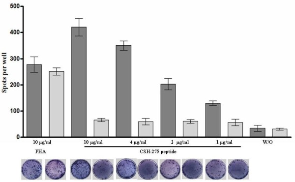
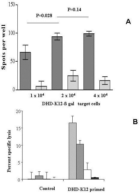
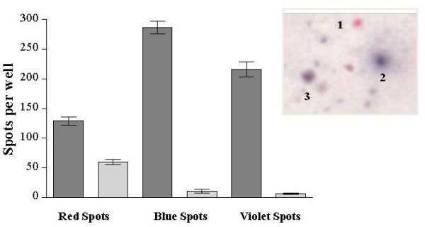
Results: Using DHD-K12 cells transfected to express Escherichia coli β-galactosidase as target cells, and by the fine setting of spot colours detection, we have developed an in vitro assay that allows the recognition of cytotoxic T lymphocytes induced in BDIX rats as well as the assessment of anti-tumour cytotoxicity. The method highlighted that in the present experimental model the tumour antigen-specific immune response was bound to killing target cells in the proportion of 55%, while 45% of activated cells were not cytotoxic but released IFN-γ. Moreover in this model by an ELISPOT assay we demonstrated the specific recognition of a nonapeptide epitope called CSH-275 constitutionally express in DHD-K12 cells.
Conclusions: The assay proved to be highly sensitive and specific, detecting even low frequencies of cytotoxic/activated cells and providing the evaluation of cytokine-expressing T cells as well as the extent of cytotoxicity against the target cells as independent functions. This assay may represent an important tool to be adopted in experimental settings including the development of vaccines or immune therapeutic strategies.
Figures
Similar articles
-
Measuring the frequency of mouse and human cytotoxic T cells by the Lysispot assay: independent regulation of cytokine secretion and short-term killing.Nat Med. 2003 Feb;9(2):231-5. doi: 10.1038/nm821. Epub 2003 Jan 21. Nat Med. 2003. PMID: 12539041
-
Host-oriented peptide evaluation using whole blood assay for generating antigen-specific cytotoxic T lymphocytes.Anticancer Res. 2004 Mar-Apr;24(2C):1193-200. Anticancer Res. 2004. PMID: 15154646
-
HIV-specific cytotoxic cell frequencies measured directly ex vivo by the Lysispot assay can be higher or lower than the frequencies of IFN-gamma-secreting cells: anti-HIV cytotoxicity is not generally impaired relative to other chronic virus responses.J Immunol. 2006 Feb 15;176(4):2662-8. doi: 10.4049/jimmunol.176.4.2662. J Immunol. 2006. PMID: 16456029
-
CTL ELISPOT assay.Methods Mol Biol. 2014;1186:75-86. doi: 10.1007/978-1-4939-1158-5_6. Methods Mol Biol. 2014. PMID: 25149304
-
Vaccination with a synthetic nonapeptide expressed in human tumors prevents colorectal cancer liver metastases in syngeneic rats.Int J Cancer. 2004 May 20;110(1):70-5. doi: 10.1002/ijc.20063. Int J Cancer. 2004. PMID: 15054870
Cited by
-
Ubiquitinated proteins enriched from tumor cells by a ubiquitin binding protein Vx3(A7) as a potent cancer vaccine.J Exp Clin Cancer Res. 2015 Apr 16;34(1):34. doi: 10.1186/s13046-015-0156-3. J Exp Clin Cancer Res. 2015. PMID: 25886865 Free PMC article.
-
Association between IL-4 -589C>T polymorphism and colorectal cancer risk.Tumour Biol. 2014 Mar;35(3):2675-9. doi: 10.1007/s13277-013-1352-4. Epub 2013 Nov 12. Tumour Biol. 2014. PMID: 24218339
-
Evaluation of cytotoxic T lymphocyte-mediated anticancer response against tumor interstitium-simulating physical barriers.Sci Rep. 2020 Aug 12;10(1):13662. doi: 10.1038/s41598-020-70694-8. Sci Rep. 2020. PMID: 32788651 Free PMC article.
-
Potential recombinant vaccine against influenza A virus based on M2e displayed on nodaviral capsid nanoparticles.Int J Nanomedicine. 2015 Apr 2;10:2751-63. doi: 10.2147/IJN.S77405. eCollection 2015. Int J Nanomedicine. 2015. PMID: 25897220 Free PMC article.
-
Association between IRS-1 Gly972Arg polymorphism and colorectal cancer risk.Tumour Biol. 2014 Jul;35(7):6581-5. doi: 10.1007/s13277-014-1900-6. Epub 2014 Apr 3. Tumour Biol. 2014. PMID: 24696264
References
-
- Kochenderfer JN, Gress RE. A comparison and critical analysis of preclinical anticancer vaccination strategies. Exp Biol Med. 2007;232:1130–1141. - PubMed
-
- Komatsu N, Matsueda S, Tashiro K, Ioji T, Shichijo S, Noguchi M, Yamada A, Doi A, Suekane S, Moriya F, Matsuoka K, Kuhara S, Itoh K, Sasada T. Gene expression profiles in peripheral blood as a biomarker in cancer patients receiving peptide vaccination. Cancer. in press . - PubMed
-
- U'Ren L, Kedl R, Dow S. Vaccination with liposome-DNA complexes elicits enhanced antitumor immunity. Cancer Gene Ther. 2006;13:1033–1044. - PubMed
-
- Darzynkiewicz Z, Bedner E, Smolewski P, Lee BW, Johnson GL. Detection of caspases activation in situ by fluorochrome-labeled inhibitors of caspases (FLICA) Methods Mol Biol. 2002;203:289–299. - PubMed
Publication types
MeSH terms
Substances
LinkOut - more resources
Full Text Sources
Medical
Research Materials
