Biomimetic poly(ethylene glycol)-based hydrogels as scaffolds for inducing endothelial adhesion and capillary-like network formation
- PMID: 22296572
- PMCID: PMC3310151
- DOI: 10.1021/bm201596w
Biomimetic poly(ethylene glycol)-based hydrogels as scaffolds for inducing endothelial adhesion and capillary-like network formation
Abstract
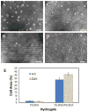
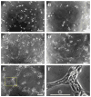
The extracellular matrix (ECM) is an attractive model for designing synthetic scaffolds with a desirable environment for tissue engineering. Here, we report on the synthesis of ECM-mimetic poly(ethylene glycol) (PEG) hydrogels for inducing endothelial cell (EC) adhesion and capillary-like network formation. A collagen type I-derived peptide GPQGIAGQ (GIA)-containing PEGDA (GIA-PEGDA) was synthesized with the collagenase-sensitive GIA sequence attached in the middle of the PEGDA chain, which was then copolymerized with RGD capped-PEG monoacrylate (RGD-PEGMA) to form biomimetic hydrogels. The hydrogels degraded in vitro with the rate dependent on the concentration of collagenase and also supported the adhesion of human umbilical vein ECs (HUVECs). Biomimetic RGD/GIA-PEGDA hydrogels with incorporation of 1% RGD-PEGDA into GIA-PEGDA hydrogels induced capillary-like organization when HUVECs were seeded on the hydrogel surface, while RGD/PEGDA and GIA-PEGDA hydrogels did not. These results indicate that both cell adhesion and biodegradability of scaffolds play important roles in the formation of capillary-like networks.
Figures


Similar articles
-
Design and synthesis of biomimetic hydrogel scaffolds with controlled organization of cyclic RGD peptides.Bioconjug Chem. 2009 Feb;20(2):333-9. doi: 10.1021/bc800441v. Bioconjug Chem. 2009. PMID: 19191566 Free PMC article.
-
Biomimetic-engineered poly (ethylene glycol) hydrogel for smooth muscle cell migration.Tissue Eng Part A. 2014 Feb;20(3-4):864-73. doi: 10.1089/ten.TEA.2013.0050. Epub 2014 Jan 9. Tissue Eng Part A. 2014. PMID: 24093717 Free PMC article.
-
Integrating valve-inspired design features into poly(ethylene glycol) hydrogel scaffolds for heart valve tissue engineering.Acta Biomater. 2015 Mar;14:11-21. doi: 10.1016/j.actbio.2014.11.042. Epub 2014 Nov 26. Acta Biomater. 2015. PMID: 25433168 Free PMC article.
-
Bioactive modification of poly(ethylene glycol) hydrogels for tissue engineering.Biomaterials. 2010 Jun;31(17):4639-56. doi: 10.1016/j.biomaterials.2010.02.044. Epub 2010 Mar 19. Biomaterials. 2010. PMID: 20303169 Free PMC article. Review.
-
Design properties of hydrogel tissue-engineering scaffolds.Expert Rev Med Devices. 2011 Sep;8(5):607-26. doi: 10.1586/erd.11.27. Expert Rev Med Devices. 2011. PMID: 22026626 Free PMC article. Review.
Cited by
-
Improved Human Bone Marrow Mesenchymal Stem Cell Osteogenesis in 3D Bioprinted Tissue Scaffolds with Low Intensity Pulsed Ultrasound Stimulation.Sci Rep. 2016 Sep 6;6:32876. doi: 10.1038/srep32876. Sci Rep. 2016. PMID: 27597635 Free PMC article.
-
Customizable biomaterials as tools for advanced anti-angiogenic drug discovery.Biomaterials. 2018 Oct;181:53-66. doi: 10.1016/j.biomaterials.2018.07.041. Epub 2018 Jul 26. Biomaterials. 2018. PMID: 30077137 Free PMC article. Review.
-
Advances in hydrogel-based vascularized tissues for tissue repair and drug screening.Bioact Mater. 2021 Jul 10;9:198-220. doi: 10.1016/j.bioactmat.2021.07.005. eCollection 2022 Mar. Bioact Mater. 2021. PMID: 34820566 Free PMC article. Review.
-
A genetically modified protein-based hydrogel for 3D culture of AD293 cells.PLoS One. 2014 Sep 18;9(9):e107949. doi: 10.1371/journal.pone.0107949. eCollection 2014. PLoS One. 2014. PMID: 25233088 Free PMC article.
-
Accelerated and scarless wound repair by a multicomponent hydrogel through simultaneous activation of multiple pathways.Drug Deliv Transl Res. 2019 Dec;9(6):1143-1158. doi: 10.1007/s13346-019-00660-z. Drug Deliv Transl Res. 2019. PMID: 31317345
References
-
- Berthiaume F, Maguire TJ, Yarmush ML. Annu Rev Chem Biomol Eng. 2011;2:403–430. - PubMed
-
- Khademhosseini A, Vacanti JP, Langer R. Sci Amer. 2009;300:64–71. - PubMed
-
- Griffith LG, Naughton G. Science. 2002;295:1009–1014. - PubMed
-
- Langer R, Vacanti JP. Science. 1993;260:920–926. - PubMed
-
- Niklason LE, Langer R. J Am Med Assoc. 2001;285:573–576. - PubMed
Publication types
MeSH terms
Substances
Grants and funding
LinkOut - more resources
Full Text Sources

