. 2012 Mar 16;287(12):9112-27.
doi: 10.1074/jbc.M111.248906. Epub 2012 Jan 25.

Cytosolic Bax: does it require binding proteins to keep its pro-apoptotic activity in check?

Affiliations

Cytosolic Bax: does it require binding proteins to keep its pro-apoptotic activity in check?

Sandra Vogel et al. J Biol Chem. .

Abstract

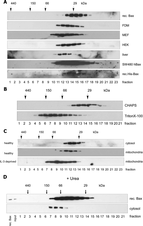
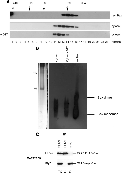
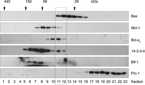
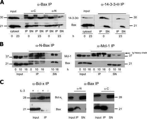
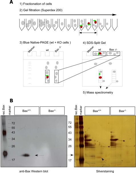
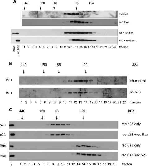
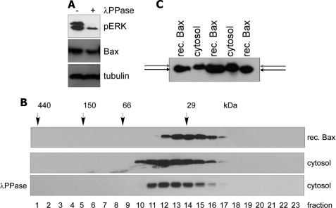
Bax is kept inactive in the cytosol by refolding its C-terminal transmembrane domain into the hydrophobic binding pocket. Although energetic calculations predicted this conformation to be stable, numerous Bax binding proteins were reported and suggested to further stabilize inactive Bax. Unfortunately, most of them have not been validated in a physiological context on the endogenous level. Here we use gel filtration analysis of the cytosol of primary and established cells to show that endogenous, inactive Bax runs 20-30 kDa higher than recombinant Bax, suggesting Bax dimerization or the binding of a small protein. Dimerization was excluded by a lack of interaction of differentially tagged Bax proteins and by comparing the sizes of dimerized recombinant Bax with cytosolic Bax on blue native gels. Surprisingly, when analyzing cytosolic Bax complexes by high sensitivity mass spectrometry after anti-Bax immunoprecipitation or consecutive purification by gel filtration and blue native gel electrophoresis, we detected only one protein, called p23 hsp90 co-chaperone, which consistently and specifically co-purified with Bax. However, this protein could not be validated as a crucial inhibitory Bax binding partner as its over- or underexpression did not show any apoptosis defects. By contrast, cytosolic Bax exhibits a slight molecular mass shift on SDS-PAGE as compared with recombinant Bax, which suggests a posttranslational modification and/or a structural difference between the two proteins. We propose that in most healthy cells, cytosolic endogenous Bax is a monomeric protein that does not necessarily need a binding partner to keep its pro-apoptotic activity in check.


Figures

FIGURE 1.
FIGURE 1.
On gel filtration chromatography endogenous Bax elutes in bigger complexes than its monomeric state. Shown is anti-Bax Western blot analysis of a Superdex 200 HR 10/30 gel filtration chromatography of recombinant (rec.) Bax or His-Bax or endogenous Bax from the cytosols of FDM, MEF, and HEK cells from the liver tissue as well as from SW480 colon carcinoma cells overexpressing human Bax (A), CHAPS- or Triton X-100-extracted total lysates of FDM cells (B), cytosolic- and CHAPS-extracted mitochondrial fractions of healthy or IL-3-deprived (16 h) FDM cells (C), or recombinant Bax and cytosol from FDM cells treated with 8 m urea buffer (D). 250 μl of 800 ng of recombinant Bax or 1 mg of subcellular fractions were loaded onto the column, and 500-μl fractions were collected. Protein standard markers (29, 66, 150, and 440 kDa) were run in parallel.
FIGURE 2.
FIGURE 2.
Bax does not form disulfide-linked or other dimers in vitro or inside cells. Shown are anti-Bax Western blot analysis of gel filtration chromatography of recombinant Bax and endogenous Bax in the absence or presence of 50 mm DTT (A) and first dimension of a BN-PAGE comparing the migration of 30 ng of recombinant (rec.) Bax after freeze/thawing to endogenous native Bax complexes from 100 μg FDM cytosol in the absence or presence of 50 mm DTT (B). Coomassie-stained molecular mass markers are shown on the left (140 and 60 kDa). Note that freeze/thawing of recombinant Bax partially generates spontaneous Bax dimers that are different in size from endogenous Bax complexes. In addition, DTT does not change the elution/migration profile of endogenous Bax on gel filtration or BN-PAGE. C, shown is and anti-FLAG or anti-myc Western blot analysis of anti-FLAG or anti-myc immunoprecipitates from the cytosol (C) of Bax/Bak DKO MEFs stably co-expressing FLAG- and myc-tagged human Bax. Note that although each anti-tag IP is very efficient, it does not contain the other tagged Bax species, indicating that FLAG- and myc-Bax do not dimerize. However, as expected, dimerization is observed in the presence of Triton (TX).
FIGURE 3.
FIGURE 3.
Reported potential Bax binding partners elute differently from gel filtration chromatography than cytosolic Bax complexes from FDM cells. Anti-Bax, anti-Mcl-1, anti-Bcl-xL, anti-14-3-3-θ, anti-Bif-1 and anti-Pin-1 Western blot analysis of gel filtration chromatography of the cytosol of FDM cells is shown. 250 μl of 1 mg cytosol in IBC-buffer was loaded onto the column and separated into 500-μl fractions. Note that whereas the elution profile of Bcl-xL, Mcl-1, and 14-3-3-θ partially overlaps with that of Bax, no such overlap is seen with Bif-1 and Pin-1 (rectangle).
FIGURE 4.
FIGURE 4.
Endogenous Bax does not co-IP with 14-3-3, Mcl-1, or Bcl-xL. 100 μg of cytosolic proteins of healthy and IL-3-deprived FDM (A) or FDC-P1 (B and C) cells were immunoprecipitated (IP) with 5 μl of the indicated antibody (Bax, N-Bax, 14-3-3, Mcl-1, Bcl-x). The IPs and resulting supernatants after IP (SN) were loaded on a 15% SDS gel and immunoblotted with anti-Bax (NT) or the indicated antibody. A, anti-N-Bax, anti-C-Bax, or anti-14-3-3 IPs are followed by anti-14-3-3 (upper row) or anti-Bax (lower row) Western blots. B, anti-N-Bax or anti-Mcl-1 IPs are followed by anti-Mcl-1 (upper row) or anti-Bax (lower row) Western blots. C, anti-Bcl-x or anti-N-Bax or anti-C-Bax IPs are followed by Bcl-x (upper row) or anti-Bax (lower row) Western blots. h, hours; Input, cytosol before IP; α-C, antibody against the C terminus of Bax (amino acids 184–192, supplemental Fig. S3); α-N, antibody against the N terminus of Bax (amino acids 2–14, supplemental Fig. S3). In B, the Ig heavy chains run just above Mcl-1. The molecular masses of the respective proteins are indicated.
FIGURE 5.
FIGURE 5.
Gel filtration/blue native/SDS-PAGE approach. A, shown is a scheme of the GF-BN/SDS-PAGE approach for mass spectrometry analysis. To ensure specificity of our purification approach, we ran WT and Bax KO samples in parallel. First, we separated cytosolic Bax complexes by Superdex 200 gel filtration chromatography (1, and 2). The anti-Bax-positive (and corresponding Bax−/−) fractions were collected, concentrated, and loaded on a first-dimensional BN polyacrylamide electrophoresis gel (3). After electrophoresis, gel pieces of both WT and Bax−/− samples corresponding to protein complexes up to 35 kDa were cut, separately overlaid on the same two-dimensional SDS gel (split gel), and run side by side by SDS-PAGE (4) followed by either anti-Bax Western blotting to localize the Bax protein spot (red) or silver staining. By this method putative Bax binding partners (green) run in the same lane as Bax (red) and can be identified by mass spectrometry after cutting this lane between 15 and 35 kDa into similarly sized rectangular pieces and digesting them with trypsin (5). B, anti-Bax Western blot (left) and silver staining (right) of the two-dimensional SDS gel containing Bax+/+ and Bax−/− samples side by side are shown. As a reference, recombinant (rec.) Bax and molecular weight markers were run in the first two lanes of the gel. Arrows indicate the Bax protein.
FIGURE 6.
FIGURE 6.
p23 hsp90 co-chaperone partially co-migrates with Bax on gel filtration but does not affect Bax localization and Bax-mediated apoptosis when over- or underexpressed. A, shown is an anti-p23 Western blot analysis of 500-μl fractions of Superdex 200 HR 10/30 gel filtration AEKTA chromatography of 1 mg of cytosol from FDM, MEF, and HEK 293 cells as well as mouse liver. On the top an anti-Bax Western blot of the same fractions of the FDM cytosol is shown. Protein standard markers (29, 66, 150, and 440 kDa) were run in parallel. Note that in FDM and MEF, but not HEK 293 or mouse liver, the elution profile of p23 overlaps in three fractions with that of Bax (rectangle). B, WT and Bak KO MEF were transfected with a p23-V5 (p23) plasmid or an empty vector (c) together with a green fluorescent protein plasmid (GFP) as a control for transfection efficiency. After 24 h, a total cell lysate prepared in IBc + 1% CHAPS was subjected to anti-V5 (detecting overexpressed p23-V5), anti-p23 (detecting both endogenous p23 and overexpressed p23-V5), and anti-Bax Western blot analysis. Anti-actin was used as a loading control. nn, non-transfected control. C, CHERRY-annexin-V FACS analysis of vector control and p23-V5 overexpressing WT and Bak−/− MEFs were treated at 24 h posttransfection with 100 μm etoposide or 1200 J/m2 UV for 0–36 h. Note that p23-V5 overexpression neither inhibited apoptosis in WT MEF, nor further blocked apoptosis in Bak−/− MEF. D, WT and Bak KO MEFs were infected with a lentivirus containing either p23 shRNA or control shRNA and selected for stable shRNA expression with puromycin. A total cell lysate prepared in IBc + 1% CHAPS was subjected to anti-p23, anti-Bak, and anti-Bax Western blot analysis. Anti-ATPase was used as a loading control. Note that the expression of p23 is greatly diminished in the p23 shRNA knockdown cells. E, GFP-annexin-V FACS analysis of untransfected (WT), control shRNA (sh ctrl), and p23 shRNA-transfected WT and Bak−/− MEFs treated with 100 μm etoposide or 1200 J/m2 UV for 0–36 h. Note that p23-V5 underexpression neither accelerated apoptosis in WT nor in Bak−/− MEF. F, anti-p23, anti-Bax, and anti-actin (loading control) Western blot analysis of total extracts of p23+/+ and p23−/− MEFs (left panel) is shown. GFP-annexin-V FACS analysis of the same cells treated with 100 μm etoposide or 1200 J/m2 UV for 0–36 h is shown. WT, another p23+/+ WT control. Note that p23−/− cells die in a similar fashion as p23+/+ cells. The data in C, E, and F are the means of three independent experiments ± S.E., p < 0.02. G, anti-Bax and anti-p23 Western blot analysis of membrane (M) and cytosolic (C) extracts of MEFs infected with control (scrambled) RNA or p23 shRNA as described under D) is shown. Note that the subcellular distribution of Bax and p23 is the same under both conditions. Tubulin serves as cytosol-specific loading marker. The molecular masses of the respective proteins are indicated.
FIGURE 7.
FIGURE 7.
No change in the elution profiles of cytosolic Bax in p23 knockdown cells or of recombinant Bax in the presence of recombinant p23 or when added to a Bax KO cytosol. Recombinant (rec.) Bax added to WT or Bax KO cytosol does not upshift the elution profile to that of endogenous Bax. A, shown is an anti-Bax Western blot analysis of a Superdex 200 HR 10/30 gel filtration AEKTA chromatography of 1 mg of FDM cytosol, 800 ng of recombinant Bax, or 200 ng of recombinant Bax added to 1 mg WT or Bax KO cytosol 30 min before applying to the column. B, analysis was as in A but of the cytosol of MEFs infected with control (scrambled) RNA or p23 shRNA as described in legend to Fig. 6D. C, anti-p23 or anti-Bax Western blot analysis of fractions from a gel filtration chromatography of 1 μg of recombinant His-tagged human p23, 1 μg of recombinant human Bax, or a combination of both. Note that the presence of recombinant p23 does not change the elution pattern of recombinant Bax and vice versa. Protein standard markers (29, 66, 150, and 440 kDa) were run in parallel. The amounts of endogenous (input) and added recombinant Bax or p23 are shown in the first two lanes.
FIGURE 8.
FIGURE 8.
Mobility of cytosolic Bax on Anderson SDS-PAGE is slightly retarded as compared with recombinant Bax, but the upshift of Bax on gel filtration is not due to a phosphorylation-dependent structural change. A, the cytosol of MEFs (prepared without phosphatase inhibitors) was treated or not with 4000 units of λ-phosphatase (λ-PPase) at 30 °C for 30 min followed by anti-phospho ERK (pERK, control for efficient dephosphorylation), anti-Bax, and anti-tubulin (loading control) Western blotting. The dephosphorylation of ERK worked efficiently as the upper phosphoprotein band was completely, and the lower markedly diminished. Thus, a putative dephosphorylation of Bax is expected to be as efficient. rec., recombinant. B, the samples in A were applied to Superdex 200 gel filtration chromatography, and the elution profiles of Bax were compared between recombinant Bax and the cytosols treated or not with λ-phosphatase. Note that λ-phosphatase treatment had no major influence on the migration behavior of endogenous Bax. C, an anti-Bax Western blot of 5 ng of recombinant Bax and 30 μg of cytosolic Bax of FDM cells loaded on an Anderson SDS gel shows the slight molecular weight difference between full-length, untagged, recombinant Bax (lower arrow) and endogenous Bax (upper arrow).

Similar articles

Cited by

References

    1. Youle R. J., Strasser A. (2008) The BCL-2 protein family. Opposing activities that mediate cell death. Nat. Rev. Mol. Cell Biol. 9, 47–59 - PubMed
    1. Willis S. N., Chen L., Dewson G., Wei A., Naik E., Fletcher J. I., Adams J. M., Huang D. C. (2005) Pro-apoptotic Bak is sequestered by Mcl-1 and Bcl-xL, but not Bcl-2, until displaced by BH3-only proteins. Genes Dev. 19, 1294–1305 - PMC - PubMed
    1. Jeong S. Y., Gaume B., Lee Y. J., Hsu Y. T., Ryu S. W., Yoon S. H., Youle R. J. (2004) Bcl-x(L) sequesters its C-terminal membrane anchor in soluble, cytosolic homodimers. EMBO J. 23, 2146–2155 - PMC - PubMed
    1. Cheng E. H., Sheiko T. V., Fisher J. K., Craigen W. J., Korsmeyer S. J. (2003) VDAC2 inhibits BAK activation and mitochondrial apoptosis. Science 301, 513–517 - PubMed
    1. Setoguchi K., Otera H., Mihara K. (2006) Cytosolic factor- and TOM-independent import of C-tail-anchored mitochondrial outer membrane proteins. EMBO J. 25, 5635–5647 - PMC - PubMed

Publication types

MeSH terms

Substances