. 2012 Feb;35(3):389-401.
doi: 10.1111/j.1460-9568.2011.07978.x. Epub 2012 Jan 25.

The influence of single bursts versus single spikes at excitatory dendrodendritic synapses

Affiliations

The influence of single bursts versus single spikes at excitatory dendrodendritic synapses

Arjun V Masurkar et al. Eur J Neurosci. 2012 Feb.

Abstract

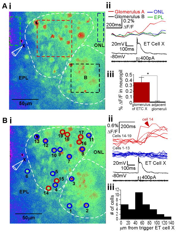
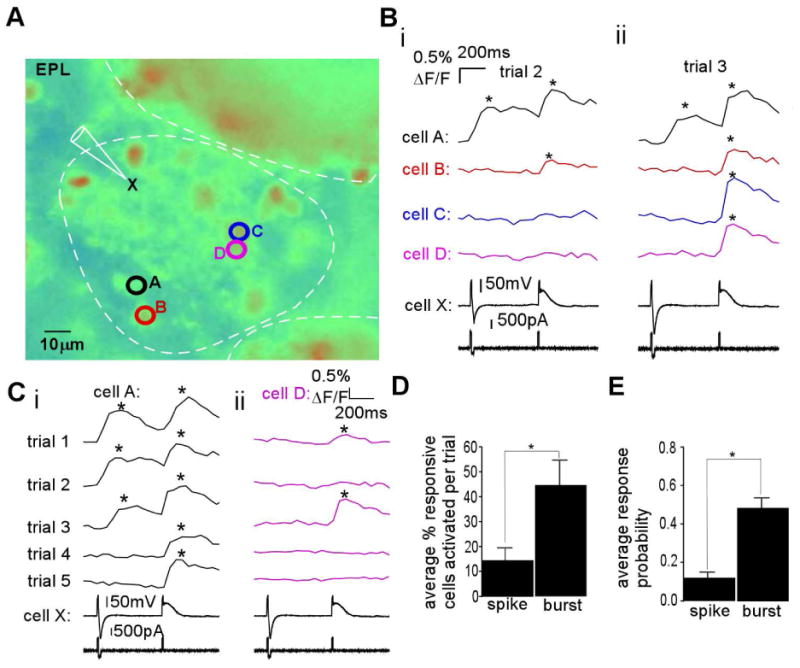
The synchronization of neuronal activity is thought to enhance information processing. There is much evidence supporting rhythmically bursting external tufted cells (ETCs) of the rodent olfactory bulb glomeruli coordinating the activation of glomerular interneurons and mitral cells via dendrodendritic excitation. However, as bursting has variable significance at axodendritic cortical synapses, it is not clear if ETC bursting imparts a specific functional advantage over the preliminary spike in dendrodendritic synaptic networks. To answer this question, we investigated the influence of single ETC bursts and spikes with the in vitro rat olfactory bulb preparation at different levels of processing, via calcium imaging of presynaptic ETC dendrites, dual electrical recording of ETC -interneuron synaptic pairs, and multicellular calcium imaging of ETC-induced population activity. Our findings supported single ETC bursts, versus single spikes, driving robust presynaptic calcium signaling, which in turn was associated with profound extension of the initial monosynaptic spike-driven dendrodendritic excitatory postsynaptic potential. This extension could be driven by either the spike-dependent or spike-independent components of the burst. At the population level, burst-induced excitation was more widespread and reliable compared with single spikes. This further supports the ETC network, in part due to a functional advantage of bursting at excitatory dendrodendritic synapses, coordinating synchronous activity at behaviorally relevant frequencies related to odor processing in vivo.


Figures

Fig. 1
Fig. 1
ETCs at the deep glomerular border fire bursts consisting of a plateau potential supporting multiple Na+ spikes. (A) Illustration of a horizontal rat OB slice, with olfactory nerve layer (ONL), glomerular layer (GL), EPL, and mitral cell layer (MCL) labeled to the left. As on the right, recordings were performed on ETCs, which were identified as large somata at the GL–EPL border. (B) Current-clamp recordings of an ETC burst (i) consisted of two separable components: (ii) fast spikes, of which the initial large spike could be separated with a hyperpolarizing pulse to block the rest of the burst, and (iii) a plateau potential that could be separated from spikes with TTX.
Fig. 2
Fig. 2
Calcium influx into ETC dendrites is more robustly mediated by bursts and plateau potentials than single spikes. (A) Fluorescence image of an ETC cell filled intracellularly with 50 μM Calcium Green-1; arrows indicate three ROIs: soma (1), secondary branch in the bottom part of the arbor (2) and secondary branch in the top part of the arbor (3). (B) (i) Bursts evoked large fluorescence signals (ΔF/F) in all three locations (top), whereas a single spike (ii) evoked a much smaller response (bottom) (n = 13). (C) Burst- and spike-induced calcium signals depreciated across the somatodendritic axis but burst-induced signals were significantly larger (n as indicated; *P<0.05, **P<0.01, ***P<0.0005). (D) With Na+ spikes blocked by TTX, a plateau potential evoked a large calcium signal in the three locations defined above (n = 6). (E) Beyond the soma, the ratio between burst- and spike-induced calcium signals (left) was close to 1 across the somatodendritic axis (n as indicated; *P<0.05; NS, P>0.4). The ratio between spike-induced and plateau potential-induced signals (right) was significantly less than 1 along the somatodendritic length (n as indicated; *P<0.02, **P<0.005, ***P<0.001). (F) The ratio between burst- and spike-induced calcium signals was reduced at each location after the addition of the ionotropic receptor blockers APV (50 μM), 6-cyano-7-nitroquinoxaline-2,3-dione (20 μM), and bicuculline (3.5-21 μM) (right) compared with control conditions (left) (n as indicated; *P<0.02).
Fig. 3
Fig. 3
Paired whole-cell recordings reveal large, robust monosynaptic excitation by ETC bursts as compared with ETC spikes. (A) Left: Illustration of a horizontal rat OB slice as before. Right: Paired electrical recordings were made between ETCs (larger circle on right) and smaller somata in the glomerular border (smaller circle on right). (B) Presynaptic burst from ETC (above) elicited a large (7.3±4.1 mV, n = 15), long (thalf-max 38±22 ms, n = 15) multicomponent depolarization in the postsynaptic interneuron with monosynaptic delay (below) (0.52±0.19 ms, n = 15). Shown is the average of many trials of a single pair. The preliminary spike (overlaid above) elicited an EPSP (overlaid below) that was similar to the first component of the burst-induced depolarization (n = 7). (C) The burst-induced depolarization was eliminated after the addition of ionotropic glutamate receptor blockers, indicating that it was an EPSP (n = 2). (D) The burst-induced EPSP was eliminated after 5 min of dialysis of presynaptic ETC with a high concentration of calcium chelator (20 mM EGTA) in the intracellular solution. (E) The ETC burst-induced EPSP amplitude, as compared with time t = 0, decayed quickly in time when dialyzed with various chelators but not in control conditions (0.2 mM EGTA). (F) The amplitude of the spike-elicited EPSP was not significantly different from the amplitude of the first component of the burst-induced EPSP (P>0.2, similar for all pairs examined, n = 7). (G) The spike-induced EPSP and burst-induced EPSP were both elicited with high probability (n = 7, P>0.3). (H) The first spike-induced EPSP peak was on average significantly smaller than the peak amplitude of the burst-induced EPSP (n = 15, *P<0.02). (I) A train of presynaptic spikes at ∼80 Hz (above) in an ETC–interneuron pair was elicited with alternating depolarizing and hyperpolarizing currents. Resulting EPSPs (below) produced by the train resembled those elicited by the ETC burst.
Fig. 4
Fig. 4
The ETC plateau potential robustly elicits large, Na+ channel-independent EPSPs. (A) ETC (above)/interneuron (below) recordings showing the contribution of the presynaptic ETC burst (ctl), plateau potential (plateau) and capacitative transient (stimulus alone) to excitation in interneurons. Blue and green traces are in the presence of 2 μM TTX (n = 4). The plateau potential produces an EPSP that is similar in duration and amplitude to the burst. The capacitative transient itself produces no EPSP. (B) Single trials of plateau potential-induced excitation show that the EPSP timing varies and shifts with the time course of plateau potential generation. (C) The average synaptic delay of EPSPs induced by bursts (plateau potential and Na+ spikes) is significantly less than the delay of EPSPs induced by plateau potential in the absence of Na+ spikes (*P<0.0003). (D) The intertrial variability of the synaptic delay of burst-induced EPSPs is significantly less than that of plateau potential-induced EPSPs (*P<0.0005). (E) The areas of burst- and plateau potential-induced EPSPs were not significantly different (P>0.5).
Fig. 5
Fig. 5
An ETC burst promotes spiking in multiple interneurons restricted to one glomerulus. (A) (i) Resting fluorescence image of a horizontal OB slice after incubation with Calcium Green-1 AM dye. The glomerular borders are indicated, and ROIs corresponding to glomerulus A and B, as well as the EPL and olfactory nerve layer (ONL) near to glomerulus A are delineated. The recorded ETC of glomerulus A is labeled (X). (ii) The ETC was stimulated via whole-cell patch (no dye in electrode) to fire one burst (bottom). The neuropil signal (ΔF/F) is shown above, demonstrating that only glomerulus A generated a measurable signal. (iii) Over all experiments, neuropil fluorescence in the glomerulus of ETC X was significantly increased over that of neighboring glomeruli (n = 11 slices, P<0.0001). (B) Cellular level analysis. (i) Same slice as in A. Neuronal somata ROIs are indicated with circles and numbers, representing responsive and some unresponsive somata. (ii) Overlay of somatic signal from neurons that were responsive (top) or unresponsive (middle) to a burst simulated in ETC X (bottom), with number identification noted. Note that responsive cells are restricted to glomerulus A. (iii) Histogram of distance between follower interneurons and trigger ETCs over several experiments (n = 6 slices). Responsive cells were restricted to distances within the typical range of glomerular diameters (100-150 μm).
Fig. 6
Fig. 6
An ETC burst promotes intraglomerular coactivity better than single spikes. (A) Resting fluorescence image of a Calcium Green-1 AM dye-loaded OB slice with trigger ETC X and responsive interneurons A-D indicated. (B) In a single trial, both a single spike and a burst were elicited in ETC X, separated by 500 ms (i and ii, “cell X”, at bottom), and fluorescence transients were monitored in interneurons A-D (i and ii, above electrical trace). (i) In trial 2, the spike triggered a response in cell A, whereas the plateau triggered a response in both cells A and B. (ii) In another separate trial (trial 3), the spike only triggered a response in cell A, whereas the burst triggered a response in all four cells. (C) (i) In multiple repeated trials, using the same stimulation protocol, and focusing on responses in cell A, the single spike triggered a response in 3/5 trials, whereas the burst triggered a response in 5/5 trials. (ii) For cell D, the burst elicited a response in 2/5 trials, whereas the spike was not successful in any of the trials. (D) In such experiments (n = 6 slices), the ability to coactivate multiple interneurons was calculated as the average percent of responding cells activated per trial. This value was significantly increased by burst firing vs. spike firing (*P<0.03). (E) For a given follower interneuron, burst firing in trigger ETC X was significantly more likely to elicit a response than a single spike (n = 32, *P<0.0001).

Similar articles

Cited by

References

    1. Abbott LF, Regehr WG. Synaptic computation. Nature. 2004;431:796–803. - PubMed
    1. Antal M, Eyre M, Finklea B, Nusser Z. External tufted cells in the main olfactory bulb form two distinct subpopulations. Eur J Neurosci. 2006;24:1124–1136. - PMC - PubMed
    1. Aroniadou-Anderjaska V, Zhou FM, Priest CA, Ennis M, Shipley MT. Tonic and synaptically evoked presynaptic inhibition of sensory input to the rat olfactory bulb via GABA(B) heteroreceptors. J Neurophysiol. 2000;84:1194–1203. - PubMed
    1. Beierlein M, Fall CP, Rinzel J, Yuste R. Thalamocortical bursts trigger recurrent activity in neocortical networks: layer 4 as a frequency-dependent gate. J Neurosci. 2002;22:9885–9894. - PMC - PubMed
    1. Cadetti L, Belluzzi O. Hyperpolarisation-activated current in glomerular cells of the rat olfactory bulb. Neuroreport. 2001;12:3117–3120. - PubMed

Publication types