. 2012;7(1):e30459.
doi: 10.1371/journal.pone.0030459. Epub 2012 Jan 20.

Protein kinase D1 maintains the epithelial phenotype by inducing a DNA-bound, inactive SNAI1 transcriptional repressor complex

Affiliations

Protein kinase D1 maintains the epithelial phenotype by inducing a DNA-bound, inactive SNAI1 transcriptional repressor complex

Ligia I Bastea et al. PLoS One. 2012.

Abstract

Background: Protein kinase D1 is downregulated in its expression in invasive ductal carcinoma of the breast and in invasive breast cancer cells, but its functions in normal breast epithelial cells is largely unknown. The epithelial phenotype is maintained by cell-cell junctions formed by E-cadherin. In cancer cells loss of E-cadherin expression contributes to an invasive phenotype. This can be mediated by SNAI1, a transcriptional repressor for E-cadherin that contributes to epithelial-to-mesenchymal transition (EMT).

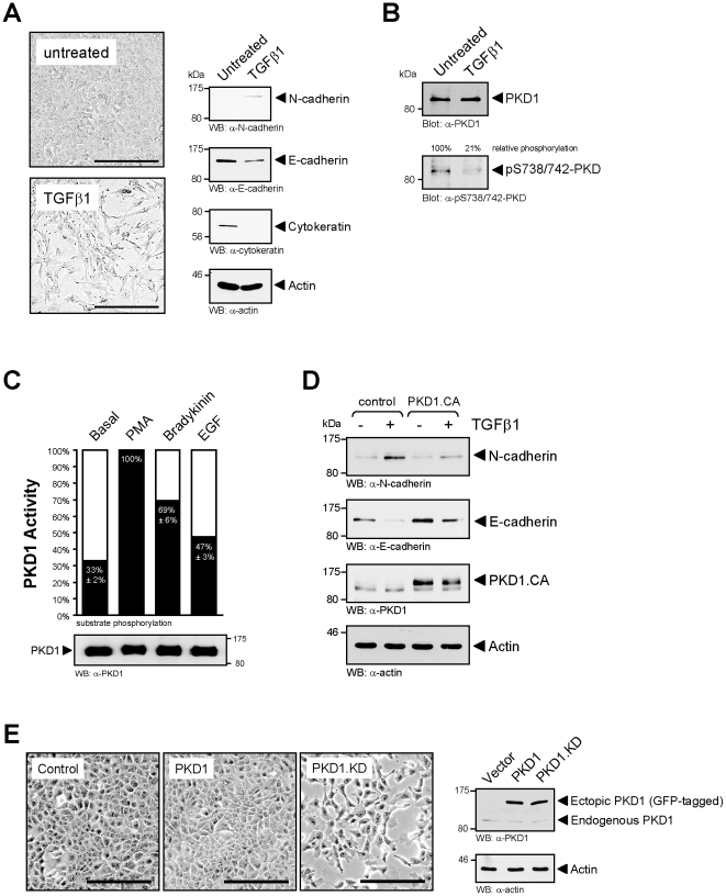
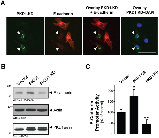
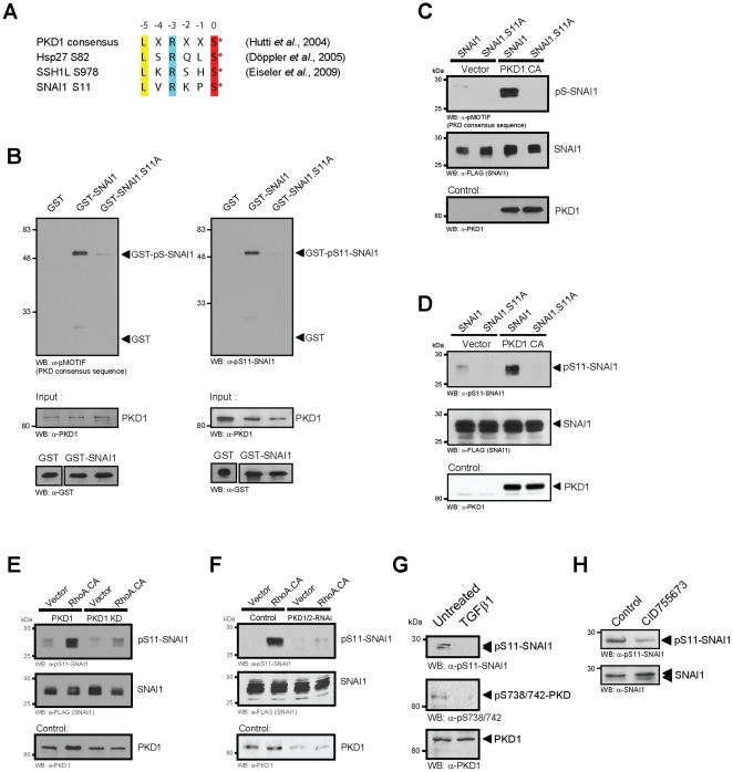
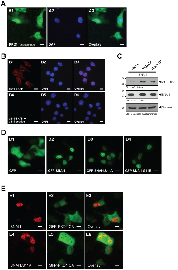
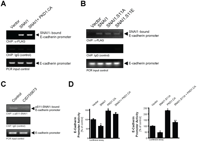
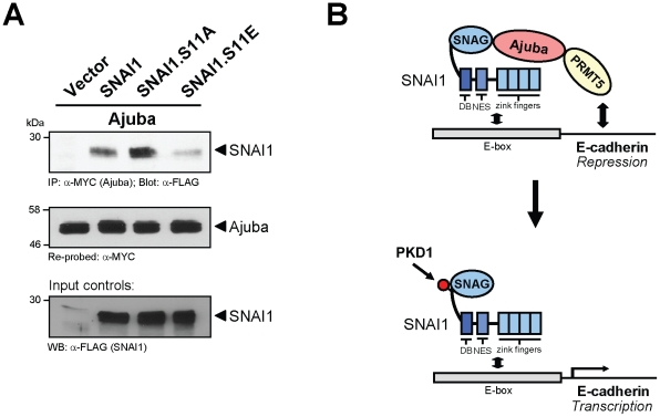
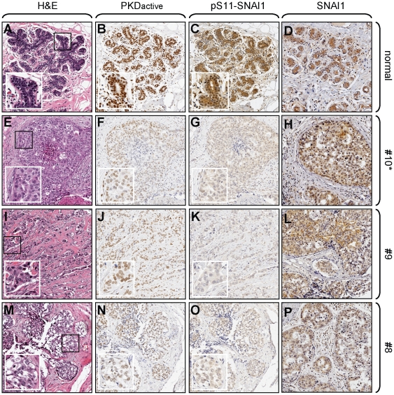
Methodology/principal findings: Here we show that PKD1 in normal murine mammary gland (NMuMG) epithelial cells is constitutively-active in its basal state and prevents a transition to a mesenchymal phenotype. Investigation of the involved mechanism suggested that PKD1 regulates the expression of E-cadherin at the promoter level through direct phosphorylation of the transcriptional repressor SNAI1. PKD1-mediated phosphorylation of SNAI1 occurs in the nucleus and generates a nuclear, inactive DNA/SNAI1 complex that shows decreased interaction with its co-repressor Ajuba. Analysis of human tissue samples with a newly-generated phosphospecific antibody for PKD1-phosphorylated SNAI1 showed that regulation of SNAI1 through PKD1 occurs in vivo in normal breast ductal tissue and is decreased or lost in invasive ductal carcinoma.

Conclusions/significance: Our data describe a mechanism of how PKD1 maintains the breast epithelial phenotype. Moreover, they suggest, that the analysis of breast tissue for PKD-mediated phosphorylation of SNAI1 using our novel phosphoS11-SNAI1-specific antibody may allow predicting the invasive potential of breast cancer cells.


Conflict of interest statement

Competing Interests: The authors have declared that no competing interests exist.

Figures

Figure 1
Figure 1. PKD1 conserves the epithelial phenotype in normal mammary gland cells.
A: NMuMG cells were either left untreated or were treated with TGFβ1 (10 ng/ml) for 48 hours. Cell morphology was photographed (bar is 200 µm) and cells were harvested and analyzed for expression of epithelial (E-cadherin, cytokeratin) and mesenchymal (N-cadherin) markers by Western blotting with anti-N-cadherin, anti-E-cadherin, or anti-cytokeratin antibodies. Staining for actin (anti-actin) served as a loading control. B: NMuMG cells were treated with TGFβ1 (10 ng/ml) for 24 hours. Endogenous PKD1 was immunoprecipitated (anti-PKD1) and analyzed for phosphorylation at its activation loop that correlates with its activity (anti-pS738/742-PKD), or samples were control stained for total PKD1 (anti-PKD1). C: Cells were stimulated with PMA (100 nM, 10 min), EGF (50 ng/ml, 10 min), Bradykinin (0.5 µg/ml, 10 min) or left untreated. Endogenous PKD1 was immunoprecipitated and subjected to an in vitro kinase assay using PKD substrate peptide. PKD1 activity is depicted relative to PMA-activated PKD1 (maximum activation). Equal immunoprecipitation was controlled by SDS-PAGE and immunoblot (anti-PKD1). D: NMuMG cells were either transfected with control vector or with active PKD1 (PKD1.CA, PKD1.S738E.S742E). 24 hours after transfection, cells were treated with TGFβ1 (10 ng/ml) for 24 hours. Lysates were analyzed for expression of N-cadherin, E-cadherin, expression of PKD1, or actin as a loading control. E: NMuMG cells were stably-transfected with vector control, wildtype PKD1 or kinase-dead PKD1.K612W (PKD1.KD) Cell morphology was analyzed by brightfield microscopy (bar is 200 µm). Expression of endogenous and overexpressed PKD1 was determined by Western blot analysis using an anti-PKD1 antibody. Immunoblotting for actin (anti-actin) served as loading control.
Figure 2
Figure 2. PKD regulates E-cadherin expression in epithelial cells.
A: NMuMG cells were transfected with GFP-tagged, kinase-dead PKD1 (PKD1.KD) and endogenous expression of E-cadherin was determined with immunofluorescence staining (anti-E-cadherin). DAPI staining served as a nuclear marker (bar is 50 µm). B: MCF-7 cells were transfected with vector control, HA-tagged PKD1 or kinase-dead PKD1 (PKD1.KD). After 48 hours, samples were analyzed by Western blot for E-cadherin expression (anti-E-cadherin) as well as expression of PKD1 (anti-PKD1). Staining for actin (anti-actin) served as loading control. C: MCF-7 cells were transfected with vector control, HA-tagged constitutively-active PKD1 (PKD1.CA) or kinase-dead PKD1 (PKD1.KD) as well as E-cadherin promoter luciferase gene reporter and renilla luciferase reporter. Induced luciferase activity was measured. Error bars shown represent standard deviations. The asterisks indicate statistical significance (p<0.05) as compared to vector control.
Figure 3
Figure 3. Active PKD1 directly phosphorylates SNAI1 at S11.
A: The amino-acids surrounding serine 11 in SNAI1 form a PKD consensus motif as it was described for S82 of Hsp27 and S978 of SSH1L. B: PKD phosphorylates SNAI1 at S11 in an in vitro assay. Bacterially-expressed and purified GST (negative control), GST-SNAI1 or GST-SNAI1.S11A were incubated in a kinase reaction with purified active PKD1. Substrate phosphorylation was detected using the pMOTIF antibody, which recognizes the phosphorylated PKD motif in PKD substrates or with the novel pS11-SNAI1 antibody specifically generated for this site. Control blots were performed for protein input (anti-PKD1, anti-GST). C, D: HeLa cells were transfected with combinations of vector control, active PKD1 (PKD1.CA) and SNAI1 or SNAI1.S11A mutant as indicated. PKD-mediated phosphorylation of SNAI1 was detected using the pMOTIF (C) or the pS11-SNAI1 (D) antibodies. E, F: HeLa cells were transfected with combinations of vector control, active RhoA (RhoA.CA) and PKD1 or PKD1.KD mutant (E) or control shRNA and shRNA specific for PKD1/2 (F) as indicated and FLAG-tagged SNAI1. PKD-mediated phosphorylation of SNAI1 was detected using the pS11-SNAI1 antibody. Samples were also control-stained for SNAI1 and PKD1 expression using anti-FLAG or anti-PKD1 antibodies, respectively. Anti-GST control staining for RhoA.CA and GST control are depicted in Figure S2. G: NMuMG cells were treated with TGFβ1 (10 ng/ml) for 48 hours. Total cell lysates were analyzed for phosphorylation of endogenous SNAI1 at S11 (anti-pS11-SNAI1) or PKD1 activity (anti-pS738/742-PKD) or total PKD1 expression (anti-PKD1) as indicated. H: NMuMG cells were treated with CID755673 (25 µM, 4 hr) or left untreated as indicated. Total cell lysates were analyzed for phosphorylation of endogenous SNAI1 at S11 (anti-pS11-SNAI1) or SNAI1 expression (anti-SNAI1).
Figure 4
Figure 4. Phosphorylation of SNAI1 by PKD1 occurs in the nucleus and does not alter its localization.
A: Immunofluorescence staining of NMuMG cells for endogenous PKD1 (anti-PKD1). The bar represents 10 µm. B: Immunofluorescence staining of NMuMG cells for S11-phosphorylated SNAI1 (anti-pS11-SNAI1) in absence or presence of competing phospho-S11-peptide and nuclei (DAPI). The bar represents 10 µm. C: HeLa cells were transfected as indicated and nuclear extracts were prepared and analyzed by Western blot for SNAI1 (anti-FLAG), pS11-SNAI1 (anti-pS11-SNAI1) and nucleolin (anti-nucleolin, loading control). D: NMuMG cells were transfected with GFP control, GFP-SNAI1, GFP-SNAI1.S11A or GFP-SNAI1.S11E mutants. Localization of GFP or GFP-tagged proteins was determined using immunofluorescence analysis (bar is 10 µm). E: NMuMG cells were transfected with FLAG-tagged wildtype SNAI1 or SNAI1.S11A mutant and GFP-tagged, active PKD1 (PKD1.CA) as indicated and localization of SNAI1 was determined by indirect immunofluorescence staining (anti-FLAG as primary antibody). The bar represents 10 µm.
Figure 5
Figure 5. PKD1-regulated SNAI1 binds to the E-cadherin promoter, but is ineffective in its function.
A: Hek293T cells were transfected with vector control, SNAI1 or active PKD1 (PKD1.CA) and SNAI1 as indicated. SNAI1/DNA complexes were immunoprecipitated (anti-FLAG) after crosslinking and precipitates were analyzed by PCR for the SNAI1-bound E-cadherin promoter. B: NMuMG cells were transfected with vector control, SNAI1, SNAI1.S11A or SNAI1.S11E mutants. SNAI1/DNA complexes were immunoprecipitated (anti-FLAG) after crosslinking and precipitates were analyzed by PCR for the SNAI1-bound E-cadherin promoter. C: NMuMG cells were treated with CID755673 (25 µM, 1 hr) or left untreated. Phospho-S11-SNAI1/DNA complexes were immunoprecipitated (anti-pS11-SNAI1) after crosslinking and precipitates were analyzed by PCR for the pS11-SNAI1-bound E-cadherin promoter. In experiments depicted in A–C, a PCR for the E-cadherin promoter using the input DNA as well as a ChIP using IgG instead of the anti-FLAG antibody served as controls. D: Hek293T cells were transfected with vector control, SNAI1 or SNAI1.S11A mutant, active PKD1 (PKD1.CA) or both and E-cadherin promoter luciferase reporter and renilla reporter plasmids. Induced luciferase activity was measured. Error bars shown represent standard deviations. P values were acquired with the t test, using GraphPad software. Asterisks indicate statistical significance.
Figure 6
Figure 6. Phosphorylation of SNAI1 decreases its binding to Ajuba.
A: HeLa cells were co-transfected with MYC-tagged Ajuba and vector control, and FLAG-tagged wildtype SNAI1, SNAI1.S11A or SNAI1.S11E mutants as indicated. Ajuba was immunoprecipitated (anti-MYC) and precipitates were analyzed for co-precipitated SNAI1 (anti-FLAG). Samples were re-stained for Ajuba (anti-MYC) and lysates were control-stained for expressed SNAI1 (anti-FLAG). B: Proposed mechanism of how PKD1-mediated phosphorylation regulates SNAI1 function as a transcriptional repressor of E-cadherin gene expression.
Figure 7
Figure 7. Loss of nuclear PKD activity and SNAI1 phosphorylation at S11 are markers for invasive breast cancer.
Tissue microarrays (TMAs) including 10 normal breast tissue samples, 40 invasive ductal carcinoma of the breast and 10 metastatic invasive ductal carcinoma samples from lymph nodes were H&E stained or analyzed for the expression of active PKD (anti-pY95-PKD), S11-phosphorylated SNAI1 (anti-pS11-SNAI1) and total SNAI1 (anti-SNAI1). Representative pictures of normal (A–D) and 3 tumor tissues (E–P) are depicted. Numbers indicate the position of the tissue on the TMA. The asterisk (sample #10) indicates tumor tissue form a region adjacent to the normal tissue (same patient). Inserts show enhanced area.

Similar articles

Cited by

References

    1. Peinado H, Olmeda D, Cano A. Snail, Zeb and bHLH factors in tumour progression: an alliance against the epithelial phenotype? Nat Rev Cancer. 2007;7:415–428. - PubMed
    1. Onder TT, Gupta PB, Mani SA, Yang J, Lander ES, et al. Loss of E-cadherin promotes metastasis via multiple downstream transcriptional pathways. Cancer Res. 2008;68:3645–3654. - PubMed
    1. Huber MA, Kraut N, Beug H. Molecular requirements for epithelial-mesenchymal transition during tumor progression. Curr Opin Cell Biol. 2005;17:548–558. - PubMed
    1. Barrallo-Gimeno A, Nieto MA. The Snail genes as inducers of cell movement and survival: implications in development and cancer. Development. 2005;132:3151–3161. - PubMed
    1. Batlle E, Sancho E, Franci C, Dominguez D, Monfar M, et al. The transcription factor snail is a repressor of E-cadherin gene expression in epithelial tumour cells. Nat Cell Biol. 2000;2:84–89. - PubMed

Publication types

MeSH terms