Whole genomes of Chandipura virus isolates and comparative analysis with other rhabdoviruses
- PMID: 22272333
- PMCID: PMC3260278
- DOI: 10.1371/journal.pone.0030315
Whole genomes of Chandipura virus isolates and comparative analysis with other rhabdoviruses
Abstract
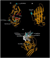
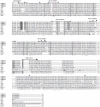
The Chandipura virus (CHPV) belonging to the Vesiculovirus genus and Rhabdoviridae family, has recently been associated with a number of encephalitis epidemics, with high mortality in children, in different parts of India. No full length genome sequences of CHPV isolates were available in GenBank and little is known about the molecular markers for pathogenesis. In the present study, we provide the complete genomic sequences of four isolates from epidemics during 2003-2007. These sequences along with the deduced sequence of the prototype isolate of 1965 were analysed using phylogeny, motif search, homology modeling and epitope prediction methods. Comparison with other rhaboviruses was also done for functional extrapolations. All CHPV isolates clustered with the Isfahan virus and maintained several functional motifs of other rhabdoviruses. A notable difference with the prototype vesiculovirus, Vesicular Stomatitis Virus was in the L-domain flanking sequences of the M protein that are known to be crucial for interaction with host proteins. With respect to the prototype isolate, significant additional mutations were acquired in the 2003-2007 isolates. Several mutations in G mapped onto probable antigenic sites. A mutation in N mapped onto regions crucial for N-N interaction and a putative T-cell epitope. A mutation in the Casein kinase II phosphorylation site in P may attribute to increased rates of phosphorylation. Gene junction comparison revealed changes in the M-G junction of all the epidemic isolates that may have implications on read-through and gene transcription levels. The study can form the basis for further experimental verification and provide additional insights into the virulence determinants of the CHPV.
Figures


Similar articles
-
Complete genome sequences of Chandipura and Isfahan vesiculoviruses.Arch Virol. 2005 Apr;150(4):671-80. doi: 10.1007/s00705-004-0452-2. Epub 2004 Dec 21. Arch Virol. 2005. PMID: 15614433
-
Complete genome sequence of Piry vesiculovirus.Arch Virol. 2016 Aug;161(8):2325-8. doi: 10.1007/s00705-016-2905-9. Epub 2016 May 23. Arch Virol. 2016. PMID: 27216928
-
Molecular detection and sequence characterization of diverse rhabdoviruses in bats, China.Virus Res. 2018 Jan 15;244:208-212. doi: 10.1016/j.virusres.2017.11.028. Epub 2017 Nov 28. Virus Res. 2018. PMID: 29196194 Free PMC article.
-
Reviewing Chandipura: a vesiculovirus in human epidemics.Biosci Rep. 2007 Oct;27(4-5):275-98. doi: 10.1007/s10540-007-9054-z. Biosci Rep. 2007. PMID: 17610154 Free PMC article. Review.
-
Fish rhabdoviruses: molecular epidemiology and evolution.Curr Top Microbiol Immunol. 2005;292:81-117. doi: 10.1007/3-540-27485-5_5. Curr Top Microbiol Immunol. 2005. PMID: 15981469 Review.
Cited by
-
Zoonotic Viral Diseases of Equines and Their Impact on Human and Animal Health.Open Virol J. 2018 Aug 31;12:80-98. doi: 10.2174/1874357901812010080. eCollection 2018. Open Virol J. 2018. PMID: 30288197 Free PMC article. Review.
-
Complete genome and molecular epidemiological data infer the maintenance of rabies among kudu (Tragelaphus strepsiceros) in Namibia.PLoS One. 2013;8(3):e58739. doi: 10.1371/journal.pone.0058739. Epub 2013 Mar 20. PLoS One. 2013. PMID: 23527015 Free PMC article.
-
Abundance of Phasi-Charoen-like virus in Aedes aegypti mosquito populations in different states of India.PLoS One. 2022 Dec 9;17(12):e0277276. doi: 10.1371/journal.pone.0277276. eCollection 2022. PLoS One. 2022. PMID: 36490242 Free PMC article.
-
Monocytes and B cells support active replication of Chandipura virus.BMC Infect Dis. 2016 Sep 14;16:487. doi: 10.1186/s12879-016-1794-6. BMC Infect Dis. 2016. PMID: 27628855 Free PMC article.
-
Chandipura virus induces neuronal death through Fas-mediated extrinsic apoptotic pathway.J Virol. 2013 Nov;87(22):12398-406. doi: 10.1128/JVI.01864-13. Epub 2013 Sep 11. J Virol. 2013. PMID: 24027318 Free PMC article.
References
-
- Bhatt PN, Rodrigues Chandipura: a new arbovirus isolated in India from patients with febrile illness. IJMR. 1967;55:1295–1305. - PubMed
-
- Chadha MS, Arankalle VA, Jadi RS, Joshi MV, Thakare JP, et al. An outbreak of Chandipura virus encephalitis in the eastern districts of Gujarat state, India. Am J Trop Med. 2005;73(3):566–570. - PubMed
-
- Gurav YK, Tandale BV, Jadi RS, Gunjikar RS, Tikute SS, et al. Chandipura virus encephalitis outbreak among children in Nagpur division, Maharashtra, 2007. Indian J Med Res. 2010;132:395–399. - PubMed
Publication types
MeSH terms
Substances
LinkOut - more resources
Full Text Sources

