

Review
doi: 10.1038/aps.2011.171.
Epub 2012 Jan 23.
Structure and activation of rhodopsin
Affiliations
- PMID: 22266727
- PMCID: PMC3677203
- DOI: 10.1038/aps.2011.171
Item in Clipboard
Review
Structure and activation of rhodopsin
Acta Pharmacol Sin.
2012 Mar.
Abstract
Rhodopsin is the first G-protein-coupled receptor (GPCR) with its three-dimensional structure solved by X-ray crystallography. The crystal structure of rhodopsin has revealed the molecular mechanism of photoreception and signal transduction in the visual system. Although several other GPCR crystal structures have been reported over the past few years, the rhodopsin structure remains an important model for understanding the structural and functional characteristics of other GPCRs. This review summarizes the structural features, the photoactivation, and the G protein signal transduction of rhodopsin.
Figures
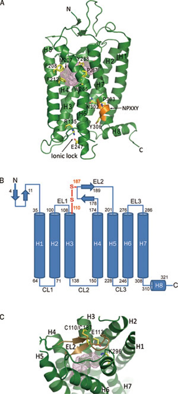
Overall structure of ground sate bovine rhodopsin and its key features (PDB: 1F88). (A) The seven-transmembrane helix domain with the retinal in gray stick and the ligand binding pocket shown as a pink mesh. Major ligand binding residues around the ligand binding pocket are shown as yellow sticks and are labeled. Other features include the ionic lock (yellow sticks) and the NPXXY motif (orange). (B) Two-dimensional sequence of bovine rhodopsin with the starting and ending residues of secondary structural elements indicated. The disulfide bond connecting EL2 to helix 3 is shown in orange. N, amino terminus; C, carboxyl terminus; EL, extracellular loop; CL, cytoplasmic loop. (C) The ligand binding pocket (pink mesh) of rhodopsin with EL2 (the lid of the pocket) shown in dark brown. The disulfide bond between C110 and C187 is labeled.
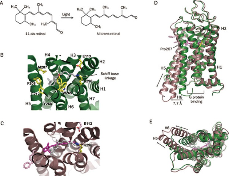
Ligand binding and conformational changes in rhodopsin. (A) Chemical structures of 11-cis- and all-trans-retinal. (B) 11-cis-retinal (in gray) in the ligand binding pocket is associated with the surrounding residues of the protein moiety (green, PDB: 1F88). (C) Conformational changes in retinal and the protein moiety of rhodopsin upon photoactivation. The photoactivated all-trans-retinal (PDB: 3PQR) is magenta and the ground-state 11-cis-retinal (PDB: 1F88, gray) is superposed on the activated all-trans-retinal for comparison. The protein moiety of activated rhodopsin (PDB: 3PQR) is dark brown. (D) The key conformational changes in rhodopsin upon photoactivation are the outward tilting of the cytoplasmic end of helix 6 (indicated by the horizontal arrow), creating a crevice for G-protein binding, and the elongation of the cytoplasmic end of helix 5 (indicated by the vertical arrow) that provides more interface for G-protein interaction. Green shows the ground-state conformation (PDB: 1F88), and brown shows the activated conformation (PDB: 3PQR). (E) Bottom view of panel D.
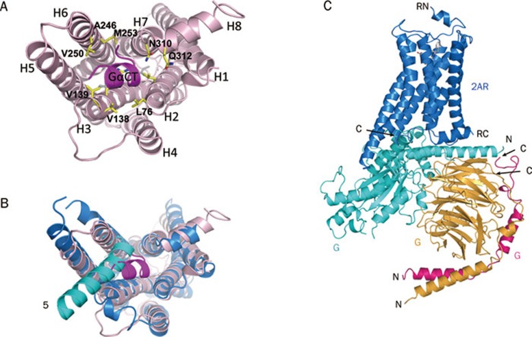
Rhodopsin-G protein interface. (A) Rhodopsin in complex with a synthetic peptide derived from helix α5 of the Gα subunit of transducin (PDB: 3PQR). The rhodopsin residues interacting with the Gα peptide are labeled. (B) Comparison of the binding mode of rhodopsin with the synthetic peptide and that of the β2-adrenergic receptor with intact G protein (PDB: 3SN6). The β2-adrenergic receptor is blue and the Gα subunit is cyan. For clarity, only helix α5 of the Gα subunit is shown. (C) The whole-complex model of the β2-adrenergic receptor with intact G protein (PDB: 3SN6). The β2-adrenergic receptor is blue, the Gα subunit is cyan, the Gβ subunit is brown, and the Gγ subunit is pink. RN, N-terminus of the receptor; RC, C-terminus of the receptor; αN, N-terminus of the Gα subunit; αC, C-terminus of the Gα subunit; βN, N-terminus of the Gβ subunit; βC, C-terminus of the Gβ subunit; γN, N-terminus of the Gγ subunit; γC, C-terminus of the Gγ subunit.
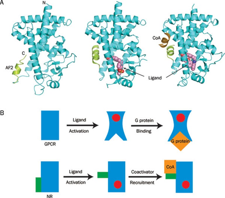
A comparison of the ligand-induced activation modes of GPCRs and nuclear receptors. (A) Ligand-induced rearrangement of the C-terminal AF2 helix and coactivator recruitment of a peroxisome proliferator-activated receptor ligand binding domain (PPAR LBD). At left is an apo PPAR LBD (PDB: 1PRG); middle, the LBD upon the ligand binding-induced conformational change in the AF2 helix and the formation of the coactivator binding site (PDB: 1I7G); at right, the LBD upon the subsequent coactivator recruitment (PDB: 1K7L). The LBD core structure is cyan; the AF2 helix, green; and the coactivator motif, brown. (B) Cartoon presentation showing that ligand activation induces conformational changes in the core domain of GPCRs but not in that of NRs. Blue are the core domains of both receptors; Red are the ligands; Orange are the G protein binding to the GPCR and the coactivator binding to the NR.
Similar articles
-
Comparative sequence and structural analyses of G-protein-coupled receptor crystal structures and implications for molecular models.PLoS One. 2009 Sep 16;4(9):e7011. doi: 10.1371/journal.pone.0007011. PLoS One. 2009. PMID: 19756152 Free PMC article.
-
Crystal structure of rhodopsin in complex with a mini-Go sheds light on the principles of G protein selectivity.Sci Adv. 2018 Sep 19;4(9):eaat7052. doi: 10.1126/sciadv.aat7052. eCollection 2018 Sep. Sci Adv. 2018. PMID: 30255144 Free PMC article.
-
Relevance of rhodopsin studies for GPCR activation.Biochim Biophys Acta. 2014 May;1837(5):674-82. doi: 10.1016/j.bbabio.2013.09.002. Epub 2013 Sep 13. Biochim Biophys Acta. 2014. PMID: 24041646 Review.
-
Activation of G-protein-coupled receptors: a common molecular mechanism.Trends Endocrinol Metab. 2003 Nov;14(9):431-7. doi: 10.1016/j.tem.2003.09.007. Trends Endocrinol Metab. 2003. PMID: 14580763 Review.
-
Restructuring G-protein- coupled receptor activation.Cell. 2012 Sep 28;151(1):14-23. doi: 10.1016/j.cell.2012.09.003. Cell. 2012. PMID: 23021212 Review.
Cited by
-
Characterizing variants of unknown significance in rhodopsin: A functional genomics approach.Hum Mutat. 2019 Aug;40(8):1127-1144. doi: 10.1002/humu.23762. Epub 2019 Jun 22. Hum Mutat. 2019. PMID: 30977563 Free PMC article.
-
Orphan G protein receptor GPR55 as an emerging target in cancer therapy and management.Cancer Manag Res. 2013 Jul 1;5:147-55. doi: 10.2147/CMAR.S35175. Print 2013. Cancer Manag Res. 2013. PMID: 23869178 Free PMC article.
-
Systematic Assessment of Protein C-Termini Mutated in Human Disorders.Biomolecules. 2023 Feb 12;13(2):355. doi: 10.3390/biom13020355. Biomolecules. 2023. PMID: 36830724 Free PMC article.
-
Advances in methods to characterize ligand-induced ionic lock and rotamer toggle molecular switch in G protein-coupled receptors.Methods Enzymol. 2013;520:153-74. doi: 10.1016/B978-0-12-391861-1.00007-1. Methods Enzymol. 2013. PMID: 23332699 Free PMC article.
-
iTRAQ Quantitative Proteomic Analysis of Vitreous from Patients with Retinal Detachment.Int J Mol Sci. 2018 Apr 11;19(4):1157. doi: 10.3390/ijms19041157. Int J Mol Sci. 2018. PMID: 29641463 Free PMC article.
References
-
- Fredriksson R, Lagerström MC, Lundin LG, Schiöth HB. The G-protein-coupled receptors in the human genome form five main families. Phylogenetic analysis, paralogon groups, and fingerprints. Mol Pharmacol. 2003;63:17. - PubMed
-
- Palczewski K, Kumasaka T, Hori T, Behnke CA, Motoshima H, Fox BA, et al. Crystal structure of rhodopsin: A G protein-coupled receptor. Science. 2000;289:739–45. - PubMed
-
- Li J, Edwards PC, Burghammer M, Villa C, Schertler GF. Structure of bovine rhodopsin in a trigonal crystal form. J Mol Biol. 2004;343:1409–38. - PubMed
Publication types
MeSH terms
Substances
Grants and funding
LinkOut - more resources
Full Text Sources
Miscellaneous
