. 2012 Apr;27(4):1149-55.
doi: 10.3892/or.2012.1634. Epub 2012 Jan 12.

Clinical significance of miR-155 expression in breast cancer and effects of miR-155 ASO on cell viability and apoptosis

Affiliations

Clinical significance of miR-155 expression in breast cancer and effects of miR-155 ASO on cell viability and apoptosis

Shu-Rong Zheng et al. Oncol Rep. 2012 Apr.

Abstract

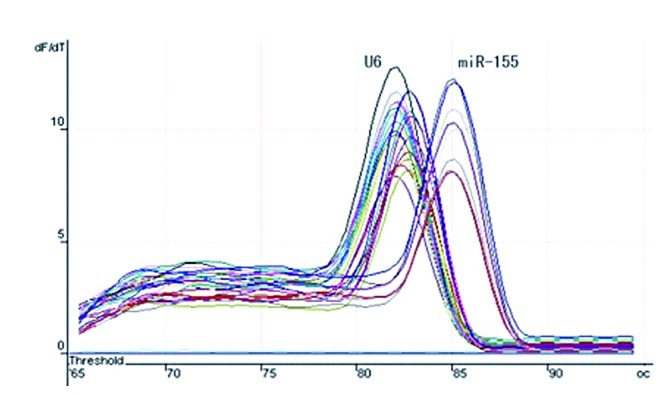
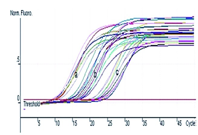
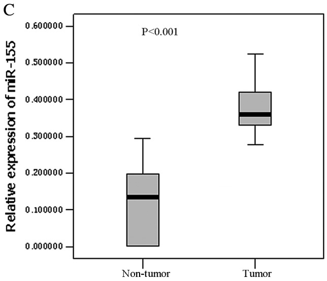
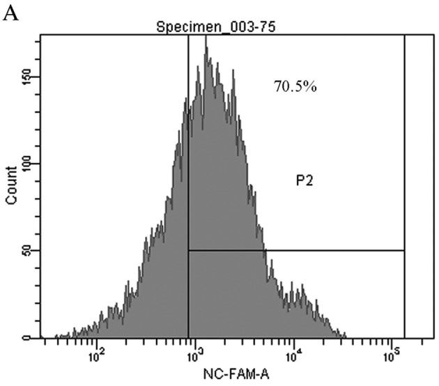
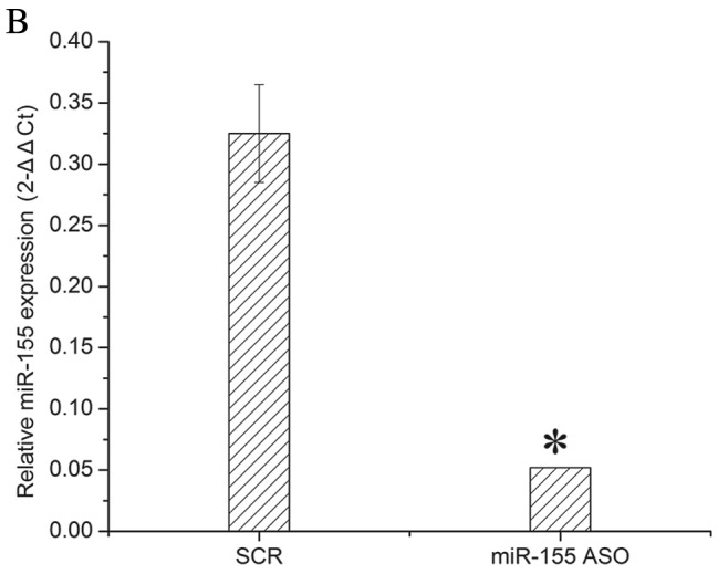
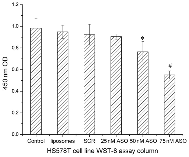
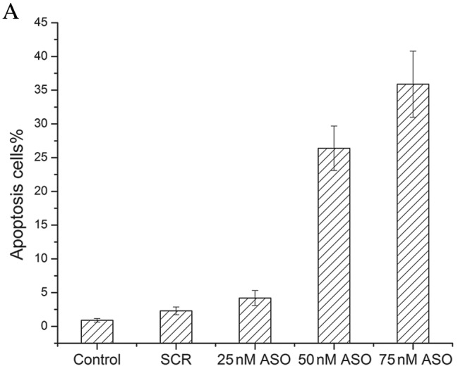
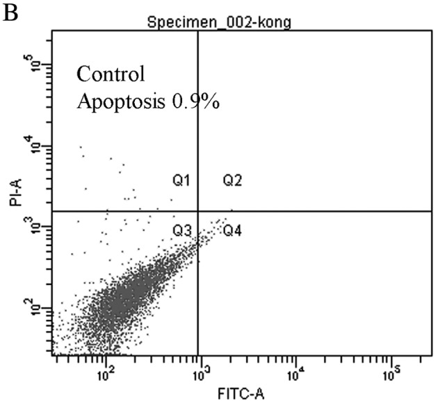
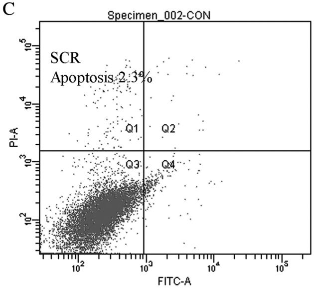
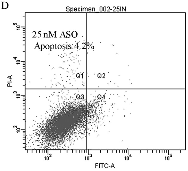
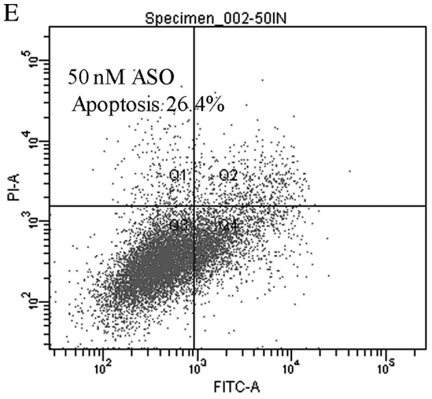
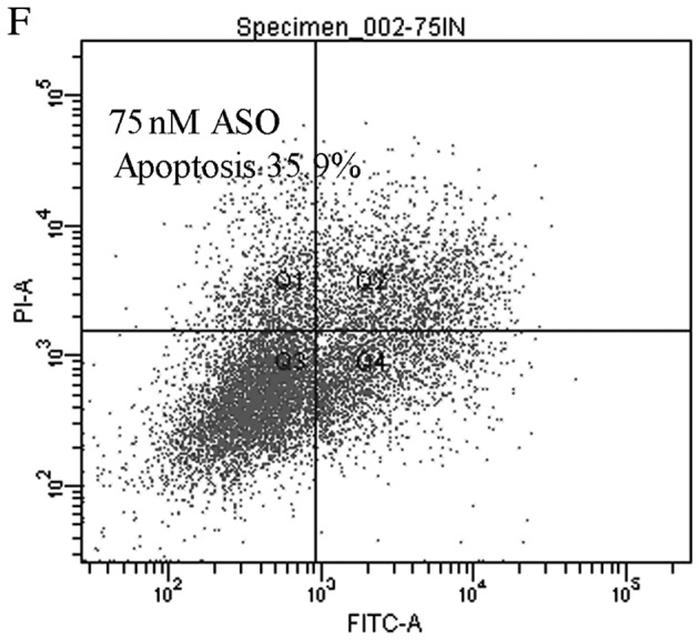
Accumulating evidence shows that mircroRNAs (miRNAs) play a vital role in tumorigenesis. miR-155 is one of the most multifunctional miRNAs whose overexpression has been found to be associated with different types of cancer including breast cancer. To further determine the potential involvement of miR-155 in breast cancer, we evaluated the expression levels of miR-155 by real-time PCR and correlated the results with clinicopathological features. Matched non-tumor and tumor tissues of 42 infiltrating ductal carcinomas and 3 infiltrating lobular carcinomas were analyzed for miR-155 expression by real-time PCR. Further, we used an antisense technique to inhibit miR-155 expression in vitro. WST-8 test was performed to evaluate cell viability and apoptosis assay was used to investigate the effect of the miR-155 antisense oligonucleotide (miR-155 ASO) on HS578T cell death. The expression levels of miR-155 were significantly higher in tumor tissues than the levels in matched non-tumor tissues (P<0.001). Up-regulated miR-155 expression was associated with lymph node positivity (P=0.034), higher proliferation index (Ki-67 >10%) (P=0.019) and advanced breast cancer TNM clinical stage (P=0.002). Interestingly, we next found that miR-155 expression levels had close relations with ER status (P=0.041) and PR status (P=0.029). Transfection efficiency detected by flow cytometry was higher than 70%, the WST-8 test showed that viability of HS578T cells was greatly reduced after transfection with miR-155 ASO compared with the scramble (SCR) group or the liposome group. The Annexin V-FITC/PI assay also indicated that transfection with miR-155 ASO promoted apoptosis.


Figures

Figure 1
Figure 1
miR-155 expression in breast cancer. (A) The melting-curves of miR-155 and U6 snRNA are presented as a single, sharply-defined melting curve with a narrow peak, indicating that pure, homogeneous PCR products were produced. (B) Representative amplification curves show that the Ct values of miR-155 in the tumor tissue are lower than in the non-tumor tissue, indicating the miR-155 expression is higher in tumor tissue than in non-tumor tissue. Reactions were performed in triplicate. (A) U6 snRNA of tumor and matched non-tumor; (B) miR-155 of tumor; (C) miR-155 of matched non-tumor. (C) Differences in the expression levels of miR-155 between the tumor tissue and matched non-tumor tissue (box-plot diagrams with median, 1st quartile, 3rd quartile and non-outlier range).
Figure 1
Figure 1
miR-155 expression in breast cancer. (A) The melting-curves of miR-155 and U6 snRNA are presented as a single, sharply-defined melting curve with a narrow peak, indicating that pure, homogeneous PCR products were produced. (B) Representative amplification curves show that the Ct values of miR-155 in the tumor tissue are lower than in the non-tumor tissue, indicating the miR-155 expression is higher in tumor tissue than in non-tumor tissue. Reactions were performed in triplicate. (A) U6 snRNA of tumor and matched non-tumor; (B) miR-155 of tumor; (C) miR-155 of matched non-tumor. (C) Differences in the expression levels of miR-155 between the tumor tissue and matched non-tumor tissue (box-plot diagrams with median, 1st quartile, 3rd quartile and non-outlier range).
Figure 2
Figure 2
Detection of transfection efficiency and miR-155 expression ratio relative to U6 snRNA in HS578T cells. (A) Eight hours after transfection with 75 nM 5′FAM SCR, the transfection efficiency was detected by flow cytometry. The P2 region stands for the number of cells that were successfully transfected with 5′FAM SCR by Lipofectamine 2000. (B) HS578T cells were transfected with 75 nM miR-155 ASO or SCR. Total RNA from treated cells was extracted by TRIzol and quantified by ultraviolet spectrophotometry. miR-155 and U6 snRNA expression were determined by quantitative real-time PCR, according to the manufacturer’s instructions. Results showed that miR-155 ASO down-regulated miR-155 levels in HS578T cells. *P<0.01, compared with the SCR group.
Figure 2
Figure 2
Detection of transfection efficiency and miR-155 expression ratio relative to U6 snRNA in HS578T cells. (A) Eight hours after transfection with 75 nM 5′FAM SCR, the transfection efficiency was detected by flow cytometry. The P2 region stands for the number of cells that were successfully transfected with 5′FAM SCR by Lipofectamine 2000. (B) HS578T cells were transfected with 75 nM miR-155 ASO or SCR. Total RNA from treated cells was extracted by TRIzol and quantified by ultraviolet spectrophotometry. miR-155 and U6 snRNA expression were determined by quantitative real-time PCR, according to the manufacturer’s instructions. Results showed that miR-155 ASO down-regulated miR-155 levels in HS578T cells. *P<0.01, compared with the SCR group.
Figure 3
Figure 3
WST-8 assay on control, liposomes, SCR and test groups.*P=0.000 compared to control group; #P=0.000 compared to control group.
Figure 4
Figure 4
Induction of HS578T cell apoptosis by miR-155 ASO. (A) HS578T cells were transfected with miR-155 ASO or SCR for 6 h and incubated in medium containing 10% FCS for another 24 h. Apoptosis of HS578T cells were double-stained with Annexin V and PI, and detected by flow cytometry. miR-155 ASO 50 and 75 nM could efficiently induced apoptosis (P<0.01 as compared with SCR). (B-F) The images of apoptosis were analyzed by double staining with Annexin V and PI.
Figure 4
Figure 4
Induction of HS578T cell apoptosis by miR-155 ASO. (A) HS578T cells were transfected with miR-155 ASO or SCR for 6 h and incubated in medium containing 10% FCS for another 24 h. Apoptosis of HS578T cells were double-stained with Annexin V and PI, and detected by flow cytometry. miR-155 ASO 50 and 75 nM could efficiently induced apoptosis (P<0.01 as compared with SCR). (B-F) The images of apoptosis were analyzed by double staining with Annexin V and PI.

Similar articles

Cited by

References

    1. Ambros V. MicroRNA pathways in flies and worms: growth, death, fat, stress, and timing. Cell. 2003;113:673–676. - PubMed
    1. Bartel DP. MicroRNAs: target recognition and regulatory functions. Cell. 2009;136:215–233. - PMC - PubMed
    1. Griffiths-Jones S, Grocock RJ, van Dongen S, Bateman A, Enright AJ. miRBase: microRNA sequences, targets and gene nomenclature. Nucleic Acids Res. 2006;34:D140–D144. - PMC - PubMed
    1. He L, Thomson JM, Hemann MT, et al. A microRNA polycistron as a potential human oncogene. Nature. 2005;435:828–833. - PMC - PubMed
    1. Tam W, Dahlberg JE. miR-155/BIC as an oncogenic microRNA. Genes Chromosomes Cancer. 2006;45:211–212. - PubMed

Publication types

MeSH terms