. 2012 Jun;166(4):1357-76.
doi: 10.1111/j.1476-5381.2012.01855.x.

Natural and synthetic modulators of SK (K(ca)2) potassium channels inhibit magnesium-dependent activity of the kinase-coupled cation channel TRPM7

Affiliations

Natural and synthetic modulators of SK (K(ca)2) potassium channels inhibit magnesium-dependent activity of the kinase-coupled cation channel TRPM7

V Chubanov et al. Br J Pharmacol. 2012 Jun.

Abstract

Background and purpose: Transient receptor potential cation channel subfamily M member 7 (TRPM7) is a bifunctional protein comprising a TRP ion channel segment linked to an α-type protein kinase domain. TRPM7 is essential for proliferation and cell growth. Up-regulation of TRPM7 function is involved in anoxic neuronal death, cardiac fibrosis and tumour cell proliferation. The goal of this work was to identify non-toxic inhibitors of the TRPM7 channel and to assess the effect of blocking endogenous TRPM7 currents on the phenotype of living cells.

Experimental approach: We developed an aequorin bioluminescence-based assay of TRPM7 channel activity and performed a hypothesis-driven screen for inhibitors of the channel. The candidates identified were further assessed electrophysiologically and in cell biological experiments.

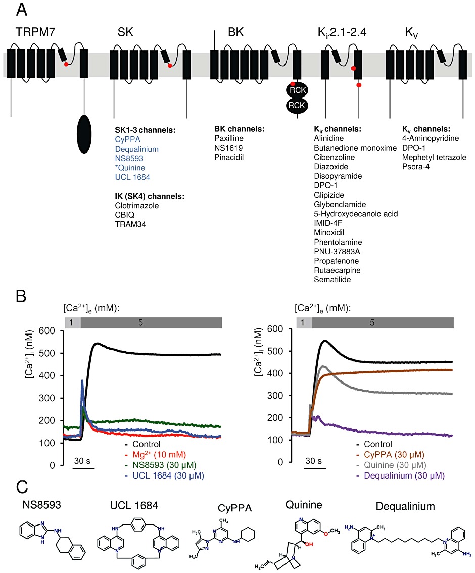
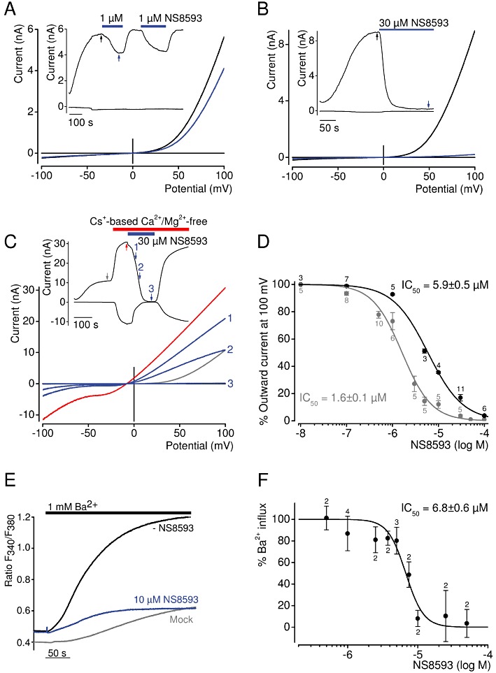
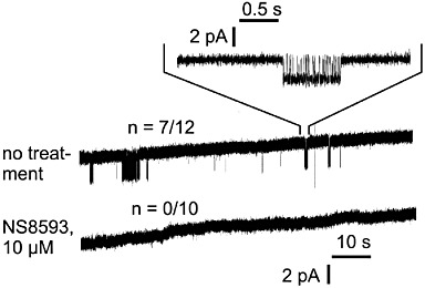
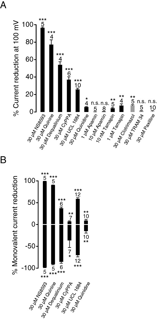
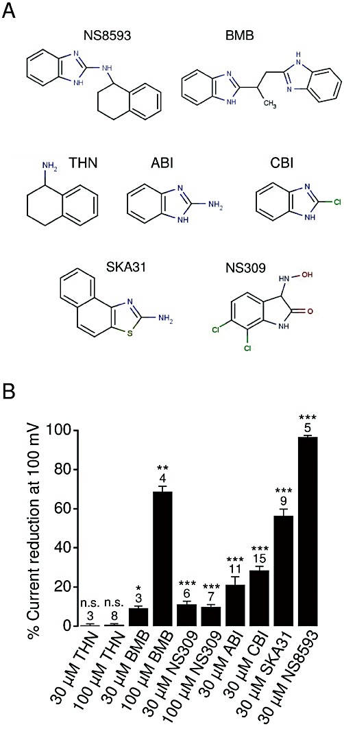
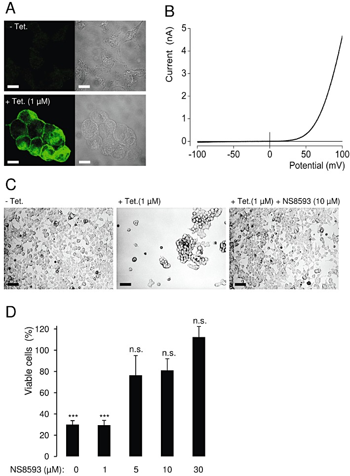
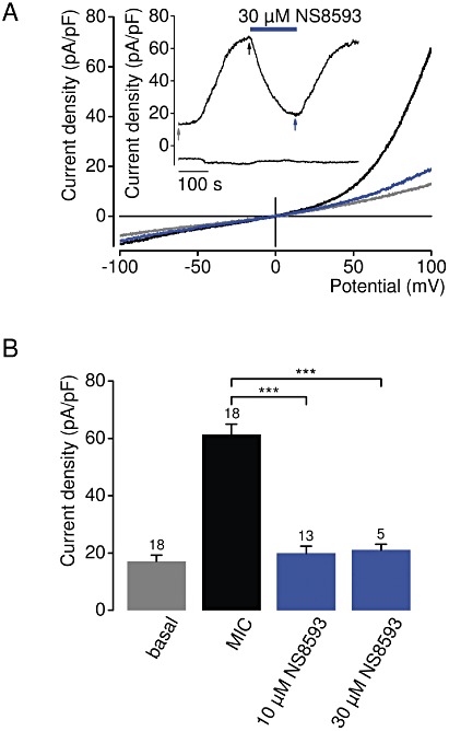
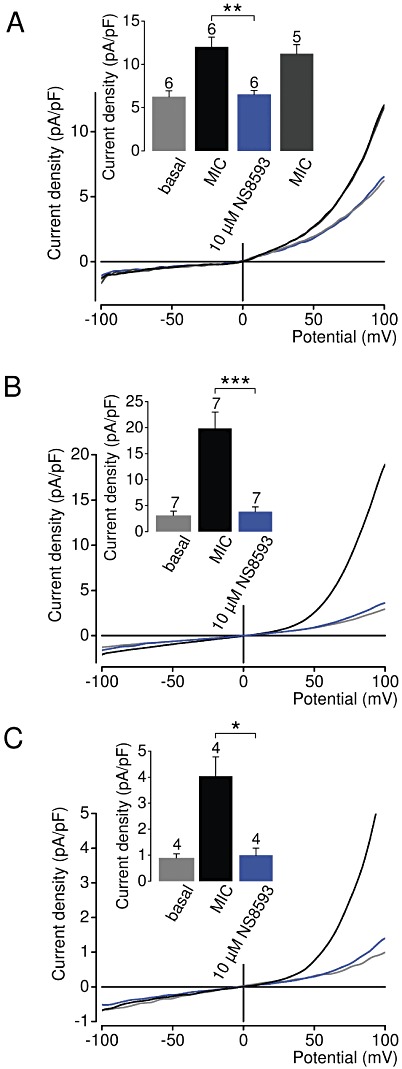
Key results: TRPM7 currents were inhibited by modulators of small conductance Ca²⁺ -activated K⁺ channels (K(Ca)2.1-2.3; SK) channels, including the antimalarial plant alkaloid quinine, CyPPA, dequalinium, NS8593, SKA31 and UCL 1684. The most potent compound NS8593 (IC₅₀ 1.6 µM) specifically targeted TRPM7 as compared with other TRP channels, interfered with Mg²⁺ -dependent regulation of TRPM7 channel and inhibited the motility of cultured cells. NS8593 exhibited full and reversible block of native TRPM7-like currents in HEK 293 cells, freshly isolated smooth muscle cells, primary podocytes and ventricular myocytes.

Conclusions and implications: This study reveals a tight overlap in the pharmacological profiles of TRPM7 and K(Ca)2.1-2.3 channels. NS8593 acts as a negative gating modulator of TRPM7 and is well-suited to study functional features and cellular roles of endogenous TRPM7.


Figures

Figure 1
Figure 1
Identification of TRPM7 channel inhibitors among known modulators of Mg2+ sensitive K+ channels. (A) Domain architecture of TRPM7, KCa2.1–2.3 (SK), KCa3.1 (IK, SK4), KCa1.1 (BK), KIR and KV channels. Locations of Mg2+-binding sites in TRPM7 (formed by E1047 and Y1049 in mouse TRPM7 (Mederos y Schnitzler et al., 2008), KCa2.1–2.3 channels [S359 in rat KCa2.2 (Soh and Park, 2002)], KCa1.1 channels (formed by E374 and E399 in human BK1) (Wu et al. 2010; Yang et al., 2008), KIR channels (D172 and E224 in mouse KIR2.1) (Stanfield et al., 1994; Wible et al., 1994; Taglialatela et al., 1995) are indicated by red dots. KD, Ser/Thr kinase domain in TRPM7; RCK, ‘regulating the conductance of K+’ domain in KCa1.1 channels. The modulators of K+ channels tested in a primary screen for TRPM7 inhibitors are listed below their known targets. Compounds labelled in blue were found to be inhibitors of TRPM7, while modulators in black showed no inhibitory effect on TRPM7. *Quinine blocks KCa1.1 and KCa3.1 channels. (B) Primary assessment of modulators using a bioluminescence-based assay of TRPM7 channel activity. Representative traces are shown from two independent experiments with similar results. The indicated compounds (30 µM) or Mg2+ (10 mM) were applied to ponasterone A-induced HEK 293 cells, and the measurements were performed in the presence of 1 or 5 mM external Ca2+ ([Ca2+]e) as indicated. (C) Chemical structures of TRPM7 inhibitors identified.
Figure 2
Figure 2
Inhibition of TRPM7 channel by NS8593. (A–B) Whole-cell TRPM7 currents measured in HEK 293 cells transiently expressing TRPM7. Representative current–voltage (IV) relationships of TRPM7 currents were acquired before and after external application of 1 µM (A) or 30 µM (B) NS8593 as indicated by arrows in the corresponding currents over time recordings (shown in inserts) at −100 and 100 mV. (C) Representative traces of whole-cell currents measured as in (A–B) except that 30 µM NS8593 were applied when cells were perfused with divalent cation-free bath solution. (D) Concentration-dependent suppression of TRPM7 currents measured with internal solutions containing 0 or 300 µM Mg2+. Numbers above dots indicate the number of cells measured. (E–F) The effect of NS8593 on Ba2+ influx in TRPM7-transfected HEK 293 cells. (E) Representative measurements of fura-2 fluorescence (ratio F340/380) in cells incubated in divalent cation-free external bath solution followed by addition of 1 mM Ba2+ without or with 10 µM NS8593. Traces obtained with mock-transfected cells are shown. (F) Concentration-dependent effect of NS8593 on TRPM7-mediated Ba2+ entry. Measurements were performed as in (E), and Δ ratio F340/380 values (calculated from datasets acquired at 25 and 250 s) were used to evaluate the inhibitory effect of NS8593. Numbers above dots indicate independent measurements.
Figure 3
Figure 3
Cell-attached recordings of TRPM7 currents in HEK 293 cells. Before as well as during recording, cells were superfused with normal bath solution or bath solution supplemented with 10 µM NS8593. Inward monovalent currents through recombinant TRPM7 are visible as intermittent deflections from baseline level in 7 out of 12 untreated patches, but in 0 out of 10 patches from NS8593-pretreated cells.
Figure 4
Figure 4
Assessment of effects of the KCa2.1–2.3 (SK) inhibitors on TRPM7 currents. (A) Whole-cell TRPM7 currents were measured in the presence of divalent cations in the same bath solutions as in Figure 2A. Inhibitory effects (%) were quantified by means of current amplitudes acquired at +100 mV before and after application of the indicated compounds. Effects of the compounds acting on KCa3.1 (IK) and KCa1.1 (BK) channels are illustrated. Numbers above columns indicate the number of cells measured. (B) Outward (upper panel) and inward (lower panel) monovalent cation currents were measured at +100 and −100 mV, respectively, in divalent cation-free bath solutions as in Figure 2C. ***P≤ 0.001; **P≤ 0.01; *P≤ 0.05; n.s., not significantly different (t-test).
Figure 5
Figure 5
Characterization of NS8593-related compounds. (A) Chemical structures of the compounds examined. (B) Whole-cell TRPM7 currents were measured in the presence of divalent cations in bath solutions as in Figure 2A. Inhibitory effects (%) were quantified by means of current amplitudes acquired at +100 mV before and after the application of the indicated compounds. Numbers above columns indicate the number of cells measured. ***P≤ 0.001; **P≤ 0.01; *P≤ 0.05; n.s., not significantly different (t-test).
Figure 6
Figure 6
Effect of NS8593 on PS-induced TRPM3 currents. (A, B) Representative traces of whole-cell TRPM3 currents measured at −100 and +100 mV over time induced by extracellular application of 10 µM PS. NS8593 10 µM (A) or 100 µM (B) were coapplied with PS as indicated. (C) Current–voltage relationships of PS-induced TRPM3 currents acquired from measurements before and after application of 100 µM NS8593 as indicated by arrows in (B). (D) Concentration-dependent suppression of PS-induced TRPM3 currents by NS8593. Numbers above symbols indicate the number of cells measured.
Figure 7
Figure 7
Effect of NS8593 on the viability of HEK 293 cells overexpressing recombinant TRPM7. (A) Tetracyclin(Tet)-induced (1 µM, 18 h) expression of the human TRPM7 protein as assessed by immunofluorescence staining using anti-TRPM7 antibody. Scale bars – 10 µm. (B) Representative IV relationship of fully developed whole-cell TRPM7 currents measured in stimulated HEK 293 cells (1 µM tetracycline, 18–24 h). (C) Effect of NS8593 (10 µM) on the morphology of tetracycline-induced (1 µM, 36 h) HEK 293 cells. Scale bars – 50 µm. (D) Viability of HEK 293 cells expressing TRPM7 in the presence of different concentrations of NS8593. Experiments were performed as in (C), and the total number of viable cells was compared with that of unstimulated cells (– tetracycline). Data from three independent experiments are shown. ***P≤ 0.001; n.s., not significantly different (t-test).
Figure 8
Figure 8
Inhibition of native TRPM7-like currents in HEK 293 cells by NS8593. (A) Representative current density–voltage relationships of native whole-cell currents (MIC) induced by depletion of internal Mg2+ in HEK 293 cells. Measurements were performed as in Figure 2A. The insert shows the corresponding currents over time measured at −100 and +100 mV. Current density–voltage relationships were acquired at time points as indicated by arrows. (B) Analysis of current density amplitudes at +100 mV acquired before and after current induction or after application of NS8593 as indicated by arrows in (A). Numbers above columns indicate the number of cells measured; ***P≤ 0.001 (t-test).
Figure 9
Figure 9
Inhibition of native TRPM7-like currents in vascular smooth muscle cells, podocytes and ventricular myocytes by NS8593. Representative current density–voltage relationships of native whole-cell currents (MIC) induced by depletion of internal Mg2+ in mouse smooth muscle cells freshly isolated from brain arteries (A), primary mouse podocytes (B) and primary human ventricular myocytes (C). Measurements were performed as in Figure 2A. The insert shows corresponding current densities at +100 mV acquired before and during current induction or in the presence of NS8593. The insert in (A) shows recovered currents, if pretreated cells were reperfused with NS8593-free solution. Numbers above columns indicate the number of cells measured; ***P≤ 0.001; **P≤ 0.01; *P≤ 0.05 (t-test).
Figure 10
Figure 10
Effects of NS8593 on motility and proliferation of HEK 293 cells. (A) Images of living HEK 293 cells cultured without or with indicated concentrations of NS8593 (24 h). In the presence of 30 µM NS8593, cell colonies frequently displayed a tight and round shape. Insert illustrates at a higher magnification the colony indicated by the red arrow. Scale bars – 30 µm. (B, C) Effect of 30 µM NS8593 on cell motility as assessed by a wound healing assay. (B) Representative images of HEK 293 cells acquired either immediately or 24 h after wounding. Outline of the wound and percentage of the closure area (%) were determined using ImageJ. Scale bars – 200 µm. (C) Data from six independent assays are shown; **P≤ 0.01 (t-test). (D) Viability of HEK 293 cells cultured (48 h) in the presence of indicated concentrations of NS8593. The total number of viable cells was compared with that of untreated cells (100%). Data from three independent experiments are shown, *P≤ 0.05; n.s., not significantly different (t-test).

Similar articles

Cited by

References

    1. Aarts M, Iihara K, Wei WL, Xiong ZG, Arundine M, Cerwinski W, et al. A key role for TRPM7 channels in anoxic neuronal death. Cell. 2003;115:863–877. - PubMed
    1. Alexander SPH, Mathie A, Peters JA. Guide to Receptors and Channels (GRAC), 5th Edition. Br J Pharmacol. 2011;164(Suppl. 1):S1–S324. - PMC - PubMed
    1. Baubet V, Le Mouellic H, Campbell AK, Lucas-Meunier E, Fossier P, Brulet P. Chimeric green fluorescent protein-aequorin as bioluminescent Ca2+ reporters at the single-cell level. Proc Natl Acad Sci U S A. 2000;97:7260–7265. - PMC - PubMed
    1. Berkefeld H, Fakler B, Schulte U. Ca2+-activated K+ channels: from protein complexes to function. Physiol Rev. 2010;90:1437–1459. - PubMed
    1. Brauchi S, Krapivinsky G, Krapivinsky L, Clapham DE. TRPM7 facilitates cholinergic vesicle fusion with the plasma membrane. Proc Natl Acad Sci U S A. 2008;105:8304–8308. - PMC - PubMed

Publication types

MeSH terms

Substances

LinkOut - more resources