

Immunolocalization of myostatin (GDF-8) following musculoskeletal injury and the effects of exogenous myostatin on muscle and bone healing
- PMID: 22205678
- PMCID: PMC3283136
- DOI: 10.1369/0022155411425389
Immunolocalization of myostatin (GDF-8) following musculoskeletal injury and the effects of exogenous myostatin on muscle and bone healing
Abstract
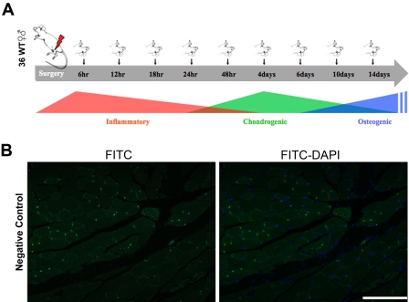
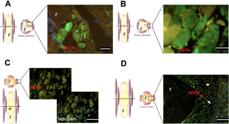
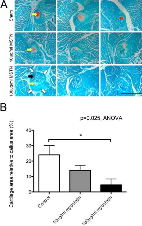
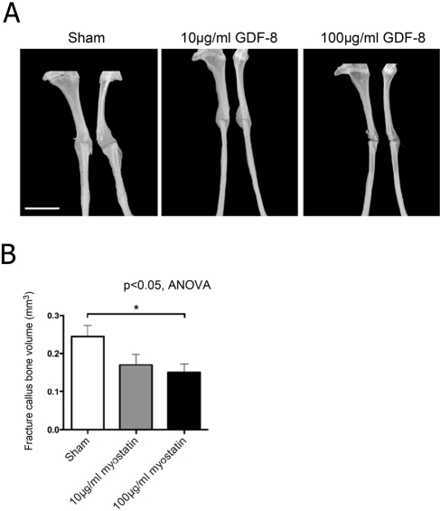
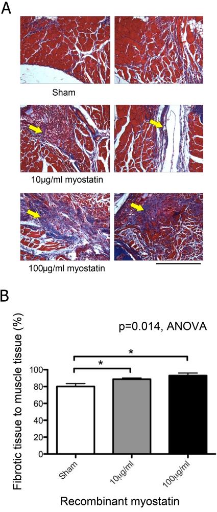
The time course and cellular localization of myostatin expression following musculoskeletal injury are not well understood; therefore, the authors evaluated the temporal and spatial localization of myostatin during muscle and bone repair following deep penetrant injury in a mouse model. They then used hydrogel delivery of exogenous myostatin in the same injury model to determine the effects of myostatin exposure on muscle and bone healing. Results showed that a "pool" of intense myostatin staining was observed among injured skeletal muscle fibers 12-24 hr postsurgery and that myostatin was also expressed in the soft callus chondrocytes 4 days following osteotomy. Hydrogel delivery of 10 or 100 µg/ml recombinant myostatin decreased fracture callus cartilage area relative to total callus area in a dose-dependent manner by 41% and 80% (p<0.05), respectively, compared to vehicle treatment. Myostatin treatment also decreased fracture callus total bone volume by 30.6% and 38.8% (p<0.05), with the higher dose of recombinant myostatin yielding the greatest decrease in callus bone volume. Finally, exogenous myostatin treatment caused a significant dose-dependent increase in fibrous tissue formation in skeletal muscle. Together, these findings suggest that early pharmacological inhibition of myostatin is likely to improve the regenerative potential of both muscle and bone following deep penetrant musculoskeletal injury.
© The Author(s) 2012
Conflict of interest statement
The authors declared no potential conflicts of interest with respect to the authorship and publication of this article.
Figures
Similar articles
-
Recombinant myostatin (GDF-8) propeptide enhances the repair and regeneration of both muscle and bone in a model of deep penetrant musculoskeletal injury.J Trauma. 2010 Sep;69(3):579-83. doi: 10.1097/TA.0b013e3181c451f4. J Trauma. 2010. PMID: 20173658 Free PMC article.
-
Myostatin (GDF-8) deficiency increases fracture callus size, Sox-5 expression, and callus bone volume.Bone. 2009 Jan;44(1):17-23. doi: 10.1016/j.bone.2008.08.126. Epub 2008 Sep 13. Bone. 2009. PMID: 18852073 Free PMC article.
-
Impairment of early fracture healing by skeletal muscle trauma is restored by FK506.BMC Musculoskelet Disord. 2017 Jun 12;18(1):253. doi: 10.1186/s12891-017-1617-y. BMC Musculoskelet Disord. 2017. PMID: 28606129 Free PMC article.
-
Myostatin (GDF-8) as a key factor linking muscle mass and bone structure.J Musculoskelet Neuronal Interact. 2010 Mar;10(1):56-63. J Musculoskelet Neuronal Interact. 2010. PMID: 20190380 Free PMC article. Review.
-
The potential role of transforming growth factor beta in fracture healing.Clin Orthop Relat Res. 1998 Oct;(355 Suppl):S294-300. doi: 10.1097/00003086-199810001-00030. Clin Orthop Relat Res. 1998. PMID: 9917649 Review.
Cited by
-
A myostatin inhibitor (propeptide-Fc) increases muscle mass and muscle fiber size in aged mice but does not increase bone density or bone strength.Exp Gerontol. 2013 Sep;48(9):898-904. doi: 10.1016/j.exger.2013.06.004. Epub 2013 Jul 4. Exp Gerontol. 2013. PMID: 23832079 Free PMC article.
-
Impact of Genetic and Pharmacologic Inhibition of Myostatin in a Murine Model of Osteogenesis Imperfecta.J Bone Miner Res. 2021 Apr;36(4):739-756. doi: 10.1002/jbmr.4223. Epub 2020 Dec 18. J Bone Miner Res. 2021. PMID: 33249643 Free PMC article.
-
Myostatin deficiency partially rescues the bone phenotype of osteogenesis imperfecta model mice.Osteoporos Int. 2016 Jan;27(1):161-70. doi: 10.1007/s00198-015-3226-7. Epub 2015 Jul 16. Osteoporos Int. 2016. PMID: 26179666 Free PMC article.
-
The skeletal muscle secretome: an emerging player in muscle-bone crosstalk.Bonekey Rep. 2012 Apr 11;1:60. doi: 10.1038/bonekey.2012.60. eCollection 2012. Bonekey Rep. 2012. PMID: 23951457 Free PMC article.
-
Smad3 induces atrogin-1, inhibits mTOR and protein synthesis, and promotes muscle atrophy in vivo.Mol Endocrinol. 2013 Nov;27(11):1946-57. doi: 10.1210/me.2013-1194. Epub 2013 Sep 3. Mol Endocrinol. 2013. PMID: 24002653 Free PMC article.
References
-
- Axelrad TW, Kakar S, Einhorn TA. 2007. New technologies for the enhancement of skeletal repair. Injury. 38 Suppl 1:S49–S62 - PubMed
-
- Bialek P, Parkington J, Warner L, St Andre M, Jian L, Gavin D, Wallace C, Zhang J, Yan G, Root A, et al. 2008. Mice treated with a myostatin/GDF-8 decoy receptor, ActRIIB-Fc, exhibit a tremendous increase in bone mass. Bone. 42:S46
-
- Borgstein CC, Yang Y, Wilson D, Haltom E, Lachey J, Seehra J, Sherman M. 2009. Preliminary results from single subcutaneous administration of ACE-031, a form of the soluble activin typeII B receptor, in healthy postmenopausal volunteers. Neuromuscul Disord. 19:546
Publication types
MeSH terms
Substances
Grants and funding
LinkOut - more resources
Full Text Sources
Miscellaneous
